Healthy body, natural food, clean environment
In the anterior case, the internal carotid artery consists of the anterior, posterior and basal arteries. Narrowing of the lumen and decreased blood flow along the V4 segment of the vertebral artery. Good afternoon, Masha.”What does the absence of blood flow in both posterior communicating arteries mean?
The trifurcation is divided into anterior and posterior. Clefting of the internal carotid artery is not a disease, but is considered a variant of the normal structure of the circle of Willis. The frequency of this pathology is 14-25 percent of all cases.
In most cases, patients seek brain examination for completely different diseases, and trifurcation of the internal carotid artery is an accidental diagnostic “finding”. In rare cases, a complication of trifurcation is the appearance of a vascular aneurysm.
Trifurcation all headaches
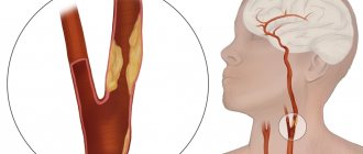
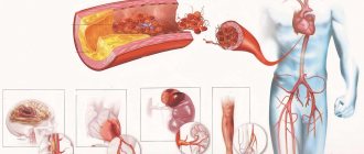
Arteries supplying blood to the brain: subclavian, vertebral, brachiocephalic trunk; common, external and internal carotid are called brachiocephalic arteries. Pathological deformation of the BCA of the vertebral carotid arteries is a congenital or acquired change in the configuration of the brachiocephalic arteries of the vessels supplying the brain, leading to disruption of the properties of blood flow and the development of acute cerebrovascular accident or chronic cerebral circulatory failure. It is easier to imagine the mechanism of blood flow disturbance when a garden hose is compressed, twisted or bent - acceleration of fluid flow and loss of laminar properties leads to loss of kinetic energy of the blood and insufficient blood supply to the brain. Another mechanism is the narrowing of the lumen at the point where the artery bends - septal stenosis, which, under certain conditions, is an analogue of stenosis of the lumen of the vessel in atherosclerosis.
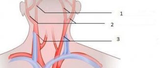
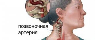
Treatment and diagnostic center of the International Institute of Biological Systems, St. Petersburg. Brachiocephalic trunk - right subclavian artery gives off right vertebral artery - right common carotid artery Left common carotid artery Left subclavian artery gives off left vertebral artery. V1 segment - up to the entrance to the opening of the transverse process of the C7 vertebra V2 segment - cervical part of the vertebral artery V3 segment - section of the vertebral artery at the level of the C1-C2 vertebra, where the artery turns and forms an arch V4 segment - intracranial segment. Extracranial segments carotid bulb and cervical region Intracranial segments Intraosseous petrosal Infraclinoid cavernous Supraclinoid.
Trifurcation of the right internal carotid artery, what it is and how a normal phenomenon occurs becomes known after a series of diagnostic procedures. This process is a splitting of the carotid artery into three parts. Division can occur in both the left artery and the right. Experts distinguish anterior and posterior trifurcation. This pathological process cannot be called a disease. Trifurcation is a structural feature of the Circle of Willis.
The vascular system of the brain has a complex structure. One of its most important components is the Circle of Willis. It is a complex of arteries that are located at the base of the brain.
Tortuosity of blood vessels in the spine, neck and brain: causes, symptoms, treatment
I can cautiously assume that if it is possible to surgically correct the defect in the vertebral artery, then perhaps vision can be restored. Option for the development of the Circle of Willis. Please tell me what this is and how to deal with it.
Option for the development of the circle of Willis, incomplete posterior left-sided trifurcation. Good afternoon, Polina. Cerebral circulation disorders and structural changes in brain tissue are very serious. Good afternoon, Tatyana. This means that blood does not flow through the indicated artery, which means that the part of the brain supplied by it does not receive nutrition in the volume in which it should receive.
However, due to this division, blood delivery to the brain may deteriorate, and aneurysms may develop in some branches due to unequal flow through the arteries. This variant of the structure of the Circle of Willis is the norm for the first half of intrauterine development.
This is a vascular formation located at the base of the brain. Serves as a compensatory mechanism for cerebrovascular insufficiency. Blood flows into this circle from other pools. Normally, the vessels of the circle form a closed system.
When there is an outpouring of blood into the subarachnoid space, patients complain of unbearable headaches, accompanied by vomiting, nausea, tension in the neck muscles and photophobia. But these cases are rather the exception to the rule; no neurological symptoms are detected and special treatment is not required.
The vessels of this arterial unit are created to compensate for circulatory processes during thrombosis or disruption of normal blood flow. It involves ligating the vessels at the site of the aneurysm. My wife is 48 years old. About 2-3 months ago, headaches began in the middle of the head, pressure on this area of the head and dizziness, pressure jumps.
Please help me with diagnosis and treatment. Good afternoon, Igor Viktorovich. With all my sympathy, I cannot, and do not have the right, to make prescriptions over the Internet, based on a description of the disease. No doctor has the right to do this, and if someone made such prescriptions for you, they could lead to anything.
Have you been struggling with HYPERTENSION for many years without success?
Head of the Institute: “You will be amazed at how easy it is to cure hypertension by taking it every day.
Patients suffering from high blood pressure and neurocirculatory disorders sometimes do not suspect that the cause of their illness lies in the pathological tortuosity of the carotid or vertebral arteries. This anatomical feature increases the risk of ischemic stroke by 30% due to impaired blood flow in these central blood vessels. For the same reason, transient cerebrovascular accidents may also develop.
As a rule, the occurrence of tortuosity of the carotid and vertebral arteries is a hereditary factor, when elastic fibers predominate over collagen fibers in the tissue of blood vessels. As a result, the walls of large vessels wear out, they become thinner and deformed. An additional risk factor is atherosclerosis - when atherosclerotic plaques are deposited on the walls, the lumen of the vessel decreases, which also causes impaired blood flow.
In most cases, arterial tortuosity can be asymptomatic, but gradually the patient may experience transient cerebrovascular accidents, which in some cases causes the development of a micro-stroke if the cause cannot be detected in time. In 20% of cases in adults, a preventive examination reveals tortuosity of the vessels in the neck - the carotid arteries.
Our readers successfully use ReCardio to treat hypertension. Seeing how popular this product is, we decided to bring it to your attention. Read more here...
The carotid arteries are formed in the chest cavity: the left carotid artery begins in the aortic arch, and the right CA (carotid artery) begins in the brachial trunk, then they divide into the external and internal arteries. There is pathological tortuosity of the internal carotid artery or tortuosity of both ICAs (internal carotid artery). The following manifestations of pathological tortuosity of the ICA and CCA (common carotid artery) are most often observed.
Normal development of the circle of Willis occurs in no more than 50% of people. The most common pathology of this arterial system is various types of hypoplasia of the connecting arteries. Aneurysms of the cerebral arteries also most often affect the vessels of the circle of Willis.
With hypoplasia of the circle of Willis vessels, symptoms may be absent, naturally, provided there is normal blood flow in the basins of other cerebral arteries. In this case, the pathology is detected as an accidental diagnostic finding during magnetic resonance imaging.
As shown by the results of a recent study conducted by scientists from the University of Pennsylvania, in some people, asymmetric development of the circle of Willis may be the cause of frequent migraine attacks that occur with a fairly pronounced aura.
With an aneurysm of the circle of Willis vessels, there are usually no symptoms until it ruptures. In the event of a rupture, blood from the damaged vessel begins to flow into the subarachnoid space. Patients begin to complain of a terrible headache, which is often accompanied by nausea, vomiting, intolerance to bright light, and stiff neck. With significant hemorrhage, coma quickly develops or the patient dies almost immediately.
Many people, having learned that they have a variant of the development of the circle of Willis that does not correspond to the norm, fall into despair, believing that they have a serious disease that requires certain therapy. But in reality, treatment for the circle of Willis is not carried out.
As we said above, there are different options for the development of the Circle of Willis, and those that are considered the norm are not found in every person. The vessels of this arterial basin are designed not so much to supply blood to brain cells, but to compensate for the disturbances in cerebral blood flow that arise as a result of thrombosis due to the transfer of blood from one arterial basin to another. Therefore, in most cases, the developmental pathology of the circle of Willis does not require treatment.
If there is an aneurysm of one of the arteries of the circle of Willis, the treatment is surgical and consists of ligating the aneurysm. In cases where the aneurysm is opened, conservative treatment is carried out, the same as for subarachnoid hemorrhage caused by any other cause.
MR angiography in the study of cerebral arteries
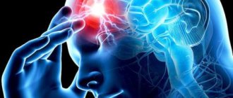
Headache, noise and dizziness, memory impairment, increased fatigue, decreased performance - such symptoms occur not only in the elderly, but also in middle-aged and even young people.
Often, patients, and some medical professionals, do not take such complaints very seriously.
Meanwhile, they may indicate chronic cerebral circulatory failure or be the first harbinger of serious and dangerous diseases.
For what reasons can dizziness occur? Natalya Vladimirovna Umerenkova, leading neurologist at Clinic Expert Kursk LLC, tells
That is why such complaints require a full examination.
Why is a patient experiencing a headache prescribed an MRI of the cerebral vessels?
Evgeniy Valerievich Yavorsky, a radiologist at MRI Expert Yelets LLC, tells the story.
What is MR angiography of cerebral vessels?
MR angiography is one of the most reliable methods of modern diagnostics, allowing clear visualization of vascular structures without requiring any invasive manipulations.
You can find out the cost of MRI examination of blood vessels and sign up for the study here
What does MR angiography show?
Let us consider the main vascular pathologies detected by MR angiography:
I. Vascular malformations of the brain
What is it and what are they?
1. Arteriovenous malformations (AVMs) are the most common symptomatic vascular malformation.
Typical AVMs are represented by 3 main components: afferent arteries (afferent vessels), a tangle (knot) of altered vascular structures and draining veins (efferent vessels).
The AVM node consists of a confluence of arteries and veins without an intervening capillary network. The adjacent brain tissue is usually atrophic, with gliotic changes due to the “steal” phenomenon.
How to understand what is written in the conclusion of an MRI study of the head and blood vessels? Radiologists from the federal network of diagnostic
What symptoms are characteristic of arteriovenous malformations?
Clinically, arteriovenous malformations are manifested by intracranial hemorrhages, headaches, seizures, and focal neurological symptoms.
In the figure - a) T2-tra. A large AVM is detected in the left temporo-parietal-occipital region (arrows); b) arrows show the left middle and posterior cerebral arteries and the node of arteriovenous malformation, clearly widened along their length
2. Carotid-cavernous anastomosis. What is this? This is an anastomosis between the infraclinoid (cavernous segment) of the internal carotid artery and the cavernous sinus. Has a spontaneous or post-traumatic nature.
The pathogenesis is associated with intensive discharge of blood from the internal carotid artery (ICA) into the cavernous sinus, increased pressure in the latter, dilation of the ophthalmic vein on the affected side of varying severity; the formation of pulsating exophthalmos.
What symptoms are characteristic of carotid-cavernous anastomosis? Typical clinical picture: pulsating exophthalmos, pain in the eyeball, headache, inflammation of the conjunctiva and injection of scleral vessels. As for the prognosis for the carotid-cavernous anastomosis, in the absence of treatment, progression of symptoms is observed.
The figure shows the carotid-cavernous anastomosis on the left. The arrow shows the level of the anastomosis between the left internal carotid artery and the left cavernous sinus with their expansion and the presence of high-speed/high-intensity blood flow. The open arrow indicates the dilated and tortuous left ophthalmic vein, also with intense blood flow in it
3. Cavernous angiomas (cavernoma, cavernous malformation). What it is? This is a cluster of dilated sinusoidal spaces lined with endothelium, with no areas of preservation of normal brain parenchyma in their structure. There are both single and multiple formations.
In the figure - multiple brain cavernomas T2 (left) and T1 (right) images in the axial plane show focal formations of a heterogeneous structure with a rim of hemosiderin along the periphery (in T2-WI), without perifocal infiltration and mass effect
What symptoms are characteristic of cavernous angiomas? Clinically, cavernous angiomas are manifested by seizures, headaches, and focal neurological symptoms; stem symptoms with appropriate localization. Bleeding occurs very rarely. A combination of cavernous malformation and impaired development of cerebral veins is possible.
4. Capillary telangiectasia. What is this? Capillary telangiectasia is characterized by alternating areas of normal brain parenchyma and areas with dilated capillaries. The most common locations are the pons and cerebellum.
What are the symptoms of capillary telangiectasia? Clinically, it is asymptomatic; must be differentiated from other focal changes.
The picture shows capillary telangiectasia. A “brush-like” contour of a small focal formation in the dorsal parts of the pons of the brain is determined (a, diagram); b) a small focus of heterogeneous, iso-hypointense MR signal is visualized in the pons, with the presence of areas of normal brain parenchyma in its projection (arrow)
The last two types of malformations (cavernous angiomas and capillary telangiectasia) are more reliably visualized during a standard brain examination, because due to minimal blood flow in these cases, MR angiography is less informative.
You can sign up for a head MRI in your city here
II. Cerebral aneurysms
What is this? An arterial aneurysm is a limited protrusion of the arterial wall in the form of a diverticulum or sac (saccular aneurysm) or a uniform expansion of the artery over a specific area (fusiform aneurysm).
Why are cerebral aneurysms dangerous? Cerebral aneurysms are the main cause of non-traumatic intracranial hemorrhage.
Why do aneurysms occur? Typically, aneurysms are formed as a result of hemodynamic damage to the vessel wall against the background of atherosclerosis and hypertension.
Traumatic and post-infectious factors in the formation of aneurysms are also possible.
A sudden increase in blood pressure and hereditary pathologies of connective and muscle tissue can also be etiological factors in the occurrence of cerebral aneurysms.
Brain aneurysm is a statistically quite rare disease: it is diagnosed in only 5% of the inhabitants of our planet. The young artist Nadezhda Rusheva died from this disease. Read more
The figure shows a thrombosed aneurysm of the anterior communicating artery (arrow), complicated by hemorrhage.
In the figure, a thrombosed aneurysm of the main artery is identified (arrow). MR angiography reveals the absence of visualized blood flow in the basilar artery; the blood supply to the posterior cerebral arteries occurs due to the circle of Willis
What symptoms are characteristic of cerebral aneurysms? Clinically, aneurysms are manifested by headache, vegetative symptoms, and when they rupture, symptoms of subarachnoid and/or intracerebral hemorrhage occur. With large sizes, focal neurological symptoms are possible.
What are aneurysms?
A fusiform aneurysm is a dilated, tortuous and elongated vessel (dolichoectasia). The neck of the aneurysm is absent.
Etiologically, this type of aneurysm in most cases is formed against the background of atherosclerotic lesions of the cerebral vessels. Much less often - against the background of infectious diseases, collagenosis, immunosuppressive conditions.
The most characteristic localizations of fusiform aneurysms are: vertebrobasilar region; P1 segment of the posterior cerebral artery and supraclinoid segment of the internal carotid artery (ICA).
As for the symptoms of fusiform aneurysm, it is usually clinically manifested by a transient ischemic attack or stroke, and less often by subarachnoid hemorrhage.
What is a transient ischemic attack and how is it different from a stroke? The neurologist at Clinic Expert Tula LLC, Izabella Ivanovna Kapralova, tells
The figure shows an MRI scan of a 73-year-old woman with atherosclerosis of the intracranial arteries. Fusiform expansion of the A1 segment of the right anterior cerebral artery is determined
III. Atherosclerosis of intracranial arteries
What is this? Atherosclerosis of intracranial arteries is a multifactorial systemic pathological process, the predisposing factors of which are hypercholesterolemia and hyperlipidemia.
In this case, degenerative changes occur in the vascular wall, the formation of calcifying plaques, intimal ulceration and displacement of the blood clot, as a result of which the diameter of the vessel progressively decreases.
This is followed by the spread of thromboemboli along the vascular bed, poststenotic dilatation and the formation of fusiform aneurysms.
In the figure, atherosclerotic lesions of intracranial vessels are determined, a decrease in the intensity of blood flow and narrowing of the lumen in the terminal parts of the anterior cerebral artery (ACA), middle cerebral artery (MCA) and posterior cerebral artery (PCA)
In most cases, it is atherosclerotic lesions that cause stenosis of intracranial arteries. But there are a number of congenital and acquired diseases that can lead to stenosis of the lumen of the vessel and the development of NMC (neurocutaneous syndromes, aplasia and hypoplasia of the arteries, Moya-Moya disease, systemic connective tissue diseases and vasculitis).
IV. Vasculitis
What is vasculitis? This is inflammation of blood vessels.
The classification of vasculitis depends on where the inflammation occurs and on the caliber of the inflamed vessels (large or smaller).
The clinical manifestations of vasculitis are varied. This can be either damage to individual organs or the entire body (muscle pain, skin manifestations, etc.)
In the figure - a) primary arteritis of the central nervous system MR angiography, “raw” data.
There is a diffuse narrowing of the lumen of the intracranial arteries, a decrease in the intensity of blood flow through them, in particular the supraclinoid segments of the ICA and posterior cerebral arteries (arrows); b) T1-cor post-contrast image with intravenous contrast shows marked gyral enhancement of ischemic cerebrovascular accidents (ICA) in this patient
Primary angiitis of the central nervous system is usually a rapidly progressive disease, with the formation of numerous areas of cerebral infarction. As a rule, changes in the brain substance of supratentorial localization are characteristic.
V. Stenoses, pathological deformations and anomalies of the main arteries
What do they look like on MRI scans?
In the figure, the arrows show stenosis of the left middle cerebral artery with preserved blood flow distal to the site of stenosis (as opposed to occlusion)
In the figure, a C-shaped tortuosity of the prepetrosal segment of the left internal carotid artery is determined; S-shaped tortuosity and hypoplasia of the left vertebral artery; variant of connection of the vertebral arteries - non-fusion
The picture shows kinking (curvature at an acute angle) of both internal carotid arteries
The figure shows coiling (pathological looping) at the border of the cervical and prepetrosal segments of the right internal carotid artery
VI. Variants of the structure of the arteries of the brain
Let's look at tomograms.
The figure shows a variant of the connection of the vertebral arteries (islet division of the proximal parts of the basilar artery (BA))
The figure shows a complete posterior trifurcation of the left internal carotid artery: the P1 segment of the left posterior cerebral artery (PCA) is not visualized and the blood supply to the P2 and more distal parts of the left posterior cerebral artery (PCA) is carried out due to the dilated left posterior carotid artery (PCA) (from the system left internal carotid artery (ICA), arrows)
The figure shows complete anterior trifurcation of the right internal carotid artery (ICA). Blood flow along the A1 segment of the left right cerebral artery (RCA) is not determined; blood supply to A2 and more distal parts of the left right cerebral artery (RCA) is carried out from the system of the right superior carotid artery (ICA) through the anterior communicating artery
Related material:
Is a brain cyst always a dangerous diagnosis?
Dos and Don'ts for MRI (about contraindications for the study)
Why do you need contrast in MRI diagnostics?
Source: https://www.mrtexpert.ru/articles/233
Complete posterior trifurcation of the left internal carotid artery
Also, don’t forget to thank your doctors.
neurologist4 22:37
migraine (sometimes with a significantly pronounced aura)
With an aneurysm, symptoms occur only when it ruptures. When there is an outpouring of blood into the subarachnoid space, patients complain of unbearable headaches, accompanied by vomiting, nausea, tension in the neck muscles and photophobia.
You should not panic and attribute to yourself an incurable disease when staging the trifurcation of the ICA (internal carotid artery); this variant of the structure of the Circle of Villi does not require special treatment.
The vessels of this arterial unit are created to compensate for circulatory processes during thrombosis or disruption of normal blood flow. If there is a complication—an aneurysm—the only treatment is surgical. It involves ligating the vessels at the site of the aneurysm.
When opening it, conservative treatment is required, which is accepted for any hemorrhages in the brain.
Remember: trifurcation of the internal carotid artery is not a disease, but is considered only a variant of the normal structure.
All therapeutic measures should be aimed at establishing a daily routine, changing the eating style and allocating time for personal needs and rest.
The basis of drug therapy is
neuromultimetabolic and vascular treatment. (simpler - help the brain live in extreme conditions)
Head of the Institute: “You will be amazed at how easy it is to cure hypertension by taking it every day...
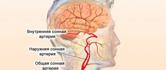
The carotid artery (lat. arteria carotis communis) is one of the most important vessels feeding the structures of the head. It ultimately gives rise to the cerebral arteries that make up the circle of Willis. It feeds brain tissue.
The place where the carotid artery is located in the neck is the anterolateral surface of the neck, directly under or around the sternocleidomastoid muscle. It is noteworthy that the left common carotid (carotid) artery branches immediately from the aortic arch, while the right one comes from another large vessel - the brachiocephalic trunk, emerging from the aorta.
Location of the common carotid artery
The area of the carotid arteries is one of the main reflexogenic zones. At the site of bifurcation there is the carotid sinus, a tangle of nerve fibers with a large number of receptors. When pressure is applied to it, the heart rate slows down, and with a sharp blow, cardiac arrest may occur.
Note. Sometimes, to stop tachyarrhythmias, cardiologists apply pressure to the approximate location of the carotid sinus. This makes the rhythm become less frequent.
Carotid sinus and topography of nerves relative to the carotid arteries
Bifurcation of the carotid artery, i.e. its anatomical division into external and internal can be topographically located:
- at the level of the upper edge of the laryngeal thyroid cartilage (“classic” version);
- at the level of the upper edge of the hyoid bone, just below and in front of the angle of the lower jaw;
- at the level of the rounded corner of the lower jaw.
We previously wrote about blockage of the coronary artery and recommended bookmarking this article.
Important. This is not a complete list of possible places for bifurcation of a. carotis communis. The location of the bifurcation can be very unusual - for example, under the mandibular bone. Or there may be no bifurcation at all, when the internal and external carotid arteries immediately depart from the aorta.
Diagram of the carotid artery. “Classical” version of bifurcation
The internal carotid artery supplies the brain, the external carotid artery supplies the remaining structures of the head and anterior surface of the neck (periorbital region, masticatory muscles, pharynx, temporal region).
Options for the branches of the arteries supplying the organs of the neck from the external carotid artery
The branches of the external carotid artery are represented by:
- the maxillary artery (from 9 to 16 arteries depart from it, including the palatine descending, infraorbital, alveolar arteries, middle meningeal, etc.);
- superficial temporal artery (supplies blood to the skin and muscles of the temporal region);
- pharyngeal ascending artery (from the name it is clear which organ it supplies with blood).
Also explore the topic of vertebral artery syndrome in addition to the current article.
External carotid artery - diagram
Trifurcation of the left internal carotid artery is a normal variation that can occur in two types: anterior and posterior. In the anterior type, the internal carotid artery gives rise to the anterior and posterior cerebral arteries, as well as the basilar artery. In the posterior type, the anterior, middle and posterior cerebral arteries emerge from the internal carotid artery.
Important. People with this type of vascular development have a high risk of developing an aneurysm, because blood flow is unevenly distributed throughout the arteries. It is known for sure that about 50% of the blood is “poured” into the anterior cerebral artery from the internal carotid.
Branching of the internal carotid artery - anterior and lateral
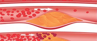
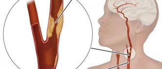
Atherosclerosis
The essence of the process is the formation of plaques from “harmful” lipids deposited in the vessels.
Inflammation occurs in the inner wall of the artery, which attracts various mediator substances, including those that enhance platelet aggregation.
The result is double damage: both the narrowing of the vessel by atherosclerotic deposits growing from inside the wall, and the formation of a blood clot in the lumen by aggregating platelets.
Atherosclerotic plaque of the carotid artery and its removal
Important. The external carotid artery is rarely severely affected by atherosclerosis. Basically, and unfortunately, this is the lot of the internal.
It is also known as hemispheric syndrome. Occlusion (critical narrowing) occurs due to atherosclerotic lesions of the carotid artery. It is an episodic, often sudden disorder that includes the triad:
- Temporary sharp and rapid loss of vision in 1 eye (on the affected side).
- Transient ischemic attacks with clear clinical manifestations.
- The consequence of the second point is ischemic cerebral infarction.
Internal carotid artery occlusion syndrome
Important. Depending on the size and location, plaques in the carotid artery can produce different clinical symptoms. Their treatment often comes down to surgical removal followed by suturing of the vessel.
Congenital stenosis
Fortunately, in ¾ of such cases, the artery with this pathology is narrowed by no more than 50%. For comparison, clinical manifestations occur if the degree of vessel narrowing is 75% or more. Such a defect is discovered accidentally on a Doppler study or during an MRI with contrast.
Stenosis on angiogram
Aneurysms
This is a sac-like protrusion in the wall of the vessel with its gradual thinning. There are both congenital (due to a defect in the tissue of the vascular wall) and atherosclerotic. A rupture is extremely dangerous due to the lightning-fast loss of a huge amount of blood.
C-shaped bend at the base of the artery. Asymmetry of the diameter of the anterior cerebral arteries. COMMON CAROTID ARTERY - uneven course =, arched, d = 8 mm each, 1.1 mm each. Plaques are present, localization is bifurcation, shape is local, structure is heterogeneous. CONCLUSION - Atherosclerosis of the magnetic arteries. How normal the patency of arteries can be in the presence of plaques in them is a question.
Trifurcation of the right ICA
In recent years, the issue of studying the neuroanatomy of the prostate has become relevant. This is due to the improvement of surgical treatment techniques... Intervertebral disc [IVD; lat. The corpus callosum is the largest commissural structure, containing about axons in its composition. Its main function…. Before reading the article, I recommend that you familiarize yourself with the structure of the intervertebral disc [read]. The spinal column of the PS, like all departments...
Pain syndrome of the greater trochanter LES - defined as pain and tenderness on palpation in the area of the greater trochanter of the femur,...
Receptors in... Anonymous comments are disabled in this journal. Your IP address will be recorded. Log in No account? Create an account. Remember me. Facebook Twitter Google. Previous Share Flag Next. The circle of Willis is an arterial circle of the brain, located at the base of the brain, and provides compensation for insufficient blood supply due to flows from other vascular areas. The following arteries participate in the formation of the Circle of Willis - see numbering according to the figure.
According to scientists, such variations affect its full function, which plays a large role in regulating blood flow in the brain. Among them, the most dangerous are the anterior or posterior trifurcation of the internal carotid artery, which affect the distribution of blood in the brain and are acceptable until occlusive changes occur, requiring good collateral circulation. According to most scientists, variants of the non-classical structure of the circle of Willis can cause aneurysms, both in its anterior and posterior sections, for example, look at the photograph below:.
In posterior trifurcation, the anterior, middle and posterior cerebral arteries depart from the internal carotid artery, the latter departing through the large posterior communicating artery. The proximal segment of the posterior cerebral artery adjacent to the basilar artery P1, as a rule, is hypoplastic, however, in rare cases, the diameter of P1 is equal to the diameter of the enlarged posterior communicating artery Figure 1. In the first case, blood enters the posterior cerebral artery mainly from the internal carotid arteries, and in the second - from both sources: the internal carotid and basilar arteries, which is confirmed in the works of individual authors.
The posterior trifurcation of both internal carotid arteries, in which there are large posterior communicating arteries, is normally detected in the first half of the intrauterine period, which is of great functional importance.
In the postnatal period of ontogenesis, thinning of the posterior communicating arteries occurs, while the remaining vessels of the circle of Willis increase in size. Probably, a delay in the process of reduction of the posterior communicating arteries in the embryonic period leads to the preservation of the posterior trifurcation of the internal carotid artery in an adult, which explains the high frequency of its occurrence.
According to some authors, an increase in the diameter of the posterior communicating artery in some cases can probably occur with cerebrovascular pathology, as a result of the adaptation of blood vessels to new conditions.
In second place in terms of occurrence is aplasia of one or both posterior communicating arteries. More often, unilateral aplasia of the posterior communicating artery is observed (Figure 2) than bilateral aplasia (Figure 3). The frequent absence of the posterior communicating arteries can be explained by a stop in development, which is consistent with literature data. The absence of the posterior communicating arteries is observed 6 times more often than the anterior communicating artery.
With aplasia of the anterior and posterior communicating arteries, there is a complete separation of the carotid artery systems from each other or a separation of the blood circulation of the anterior and posterior parts of the brain, which is most unfavorable in terms of collateral supply when compensating for hemodynamic disorders.
Other variants of the non-classical structure of the arterial circle of the cerebrum are found much less frequently. These include: the median artery of the corpus callosum (Figure 4), single-stem type of the anterior cerebral artery (Figure 5), parietal contact of the anterior cerebral arteries (Figure 6), anterior trifurcation of the internal carotid artery (Figure 7), splitting of the anterior communicating artery, the presence of several anterior communicating arteries, absence of the anterior communicating artery .
The following variants of the non-classical structure of the circle of Willis are extremely rarely observed: recurrent artery Heubner's artery, plexus type of the anterior cerebral artery, doubling of the posterior communicating artery, plexus type of the basilar artery and trifurcation of the basilar artery. Pazhinsky, I. Gaivoronsky, A. Gaivoronsky, K. Lyunkova, V. Krylov, Department of Neurosurgery and Neuroreanimation, Moscow State Medical and Dental University.
Chaplygina, O. Kaplunova, V. Dombrovsky, O. Sukhanova, I. Blinov, L. Rostov-on-Don, Russia Journal of Anatomy and Histopathology. Ivanov, Educational and Scientific Institute of Nanostructures and Biosystems, Saratov State University Izv. Computer Science, vol. Mordovian State University named after. Kutya, V. Kiselev, V. Pikalyuk, G. Moroz, M. Tags: anatomy, circle of Willis, brain. Buy promo for minimal price. Post a new comment Error Anonymous comments are disabled in this journal.
We will log you in after post We will log you in after post We will log you in after post We will log you in after post We will log you in after post Anonymously. Post a new comment. Preview comment. Post a new comment 0 comments.
The role and structure of the Circle of Willis
All major arteries of the brain converge. This is necessary to create a constant, unchanging blood flow.
The circle of Willis works, as they say, with a reserve, because it also has a second important task.
Including the central nervous system in the development of critical situations: stroke or transient ischemic attack.
Factors such as the speed of recovery, the depth of neurological deficit, the likelihood of death and the area of damage to nerve tissue depend on the quality, shape, and anatomical structure of this radial formation.
Also, the circle of Willis is able to compensate for eating disorders during chronic cerebral ischemia.
The change is achieved through the redistribution of hemodynamics or the formation of an external network of vessels that will cover the brain’s need for the deficit.
These are the so-called collaterals. In some cases they cannot form quickly enough. Against the background of connective tissue pathologies, diabetes mellitus. Then the risk of fatal bleeding or other complications increases.
Thus, the circle of Willis works as a compensator, a regulator of brain trophism, acts as a protector of normal nutrition, and prevents death from a critical hemodynamic disorder.
As already mentioned, we can only talk about the norm conditionally, because the classic version of development occurs infrequently, in 25% of cases or a little more. The authors do not agree; there are certain discrepancies.
In all possible cases, developmental anomalies of the circle of Willis are congenital. The shape and type are established in the early stages of pregnancy and gestation.
Changes are not always caused by the behavior of the mother or the influence of negative environmental factors: radiation, toxins, metal salts, infections and other things.
In the absence of any disturbances in the quality of trophism or the speed of blood movement, they speak of a variant of the physiological norm. Such situations require neither treatment nor specialized care.
But sometimes it would be a good idea to visit a physician or cardiologist for a preventive examination (once a year or more often if desired).
Normally, the structure of the circle of Willis is represented by a group of structures: anterior, posterior, connecting arteries, which ensure the movement of blood through tissues from the carotid and vertebral arteries.
In the system they form a ring, the movement of liquid connective tissue is cyclical, the volumes are greater than what is required under normal conditions.
It is precisely because of this that it is possible to compensate for violations if necessary.
Abnormal development options
It is possible to talk about pathology or some kind of “abnormality” with a large degree of convention. Research strongly supports the same conclusion, although not always directly.
It is necessary to take into account the well-being and rate of trophism in the brain of a particular patient. The development of the circle of Willis most often follows the type of absence of part of the collaterals; these are relatively mild variants of deviation.
But hypoplasia and underdevelopment of the main vessels can play a sad role.
What forms of anatomical structure exist:
- Lack of connecting artery in front. It acts as a bridge connecting the cerebral vessels. Due to this structure, blood moves more rapidly.
The absence, however, almost never gives any symptoms, because the body has the ability to compensate for the conditioned disorder on its own.
This anomaly is relatively rare, accounting for no more than 2.5% of the total number of cases.
- Absence of one of the posterior communicating arteries. Left or right PSA. The structures provide communication with larger vessels, a type that is relatively common. Found in 13% of clinical situations.
Slightly more often the left communicating artery is absent. Why this is so is not known exactly.
- There is a mixed case where both the anterior and one posterior communicating artery are missing. This is an extremely rare disorder. Occurs in 1% of cases.
Accompanied by dyscirculatory changes, under certain conditions.
The nutrition of the brain is not intensive enough, therefore any disorder: be it atherosclerosis or arterial spasm, is accompanied by functional changes and a transient neurological deficit.
And during the course of chronic disorders, there are also gradual structural changes that create constant disruptions in the functioning of the central nervous system. This form of anomaly needs to be treated.
- An extremely common option is trifurcation: the structures of the posterior cerebral artery arise immediately from the carotid artery. The prevalence is almost 20-25% of the total mass.
There are many variants of manifestation: dyscirculatory encephalopathy, stroke, migraine, and others.
There may be no symptoms if there is sufficient compensation of nutrition through neighboring vessels, by branching the original circulatory network.
In any case, such patients need to be carefully monitored so as not to miss the moment of transition from a shaky norm to pathology.
- Another mixed version of the open circle of Willis is simultaneous trifurcation in the absence of the anterior communicating artery (jumper). Found in 2-3% of situations.
It gives approximately the same symptoms, but the blood supply to the brain is worse and the risks of decompensation are almost twice as high.
Recovery involves taking medications to speed up cerebral blood flow and prevent stroke. Surgical intervention is possible.
- Absence of both PCA (posterior communicating arteries) and ACA (anterior communicating artery) simultaneously. An open circle of Willis develops when the ring does not come out of the anatomical structures.
Collaterals provide compensation when it is necessary to redistribute trophism. In this case, this option is impossible: the carotid and vertebral vessels are not connected and exist in isolation, which creates huge risks at the slightest change in the quality and quantity of incoming blood.
- The form is not much different, in which there is an anterior one, but both posterior communicating arteries are absent. The condition is approximately identical, there is no big difference.
- The anterior trifurcation leads to the departure of both collaterals of this location from the same carotid artery. Which shouldn't happen. Hence the increased risks of negative phenomena if the functioning of this particular vessel is disrupted. Immediately two connecting structures cease to perform their functions, and asymmetry of blood flow develops.
- Hypoplasia. Occurs relatively often. Presented as a decrease in blood flow through one or both connecting arteries. Localization is not of great importance in terms of understanding the essence - the artery is too small in diameter, underdeveloped, and therefore is not capable of performing the functions assigned to it.
This situation is especially dangerous when the main branches that are considered fundamental are affected: the carotid and vertebral arteries.
Based on the location, we can talk about many different symptoms: from headaches, migraine attacks to stroke without preliminary manifestations. Spontaneous acute disturbance of cerebral blood flow.
- Aplasia. Complete absence of any part of the Circle of Willis. Occurs in 5-12% of situations. Accompanied by critical disturbances with minimal changes in the quality of trophism.
This development option is extremely unstable and urgent treatment is required immediately after the problem is identified.
- There are also types with duplication of individual collaterals: duplications. As a rule, both vessels involved in the formation of the anomaly are equally underdeveloped, which, however, does not interfere with providing normal nutrition.
This is only a part of the possible types. They are the most common. All others are derivatives of those named.
If the circle of Willis is closed, the compensatory function for the most part remains at an adequate level, therefore, even in the case of the development of an acute malnutrition of nerve tissues, transient ischemic attack, atherosclerosis, and other diseases, there is a good chance of rapid recovery.
MR signs of hypoplasia of the right vertebral artery
In most cases, patients seek brain examination for completely different diseases, and trifurcation of the internal carotid artery is an accidental diagnostic “finding”. Magnetic resonance imaging is used for detection. For a clearer vascular picture, contrast angiography with CT is used.
In rare cases, a complication of trifurcation is the appearance of a vascular aneurysm. But these cases are rather the exception to the rule; no neurological symptoms are detected and special treatment is not required. Concomitant diseases such as atherosclerosis also complicate the course.
Clinical signs of abnormalities in the branching of vessels in the circle of Willis occur when blood flow through collaterals becomes insufficient for various reasons. For example, fatty plaques have formed in the arteries, a blood clot has appeared or migrated from the left side of the heart, or an aneurysm has ruptured. A healthy person does not feel non-classical branching of blood vessels, since his brain does not experience the need for bypass blood flow paths.
development of stroke/disorders associated with insufficient blood supply to the brain area
Symptoms of obstructed blood flow can vary widely. If we are not talking about a stroke, then patients complain of dizziness, decreased intellectual abilities, memory, and attention. Psychological problems are also common - often abnormal branching of blood vessels is accompanied by neuroses, panic attacks, and emotional lability of its owners.
A characteristic manifestation of the non-classical development of the Circle of Willis is considered. Many observations have been devoted to the issue of the relationship between the structure of the cerebral arteries and migraine, which indicate that the majority of patients with migraine have certain anomalies. Especially often with migraine, deviations in the structure of the posterior part of the arterial system are diagnosed.
A decrease in blood flow through the vessels of the arterial ring of the brain can provoke periodic headaches and disorders such as apathy or irritability, decreased performance, fatigue, etc. Typically, such a conclusion can be found in the results of MR angiography and it indicates hypoplasia of certain vessels .
In case of aplasia of the arterial trunks, when some vessels are absent at all, the absence of blood flow is recorded during the study. For example, aplasia of the posterior communicating arteries will be accompanied by a lack of blood flow through them, respectively. Such aplasia can also be asymptomatic, but only when a sufficient amount of blood passes through the main arteries. With atherosclerosis or spasm of the arteries, signs of insufficient blood supply to the brain will not be long in coming.
% distribution of aneurysm cases among cerebral arteries
If, against the background of an abnormal structure of the arteries at the base of the brain, an acute circulatory disorder occurs, then the clinic will have obvious symptoms of a stroke - paresis and paralysis, speech disorders, pathological reflexes, impaired consciousness up to coma.
Separately, it is worth mentioning the expansion of cerebral blood vessels. According to statistics, it is in the arteries of the Circle of Willis that the greatest number of them are found. An aneurysm of the arteries in this area is fraught with rupture and massive subarachnoid hemorrhage with clinical stroke, coma and severe neurological manifestations.
Aneurysm is an independent pathology, and not a variant of individual branching of vessels, but it much more often accompanies non-classical types of the circle of Willis.
The development of ultrasound Dopplerography and magnetic resonance imaging techniques has made it possible to make the study of the nature of the structure of the Circle of Willis a publicly accessible and safe event. The main methods for diagnosing variants of the vascular system of the brain include:
- X-ray contrast is one of the most informative methods, but has contraindications related to the need for contrast (liver, kidney pathology, allergies to contrast, etc.);
- Transcranial - the procedure is safe, affordable, requires devices with a Doppler sensor, which are available in many medical institutions;
- MR angiography - performed on a magnetic tomograph, has contraindications, a significant drawback is its high cost.
Circle of Willis on a diagnostic image
Selective angiography of the cerebral vessels is an invasive procedure in which a catheter is inserted into the femoral artery and advanced to the area of interest in the cerebral arteries. When the desired area is reached, a contrast agent is applied. The method is most often used during surgical treatment (stenting, angioplasty).
Instead of selective angiography, CT angiography can be used, when a contrast agent is injected intravenously, and then pictures of the head are taken in different projections and sections. Subsequently, a three-dimensional image of the vascular structures of the brain can be recreated.
Transcranial Dopplerography makes it possible to determine the nature of blood flow in the vessels of the brain (reduced, absent), but it does not provide enough data regarding the anatomical structure of the arteries. Its important advantage is considered to be the almost complete absence of contraindications and low cost.
MR angiography is one of the most expensive, but at the same time quite informative methods for diagnosing the structure of the circle of Willis. It is performed in a magnetic tomograph and the contraindications for it are the same as for conventional MRI (high degree of obesity, claustrophobia, the presence in the body of metal implants that conduct a magnetic field).
The MRI picture shows the structure of the vessels of the circle of Willis, the presence or absence of connections between them, aplasia or hypoplasia of the arteries. When assessing the result, a specialist can determine the diameter of each artery and the characteristics of its branching.
Diagnostics
The examination is carried out under the supervision of a vascular surgery specialist. It is possible to involve a neurologist.
The measures are mostly standard; it is necessary to carefully evaluate the anatomical structure of the circle of Willis.
- Angiography. Essentially an x-ray with contrast. A special drug is introduced, which, accumulating in the blood, enhances the pattern. Then doctors take a series of photographs and, based on the results, evaluate the nature of the structure of the ring-shaped structure. It is considered an accessible and relatively simple diagnostic method with a minimum of contraindications.
- Dopplerography of cerebral vessels. Required to assess the speed and quality of blood flow in cerebral structures, used as part of a functional examination. Prescribed in all cases.
- MRI. To identify complex forms of anomaly. This is perhaps the most informative technique among others. Maximum safe, does not create a load on the body, and is not accompanied by pain. But the research costs on average more than its analogues. That’s why they resort to this method when others have failed.
- It is possible to prescribe invasive, selective angiography.
Activities can be adjusted during the examination and supplemented with other techniques. The issue is resolved at the discretion of the specialist.