What is carotid artery stenosis
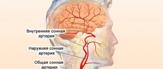
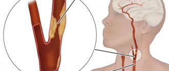
The brain is supplied with blood by paired vertebral and carotid arteries, the largest volume passing through the latter. The common carotid arteries (CCA), after branching from the aorta, run through the tissues along the anterolateral surface of the neck on both sides. There they are divided into internal and external branches. They supply oxygenated and nutrient-rich blood to the soft tissues of the face, eyes, thyroid gland and brain. Deterioration of blood flow due to vascular stenosis entails disruptions in the functioning of these organs and systems, the most dangerous of which is considered to be cerebrovascular accident.
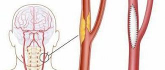
The process usually develops in both arteries, but most often the most pronounced stenosis is on one side. Despite the fact that blockage of blood vessels can form in any area, in most cases this process is observed in places of anatomical narrowing, where division into branches or at the mouth occurs. Stenosis of the carotid arteries develops as a result of the deposition of cholesterol plaques on the vascular walls or the actual narrowing of the lumen of the vessel. Pathology may appear against the background of spasm as a result of taking certain medications, diseases of the endocrine glands, autoimmune diseases, long-term smoking, abuse of alcoholic beverages, and drugs. Stenosis is most often diagnosed in men over 60 years of age.
Important! Late diagnosis and untimely treatment can lead to memory, vision, and brain disorders. In severe cases, death is possible.
Reasons for narrowing
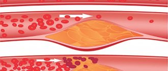
The main reason for the narrowing of the lumen of the carotid arteries is considered to be atherosclerotic lesions, expressed in the appearance of cholesterol plaques on their inner surface.
It is plaques formed in the area of the bifurcation of the carotid artery, or the mouth (division into external and internal branches), that most often become the cause of stenosis.
Other reasons include:
- Collagenosis caused by the proliferation of connective tissue.
- Inflammation of the vascular wall (arteritis) or its dissection.
- Congenital anomaly of the SA structure, the presence of pathological valves (pathology can manifest itself in both an adult and a child).
- Development of fibromuscular tissue dysplasia.
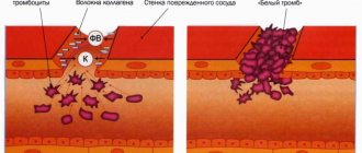
- Diseases accompanied by increased blood clotting.
The result of the above processes is a change in the direction of blood flow, increasing the risk of blood clots, which become harbingers of acute cerebrovascular accident.
The likelihood of negative symptoms increases if provoking factors are present:
- Diabetes.
- Violation of fat metabolism.
- Arterial hypertension.
- Injury to the vessels of the cervical spine.
- Elderly age.
- Long-term use of oral contraceptives.
- Smoking.
- Insufficient physical activity or physical inactivity.
Increased attention to one’s own health should also be present in the case of a hereditary predisposition to vascular pathology.
Classification
You can assess how far the process has progressed with carotid artery stenosis by the severity of vasoconstriction. Pathology is classified as follows:
- 1st degree. The lumen is narrowed within 30-50%, blood supply is carried out through the collateral vascular network. The disease is asymptomatic, there are no signs of cerebral ischemia. Carotid artery stenosis may be detected during diagnostic testing.
- 2nd degree. Vessel narrowing is 50-70%. There are signs of periodic disturbances of cerebral circulation, as a result of which transient ischemic attacks develop. Attacks of focal neurological deficit are stopped within 24 hours.
- 3rd degree. The narrowing of the lumen of the vessel reaches 70-80%. The hemodynamic process becomes more significant, and the risk of cerebrovascular accident increases significantly. This degree is characterized by severe neurological symptoms and a chronic course of cerebrovascular insufficiency.
- 4th degree. Vascular obstruction reaches 80% or higher, stenosis becomes critical. Most often, a stroke is diagnosed with signs of focal neurological symptoms persisting for more than 24 hours.
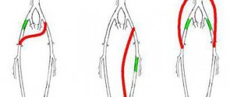
Carotid artery stenosis is also classified by location. There are:
- Stenosis of the internal carotid artery (ICA). Most often located in the brain area. It can be right- or left-handed. It is characterized by nonspecific symptoms, difficult access, difficult diagnosis and a low probability of a successful outcome.
- Stenosis of the common carotid artery (CAA). Narrowing is observed in the outer tissues of the vessel on the right or left side of the neck.
Symptoms of pathology
Symptoms of carotid artery stenosis are absent in the initial stages, when the narrowing is just beginning. As it progresses, due to chronic oxygen deficiency, neurological general and focal signs of circulatory disorders in the brain appear. The first group of symptoms include:
- Deterioration in sleep quality. Complaints appear at the very beginning of the disease, progressing slowly but steadily. Disturbing sleep that is shallow or with frequent awakenings. Sleeping pills help in the short term.
- Headache. Has a paroxysmal character. The intensity is medium, lasting up to a couple of hours. Pain is noted in the back of the head or temples, sometimes covering the entire skull, often spreading to the face, neck and eyes.
- Dizziness. Resulting in the inability to stand on one's feet and loss of orientation in space. It appears in the form of attacks and does not last long. Sometimes the dizziness becomes constant, but not severe.
- Lability of the emotional background. Euphoria can instantly give way to irritability and a fit of anger. In some cases, apathy, tearfulness, reluctance to perform any actions, and a depressive state appear.
- Inhibition in the sphere of intellectual activity. It becomes impossible to quickly answer a question or solve a problem. At the same time, cognitive and mnestic functions remain at the same level.
- Inadequate perception and poor memory of information.
Important! If such complaints occur, it is recommended to conduct an examination. This will help to identify signs of narrowing of the carotid arteries and take timely measures to eliminate the pathology.
The appearance of focal symptoms indicates the development of a transient ischemic attack. Pathology can be suspected if the following symptoms are present:
- Visual impairment in one or both eyes. Narrowing or loss of the field of view, altered perception of colors, darkening of the eyes, blurred contours, flickering spots or dots.
- Unsteadiness when walking. Appears when coordination is impaired, dizziness, or severe muscle weakness.
- Fainting state. Regular but quickly passing attacks of loss of consciousness indicate negative dynamics in the development of stenosis and a high risk of developing a cerebral stroke.
- Slurred speech. In addition to the fact that the patient cannot clearly pronounce words, he poorly understands speech addressed to him.
- Asthenia. They are worried about weakness, apathy, drowsiness, total fatigue, and the inability to take care of themselves in everyday life.
- Vomiting or nausea. The symptom appears and disappears suddenly, without any intervention. Classical treatment methods do not bring relief.
- Difficulty swallowing. Dysphagia indicates damage to the brain stem, which may result in cardiac and respiratory arrest and a critical increase in body temperature. This often leads to death.
The duration of the attacks does not exceed an hour; the patient’s condition returns to normal within 24 hours. Even a single transient ischemic attack is a good reason to visit a doctor, since such a condition is considered a harbinger of a cerebral stroke.
If measures to eliminate carotid artery stenosis have not been taken, the patient will experience the following signs of chronic cerebral ischemia:
- weakening of memory;
- decreased concentration;
- violation of tolerance to mental stress;
- change in character;
- inability to perform normal activities.
Important! When the blood flow is completely blocked, an ischemic stroke develops, the first signs of which include loss of consciousness, paralysis or paresis, impaired sensitivity, speech, and swallowing.
Symptoms and stages of development
The clinical picture of carotid artery blockage is determined by the severity of pathological changes. There are 4 stages of occlusion:
- The initial stage does not have typical manifestations. When examining the patient, the following are detected: stenosis of the ICA, impaired blood flow through the ophthalmic artery, and mild neurological disorders.
- In the second stage, occlusion begins to manifest itself. Muscle paresis occurs, lasting several minutes. Transient ischemic attacks are present, accompanied by dizziness, headache and stupor. The patient's gait is impaired, visual acuity and hearing are reduced. At this stage, blood clots appear in the brain vessels, which increases the risk of ischemic stroke.
- At the third stage, most of the lumen of the vessel is blocked. Fainting often occurs. Against the background of a severe course of the disease, an apoplexy stroke develops. The patient's intellectual abilities decrease, memory and speech deteriorate. The development of muscle weakness up to paralysis is observed.
- The fourth stage is characterized by the presence of severe neurological disorders, due to which the patient loses the ability to self-care.
Important information: How to treat dyscirculatory encephalopathy of mixed (vascular) origin
The following procedures are used for diagnostics in neurology:
- Doppler ultrasound of the vessels of the cervical region and head (reverse blood flow in the external carotid artery is detected);
- angiography (allows you to determine the degree of blockage of the lumen of the vessel);
- magnetic resonance angiography (in combination with MRI of the brain is an informative method for diagnosing ICA occlusion);
- CT scan of the brain (aimed at identifying signs of stroke; the presence of white ischemia indicates the atherosclerotic origin of the pathology);
- blood test for cholesterol levels.
Diagnosis of the disease
If carotid artery stenosis is suspected, a thorough examination is performed, and the following signs of pathology may be detected:
- uncharacteristic vascular murmur above the site of branching into branches (bifurcation zone);
- pulsation in the temporal and carotid arteries of different strength and symmetry;
- decreased pressure in the central retinal artery on the side of the stenosis.
In addition, the doctor prescribes an ECG, blood and urine tests, blood biochemistry and coagulogram. The most accurate information can be obtained by performing the following diagnostic methods:
- ultrasound examination of the carotid arteries, supplemented by Doppler ultrasound (USDG);
- Magnetic resonance imaging;
- CT scan;
- angiography with contrast.
The examination reveals the structure of the vascular walls, features of blood flow, localization, extent and degree of stenosis.
Treatment of pathology
Treatment tactics for carotid artery stenosis are selected depending on the size of the lumen in the arteries. In cases where the stenosis is minor and normal blood flow is maintained, the patient is advised to adhere to a healthy lifestyle, eliminate bad habits, eat right, and establish a work and rest schedule. If necessary, the following groups of drugs are prescribed:
| Group | Action | Drugs |
| Hypotensive | Stabilization of blood pressure | Lisinopril, Indapamide, Lozap, Losartan |
| Statins | Cholesterol correction | Atorvastatin, Rosuvastatin, Roxera |
| Anticoagulants | Dissolving existing blood clots and preventing their occurrence | Warfarin, Heparin, Marevan |
| Antiplatelet agents | Reduced blood viscosity | Clopidogrel, Aspirin Cardio, Acecardol |
| Nootropics | Improves cerebral circulation, protection against hypoxia | Mildronate, B vitamins, Piracetam |
Important! At the same time, treatment of diseases that can lead to the development of carotid artery stenosis is carried out.
If the vessel lumen is narrowed by more than 50%, there is a history of transient ischemic attacks, or signs of cerebral disorders appear, surgical treatment is recommended to prevent the development of cerebral ischemia and stroke. Blood flow is restored in the following ways:
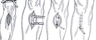
- Carotid endarterectomy. Open access is provided to the site of localization of stenosis, where the removal of an atherosclerotic plaque or other formation that impedes normal blood flow is carried out. Surgery is necessary for patients after TIA or stroke when the lumen of the vessel is narrowed by more than 50%. It is also indicated for asymptomatic cases when stenosis reaches over 60%.
- Artery stenting. The minimally invasive intervention is aimed at widening the vessel using a mesh stent tube. The operation is performed using an angiographic unit. The function of the inserted device is to maintain the artery in a straightened state. The operation is performed in cases where carotid endarterectomy is contraindicated, repeated stenosis has formed, or the arteries are narrowed as a result of radiation therapy for malignant neoplasms of organs and tissues of the neck.
- Bypass surgery or extra-intracranial microarterial anastomosis (EICMA). During surgery, additional blood flow is formed, bypassing the blocked vessel. Indicated for occlusion (100% stenosis) of the carotid arteries. The patient's vein or artery is used as a shunt.
- Arterial prosthetics. In cases where the area of vascular damage is significant, part of the wall is replaced with a special prosthesis to restore normal blood supply.
Neurologists and vascular surgeons treat patients suffering from narrowing of the carotid arteries.
Postoperative period
The patient spends the first day after surgery in the anesthesiology and intensive care unit or in the intensive care ward. For the next three days, you must remain in bed.
On the fourth day, you are allowed to slowly get out of bed and take short walks on level ground under the supervision of a doctor.
Within two weeks the following are excluded:
- physical exercise;
- bends;
- squats;
- any other sudden movements;
- consumption of alcohol and alcohol-containing drinks;
- smoking.
Since stenting is performed using a radiopaque contrast agent, in order to remove it from the body as quickly as possible, it is recommended to increase the amount of fluid you drink.
When the postoperative period ends, the patient goes home.
During the year, he must attend a consultation and preventive examination of a specialist at least twice.
Daily measurement of blood pressure is recommended, the rise of which can cause dangerous and adverse reactions.
It is mandatory to take prescribed medications.
Turns in the cervical spine are also done with caution. It is recommended to take showers rather than baths.
In order to prevent the formation of new atherosclerotic plaques, you should adhere to a rational hypocholesterol diet.
Its main parts are:
- minimum salt content;
- Take 5-6 times a dose at regular intervals in small portions;
- baked or steamed dishes.
Prognosis for stenosis
The sooner the disease is diagnosed and treatment is started, the more optimistic the prognosis. Until persistent organic damage appears, complete recovery is possible. In the absence of treatment, an increase in symptoms, and the appearance of attacks of transient ischemic attacks, the prognosis becomes less and less optimistic. The outcome of a stroke will depend on the timeliness of diagnosis, the speed of specialized medical care and the degree of brain damage.
Important! Carotid artery stenosis is a progressive disease, so re-formation of areas of vascular narrowing is possible. Relapse can occur even after surgery.
Possible complications after stenting
A serious complication of such surgery is embolism. Distal antiembolic protection helps prevent stroke. In addition, postoperative aggravation of the patient's condition can be caused by the formation of a blood clot along the stent. Pathological processes such as restenosis (re-clogging of the artery) also occur. In addition, there may be toxic effects of contrast on the kidneys. Such problems are most often observed in patients with pathologies of these organs.
Many patients are looking for how to cure stenosis using alternative medicine. In cases where plaques are diagnosed in the carotid artery, treatment with folk remedies can improve blood circulation and slightly expand the lumen of the arteries. But such treatment should be carried out only after consultation with a doctor.
Possible complications and prevention of the disease
As the pathology progresses, dementia and disruption of the central nervous system may develop. The most serious complication is ischemic or hemorrhagic stroke. As a result, the patient may become disabled or die.
To slow down the development of pathology or prevent recurrent lesions, it is recommended to give up bad habits, lead a healthy lifestyle and be active. It is equally important to adhere to the principles of proper nutrition; fatty, fried, and spicy foods should be excluded from the diet. It is also necessary regularly:
- visit your doctor;
- control cholesterol and blood sugar levels;
- maintain normal blood pressure;
- undergo ultrasound scanning to monitor the dynamics of the process.
Carotid artery stenosis is a dangerous and unpredictable disease. Pathology can lead to serious health problems, including death. The absence of specific symptoms makes diagnosis difficult, so it is necessary to pay attention to even minor signs of brain dysfunction. Early diagnosis and timely treatment will help avoid complications.