Causes of blood clot formation
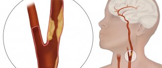
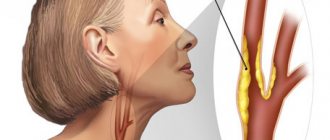
Most often, the triggering factor for the process is atherosclerosis. This artery is one of the large vessels most susceptible to the accumulation of cholesterol and calcium formations, often leading to severe narrowing of the artery lumen.
At the same time, blood flow is hampered, which leads to a significant deterioration in the blood supply to brain tissue. Plaques deform and injure the walls of blood vessels, creating conditions for blood cells to settle in the thickness of the damaged walls.
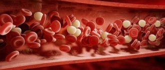
A blood clot often occurs when clumps of platelets (a type of blood cell responsible for clotting) adhere to lipid deposits.
Such formations are relatively inert, but they interfere with normal blood flow and can completely block the lumen. The symptoms of the condition greatly depend on where the blood clot has formed.
If a blood clot grows quickly, it can cause a cerebral infarction, or ischemic stroke. If the process develops slowly, you can reduce the risk of developing life-threatening conditions if you consult a doctor in a timely manner.
Factors that increase the likelihood of thrombosis are:
- heart disease, in particular ischemia, atrial fibrillation;
- heart valve defects;
- vasculitis, including Horton's syndrome;
- malignant or benign tumors;
- circulatory disorders, tendency to blood stagnation;
- alcoholism;
- diabetes mellitus (it is well known that patients with this disease have increased blood density, which reduces the speed of blood circulation);
- traumatic brain injuries;
- the patient is overweight;
- stenosis of certain vessels (especially if the lumen between the walls narrows by half or more);
- vascular spasms due to stress;
- a history of blood clots, especially in the vessels of the heart, pulmonary and subclavian arteries;
- individual anatomical features of the artery;
- problems with blood clotting, as well as (in combination with other factors) taking drugs that increase clotting.
In most cases, pathology is detected in elderly men. But in some cases, thrombosis can be detected in young people, which is often associated with head injuries or hematomas of the vessel walls.
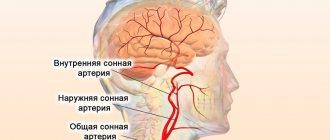
Depending on which part of the artery is affected by the disease, thrombosis of the internal carotid artery and the external one are distinguished.
Root Causes
Why does such a disease appear? Thrombosis of the internal carotid artery can be divided into types based on the area of localization of the blood clot. The main reasons for the appearance are listed below:
- Formation of a blood clot in the subclavian vessel.
- Pulmonary vein thromboembolism.
- Thrombosis in the veins of the heart.
- Acute atherosclerosis.
- Vascular stenosis.
- Age above sixty years.
- Having diabetes of any type.
- Active smoking.
- Predisposition to blood clots.
The appearance of a blood clot in a vein without any reason is extremely rare, in about three cases out of a hundred. According to statistics, thrombosis occurs due to atherosclerosis in 90 percent of cases. We should not exclude a situation where a blood clot in the carotid artery, which appeared in a vein, breaks off and quickly reaches the vessels in the head.
Symptoms
Symptoms of the pathology are determined by which part of the vessel the blood clot has formed.
Stand out:
- external artery - responsible for feeding structures external to the skull, including the dura mater of the brain, as well as tissues of the head and face;
- internal - it is she who primarily provides nutrition to the brain tissue (both cortical and subcortical), as well as the pituitary gland and visual organs.
When a blood clot blocks an internal artery, the patient experiences the following symptoms:
- severe, often migraine-like headaches;
- convulsions, paresis of the limbs on the side opposite to the one where the thrombus occurred (that is, if the left artery is damaged, dysfunction of the right limbs is observed);
- loss of consciousness;
- visual impairment, sometimes blindness or atrophic phenomena in the optic nerve;
- nausea and vomiting.
When a blood clot has formed in the external artery, the following phenomena are observed:
- weakness of the masticatory muscles;
- speech disorders;
- migraine attacks;
- feeling of dry mouth;
- pain in the neck area.
It should be noted that the intensity of the increase in symptoms depends on the rate at which the blood clot increases in size.
Sometimes it happens that for several days the condition is almost asymptomatic (slight dizziness or a feeling of numbness in the hands may appear), after which the symptoms sharply worsen, and obvious signs of impaired blood supply to the brain appear.
In this case, if you do not call a doctor, death may occur within a few hours. In most cases, however, symptoms develop smoothly over several days (up to two weeks).
It often happens that the patient notices a noticeable deterioration in his condition in the morning, after waking up.
How does thrombosis manifest?
There are external and internal carotid arteries. Symptoms of thrombosis of the general channel and the internal branch may differ, since parts of the carotid artery provide blood supply to different organs and parts of the brain.
With thrombosis of the common carotid artery, the patient may experience:
- fainting when rising from a horizontal position;
- short-term loss of consciousness without obvious reasons;
- attacks of severe headache;
- pain in the neck;
- weakened chewing muscles;
- visual impairment (nerve or retinal atrophy, cataracts, periodic temporary blindness);
- tinnitus at high or low frequency.
If thrombosis of the internal carotid artery, which supplies parts of the brain, occurs, the symptoms will be as follows:
- severe paroxysmal headache, sometimes combined with vomiting;
- speech disorder (aphasia, impaired ability to count, write);
- convulsions, paresis, paralysis of limbs;
- violation of orientation in space;
- unilateral visual impairment (blurred image, loss of peripheral field);
- pain when lightly tapping the skull on the affected side;
- unusual mental manifestations (irritability, euphoria, memory impairment, hallucinations or delusions).
If you try to feel the pulse in the patient’s neck, then with thrombosis its weakening or complete absence will be noticeable. Even at home, one can assume a diagnosis based on the following signs: blurred vision, absence of pulsation of the carotid artery on one side of the neck, the occurrence of fainting and convulsions when pressing on a vessel on the other side of the neck.
These manifestations should be a reason to immediately call an ambulance.
Depending on the speed of development of the disease state, the following forms are distinguished:
- Asymptomatic. Characterized by a mild cerebrovascular accident with minor signs of dizziness, numbness of the extremities, etc.
- Chronic. With this form, symptoms increase gradually over 1-2 weeks.
- Subacute. The thrombus closes the lumen of the artery and can then be moved (blood circulation is partially restored). Symptoms appear and develop within 8-48 hours.
- Spicy. It appears suddenly and develops very quickly. With this course of thrombosis, the probability of death is high.
It is advisable to seek help in any of these cases as soon as possible. It is unknown how exactly the symptoms of the disease will develop.
Possible complications and consequences
The main danger of this condition for human life is a significant deterioration in blood circulation in the brain, which leads to disruption of its functioning and is fraught with the development of ischemic stroke.
Other possible consequences are determined by where the embolus is located.
Since the internal arteries (left and right) are responsible for the blood supply directly to the brain tissue, a blood clot formed in these vessels is fraught with the following consequences:
- incoordination of hand movements;
- convulsions;
- complete or incomplete loss of speech;
- fainting conditions;
- visual impairment;
- paralysis of the muscles of the face, or, more often, of the left or right half.
When the external carotid artery is blocked, the following phenomena are possible:
- migraine-type headaches;
- paralysis or paresis of facial muscles;
- fainting;
- speech disorders of varying severity.
One way or another, if the patient has the indicated symptoms, he should immediately call an ambulance.
The sooner treatment measures are carried out, the greater the patient’s chances of survival and successful rehabilitation with a subsequent return to normal life.
Features of the disease and ICD classification
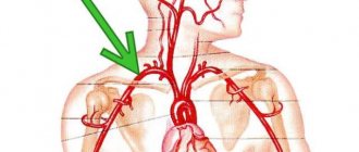
The jugular venous system simultaneously includes several cervical vessels, which are necessary for the outflow of blood from the head and neck.
If we consider in detail the anatomical structure of a person, we can distinguish 3 main pairs of jugular veins in this system:
- internal;
- external;
- front
The first of the vessels is the largest and allows most of the blood to exit the skull. The vein connects with the subclavian artery. The external vein is significantly smaller in size and is located in the front of the neck. It is clearly released during singing, screaming or coughing. The last vessel is considered the smallest of the above. These are the saphenous veins, which create the so-called jugular venous arch.
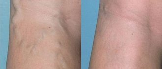
Thrombosis can occur in any of the above veins, but statistics show that most often it develops in the external jugular vein. Over time, this process leads to mechanical damage to the vessel wall, and the body begins to intensively produce fibrin and platelets. As a result of this process, a blood clot occurs. Some chronic diseases can contribute to the appearance of blood clots even without vascular damage.
This disease is a pathological condition caused by blockage of the vessel cavity with a blood clot, which causes blood flow disturbances and, as a result, some organs and systems suffer. The lack of adequate therapy can cause the development of a number of rather dangerous complications, so it is extremely important to stop not only its manifestations, but also the cause itself.
It is impossible to unambiguously answer the question about the age category of patients suffering from this disease. Both young people and elderly people are susceptible to it, but in the latter the disease is somewhat more common. Doctors also note that gender may be a risk factor. Thus, among patients who come to medical institutions with a similar problem, the majority are men.
According to the international classification of diseases, created to systematize the statistical information obtained, this pathology has ICD 10 code I82.
Diagnostics
To diagnose thrombosis, hardware tests are needed in a clinical setting.
Direct or indirect signs of a blood clot can be detected using the following methods:
- magnetic resonance angiography – reveals circulatory disorders and narrowing of arterial walls;
- computed tomography – visualizes cervical and cerebral vessels;
- duplex scanning of blood vessels - determines the location of blood clots and the speed of blood flow in these places.
Detection methods
Since a thrombus in the jugular vein is a serious pathology, it is advisable to diagnose it in the initial stages. True, it’s quite difficult if he doesn’t make himself known in any way. But, if you still have some suspicions, it is better not to refuse to visit the doctor.
READ MORE: How to find out if the spine is curved
In order to detect a blood clot, modern instrumental and laboratory research methods are used:
- Ultrasound of the area where the jugular vein runs.
- Duplex scanning of vessels, which will allow you to examine possible pathologies of the veins in a two-dimensional image.
- MRI – makes it possible to examine in detail centimeter by centimeter the area under study.
- Thrombodynamics is a study that allows you to identify blood clotting indicators.
- Thrombosis time tests – aimed at assessing the extrinsic clotting pathway.
- Thromboelastography. With this study, the doctor receives a complete picture of the elastic properties of blood clots.
Since thrombosis of the veins of the upper body is rarely complicated by thromboembolism, treatment is predominantly conservative. The patient does not have strict bed rest, but physical activity should be prohibited. The following treatment methods are used:
- Taking direct anticoagulants - Heparin, Fibrinolysin, Fraxiparin. In the acute stage of the disease, these drugs are administered intravenously in the hospital. The course of therapy with these drugs continues until fibrinogen disappears in the plasma and until the level of the prothrombin index normalizes. Subsequently, indirect anticoagulants are prescribed, for example, Aspirin Cardio, Cardiomagnyl.
- Taking or administering nicotinic acid to activate blood thinning and blood clot resorption.
- The use of venotonics - Detralex, Troxevasin, Aescusan, Glivenol. These drugs are needed to speed up metabolism in the walls of the veins, relieve inflammation and relieve pain.
- Introduction of antispasmodics to relax the muscle wall - No-Shpa, Papaverine.
- Local application of heparin ointment, troxevasin ointment for additional effect on the vessel walls.
Surgery for jugular vein thrombosis is extremely rare. As a last resort, minimally invasive techniques are used - percutaneous endovascular thrombolysis, transluminal aspiration thrombectomy. These methods involve dissolving the clot. or its removal using a balloon catheter. It is imperative to influence the factors that led to the development of the disease, for which you should eliminate bad habits and undergo cancer treatment in a specialized hospital.
Treatment of carotid artery thrombosis
The treatment method for carotid artery thrombosis is chosen by the doctor after taking a medical history and conducting proper diagnostic procedures.
To restore blood supply to brain tissue and prevent the development of irreversible changes in the cortex or subcortical layers, the following drugs are prescribed:
- thrombolytics - they act directly on existing blood clots, promoting their resorption;
- anticoagulant - they help accelerate blood flow, prevent blood clotting and stagnation, and thin it.
It is especially advisable to take them for patients with diabetes.
For diseases of the gastrointestinal tract (especially those accompanied by ulcerations of the mucous membranes), as well as for certain diseases of the urinary system in the anamnesis, such medications are prescribed with caution so as not to provoke bleeding.
In some cases, the doctor decides whether surgery is necessary.
Indications for it are:
- ulcers, acute colitis and other diseases for which taking medications that reduce blood clotting is not recommended;
- aneurysm of the heart muscle;
- many diseases of the hematopoietic system, for example, anemia, leukemia;
- severe thrombophlebitis, when more than four to five hours have passed since the formation of a blood clot.
The following types of operations are practiced:
- endarterectomy - during which a small incision is made in the neck, through which the vessel is accessed. The section of the wall where there is damage is removed, and then the blood clot itself is eliminated, then the incision is sutured. Typically the procedure lasts about 100-120 minutes. It is performed under anesthesia (local or general). It is not cheap, but it allows you to completely remove the blood clot. The rehabilitation period that the patient spends in the hospital is about twenty days;
- balloon dilatation - a filter element is inserted into the artery, on which thrombotic formations and plaques are deposited, and a balloon catheter is inserted. When the catheter is inflated, the vessel expands. Finally, a stent is placed in the artery. This procedure is half as long as the previous one, but rehabilitation after it lasts about the same;
- It is also widely practiced to replace the damaged artery fragment with a prosthesis using a special shunt. Essentially, an artificial blood supply is created to replace the affected artery. However, if the lesion is extensive, the procedure may be fraught with thromboembolism of a pulmonary vessel, so in this case they resort to another type of intervention;
- another form of surgical intervention: novocaine is injected into the stellate node, which has a strong analgesic effect, and then the node is removed along with a fragment of the affected artery. The supply of blood to the brain in such a patient will occur due to the work of a paired vessel on the other side.
Balloon dilatation and neurosurgery
Another surgical method that can be performed is balloon dilatation. This operation is performed under local anesthesia. A special filter is installed in the damaged vessel to catch blood clots and plaques. A balloon catheter is inserted into the vessel, which uses pressure to move the blood clots towards the filter. The process lasts about an hour. The duration of rehabilitation after surgery depends on the complexity of the operation and the patient’s health status after it. On average, the duration is from 2 weeks to 1 month.
The most reliable type of surgical intervention is the replacement of damaged areas of blood vessels with prostheses. A bypass is created for blood flow due to the operation using an installed shunt, which is located over the damaged areas. Sometimes neurosurgical operations are used, as a result of which nodes, clogged vessels and plexuses are removed.
After surgery, blood circulation to the brain significantly improves, and a slowdown in the atherosclerotic process is observed. The most effective result after surgery is possible if there is an embolic form of vascular damage. Any treatment for carotid artery thrombosis should be carried out under the supervision of the attending physician, after passing all the necessary examinations.
Forecast
The prognosis for treatment of thrombosis depends on the following factors:
- competently prescribed therapy and the patient’s adherence to the doctor’s recommendations, lack of self-medication and ignoring symptoms;
- how quickly medical care was provided after a blood clot formed;
- the presence of concomitant diseases, including those associated with blood vessels (especially cerebral);
- patient's age;
- the degree of severity and nature of changes in brain tissue caused by deterioration of blood supply due to thrombosis (if any).
The timeliness and correctness of medical care is very important.
Drug treatment
The basis of therapy for the formation of blood clots in the circulatory system is the use of direct and indirect anticoagulant drugs. They are aimed at thinning the blood by reducing the level of fibrinogen, a substance whose concentration increases to form clots.
Direct anticoagulants include:
- Fibrinolysin,
- Fraxiparine,
- Heparin.
After a decrease in the prothrombin index, they switch to medications with an indirect anticoagulant effect, such as Curantil or Aspirin.
Another important aspect in complex therapy is the prescription of phlebotonics, whose action is aimed at improving metabolic processes and eliminating inflammation.
Remember! The dosage of any medications and the method of their use are prescribed exclusively by a competent medical professional.
If necessary, antispasmodics are prescribed to reduce the tone of the vascular walls and relieve pain. The addition of sepsis indicates the need for antibacterial therapy.
You can find out what varifort for varicose veins is and the opinion of Elena Malysheva from our article.
Prevention
In order not to encounter blockage of the main arteries, it is important to pay due attention to following the rules of a healthy lifestyle:
- Diet is of decisive importance in prevention: it is necessary to minimize the content of animal fats in the diet, avoid trans fats, include in the menu foods containing organic acids that strengthen blood vessels (there are many of them in apples, as well as blue berries and fruits), and buckwheat porridge. If the patient is overweight, he needs to lose weight;
- To prevent blood from stagnating in the vessels, regular physical activity is needed;
You should also stop smoking - it leads to vascular spasms in the upper half of the body and deterioration of blood supply to tissues.
siteboss
General practitioner with 10+ years of experience. Graduated from Bukovina State Medical University in 2008 with a degree in general medicine.
The main causes of the disease
Thrombosis in the jugular veins can begin for 3 main reasons. First of all, this is the peculiar composition of the blood. Many people's blood is a little thicker than it should be normally. This is due to special congenital pathologies or chronic diseases. Blood thickening can be caused by chemotherapy, excessive radiation, and other factors.
IJV (internal jugular vein) may begin due to damage to endothelial cells. If the vessel wall has been mechanically damaged due to surgery, trauma or infection, a blood clot may occur in the jugular vein. The cause of a blood clot may be a change in blood flow. Blood stagnation, heart disease and oncology can slow down the blood flow process, thereby triggering the formation of a blood clot.
Much less often, pathology develops after intravenous administration of narcotic drugs. There are also factors that only aggravate the problem and contribute to the faster development of blood clots. These include bad habits (smoking, alcohol), old age, frequent air travel, excess weight, hormonal imbalances, long-term wearing of a cast and taking strong hormonal contraceptives.
Atherosclerosis of the cervical vessels: symptoms and treatment
The brain contains control centers for all body systems. He orchestrates the work of every organ and every cell. Its normal functioning depends on adequate blood supply, which is provided by the arterial trunks arising from the aortic arch. With a disease such as atherosclerosis of the neck vessels, these vessels are affected by cholesterol plaques.
Today we will talk about this pathology - how it is dangerous, why it can occur and what its leading clinical symptoms are.
General description of the disease
Atherosclerosis of the main arteries of the neck is a disease of the vessels located at the level of the cervical spine.
The mechanism of cervical vascular disease is typical, as for atherosclerosis of any other location. Due to long-term and persistently elevated cholesterol in the blood, it infiltrates the walls of blood vessels. A local limited inflammatory process develops, as a result of which foam cells .
By their structure, foam cells are macrophages that cannot phagocytose excess cholesterol. Their number increases exponentially, and a sclerotic process develops in this area of the vessel wall. Stenosis of the vessel occurs, cholesterol tubercles protrude into the lumen, on which micro-thrombi, large cells and molecules get stuck. This is how atherosclerotic plaques form.
With age, a lot of trigger factors appear for the development of this pathology. Reduced elasticity of the walls of blood vessels, hypertension, underlying diseases and bad habits - all this accelerates and aggravates the atherosclerotic process. When the affected vessel is stenosis by more than half, the disease can acquire an occlusive form. Blood circulation is impaired and the risk of severe complications increases due to low perfusion of brain cells.
The most dangerous complication is ischemic stroke of the brain.
What it is?
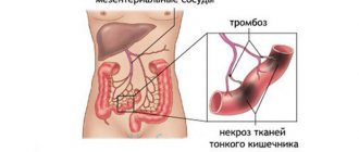
Thrombosis
is a pathological condition in which dense blood clots (thrombi) form in the vessels, slowing down or completely stopping the normal flow of blood. As a result, a lack of organ nutrition (ischemia) may occur, which in turn can lead to tissue death (necrosis, infarction) and death. There are two types of thrombosis: venous and arterial. From the names it is clear where the formation of blood clots occurs. In the first case - in the veins, in the second - in the arteries. The disease can occur in acute and chronic form. Arterial thrombosis is the most dangerous.