From the article you will learn what phleboliths are, pathological causes, clinical manifestations, features of treatment and prevention, prognosis.
general information
Phleboliths (venous stones) are pathological inclusions in venous vessels, calcified blood clots. Essentially an asymptomatic pathology. A similar finding is diagnosed by chance, when examining a patient for another disease. But sometimes blood clots saturated with calcium cause blood flow disturbances, lead to stagnation, and provoke complications and pain.
They are found in half of the cases in the pelvis, but can be located everywhere. With age, the frequency of such inclusions increases, which is explained by involutive changes in the blood coagulation system and structural degeneration of the vascular wall. Phleboliths of the pelvis are more often formed in women; in other cases there are no gender differences.
Causes
There are many root causes of phlebolithiasis; calcified blood clots in the pelvis deserve special attention. Pathology triggers are:
- Physiological or genetic features of the blood coagulation system: antiphospholipid syndrome, hereditary thrombophilias, life-threatening without timely medical intervention.
- Inflammatory changes in the pelvic organs, especially common in women of reproductive age: salpingitis, salpingoophoritis, endometritis, metroendometritis, which destroy the vascular wall and trigger thrombus formation.
- Vascular anomalies: malformations, hemangiomas, venolymphatic malformations with multiple stones.
- A sedentary lifestyle (the work of programmers, accountants, secretaries, drivers, cashiers) is an obstacle to the venous outflow of the legs and pelvic organs, which contributes to the thickness of the blood.
- Long-term varicose veins of the lower extremities and pelvis.
- Pregnancy is a trigger for hypercoagulation.
- Taking oral contraceptives, when estrogen triggers a cascade of abnormal blood clotting, thrombus formation in the lungs, brain, and liver.
- Lack of sexual activity and anorgasmia are the causes of venous stagnation, and the occurrence of calcifications is the process of development of intravascular blood clots against the background of slowing venous blood flow and damage to the endothelium.
- Thrombophlebitis and postthrombophlebitis syndrome, when mineralization of blood clots becomes the result of chronic inflammation of the venous wall, occurring under conditions of local hypercoagulation.
- Increased pressure inside the pelvic area: straining during bowel movements, severe coughing, heavy lifting, during childbirth with damage to the endothelium and the development of stagnation.
- Liver diseases with portal hypertension, stasis in the portal vein system, varicose deformation of the vessels of the stomach wall, the formation of calcified blood clots.
- Benign and malignant neoplasms.
- Vein injuries.
- Defects of the valve apparatus.
- Physical inactivity, passive lifestyle, excess weight.
- Intestinal diverticulosis.
Pathogenesis
The mechanism of calcium impregnation of blood clots is not completely clear. Phleboliths are the result of a slowdown in local blood flow, lymphatic drainage, and vascular dilatation. The onset of pathology is marginal intravascular thrombosis with endothelial defects, impaired coagulation and fibrinolysis. Then, fibrosis, mineralization as natural healing.
Passive precipitation of calcium crystals forms the core of stones; connective tissue fibers undergo secondary calcification. Each repetition of the pathological process provokes the growth of layers with the formation of phleboliths of the small pelvis or venous calculi. Phlebolithiasis is explained by a high concentration of phosphatase in the tissues around the bladder with deposition of the enzyme on the surface of the blood clot.
Phleboliths have an oval shape with the diameter of a vein and a multilayer membrane. According to the chemical composition, the stones contain calcium, magnesium, iron, which have high radiopacity. Therefore, shadows of phleboliths are the basis for x-ray diagnosis of the disease.
Classification
There is no generally accepted classification of phleboliths. Calcified thrombi are distinguished by location, number, size, and degree of contact with the endothelium. There are stones:
- Pelvic and suprapelvic: paraprostatic, perivesical in men, perivaginal in women. In other places, phleboliths are much less common.
- Fixed and free, capable of migrating into the inferior vena cava, creating a risk of embolism.
- Single and multiple.
- Small (1–5 mm), medium (5 to 10 mm), large (more than 1 cm).
- By chemical composition: radiopaque or radiotransparent.
Symptoms of pathology
Phleboliths are practically asymptomatic. Few clinical symptoms manifest differently according to the localization zone. In the pelvic area - symptoms of gynecological or urological pathologies:
- spontaneous appearance of a vascular network on the buttocks and thighs;
- pain during sexual intercourse;
- discomfort in the lower abdomen when standing for a long time;
- Irregular menstruation, heavy discharge.
More dangerous is the development of symptoms of varicose veins of the lower extremities, provoked by stones as obstacles to regional blood flow:
- heaviness in the legs, chronic fatigue syndrome;
- swelling;
- cramps of the calf muscles;
- pain syndrome;
- aesthetic discomfort in the form of swollen veins;
- prodrome syndrome with fever and malaise – rare.
But the most dangerous is infertility due to pelvic varicose veins.
Complications
Doctors consider phleboliths harmless to humans, except in cases of risk of relapse of occlusion. Varicose veins complicated by phlebolithiasis increase the likelihood of recurrent thrombosis. Migration of stones into the portal vein can become a trigger for pulmonary embolism with a fatal outcome.
Diagnostics
Since phleboliths do not have classical symptoms, their diagnosis is based on instrumental examination methods. The algorithm includes:
- History taking, physical examination.
- UAC, OAM, biochemistry.
- Analysis of plasma coagulability, prothrombin index. coagulogram.
- Fundus examination.
- Blood for hormones.
- Ultrasound scanning of veins, which makes it possible to assess the anatomical and hemodynamic features of the affected segments: phleboliths are defined as echogenic oval foci with a flickering artifact.
- Radiography reveals phleboliths in the projection of the small pelvis in the form of rounded shadows with many layers. An important diagnostic sign is the presence of radiotransparent).
- Computed tomography with contrast visualizes intravascular calcifications with multiple voids and branches.
- MRI of soft tissues visualizes the condition of nearby tissues and the degree of vascular damage (the “void flow” phenomenon).
- Biopsy with histological analysis of stones.
- Excretory urography.
- Sialography is a radiopaque or radionuclide study of the major salivary glands.
In order not to confuse phleboliths with stones of the kidneys, bladder, ovaries, prostate, you need to focus on their distinctive features:
- after filling the bladder, phleboliths do not change position;
- not associated with contrast enhancement of the kidneys or ureters;
- close to the pubis.
Features of therapy
Phleboliths in the pelvis and stones in other locations do not require active treatment. Therapeutic correction is necessary when symptoms appear. For this purpose, the following methods are used in clinical phlebology:
- Sclerotherapy is the exclusion from the general blood flow of deformed veins with branches and malformations, with which phleboliths are in direct contact, by introducing into the vein a special sclerosant substance that glues the walls of the veins with subsequent death and desolation. Classic sclerotherapy and precision sclerotherapy are used under the control of visualization tools.
- Endovascular laser coagulation as monotherapy or in combination with vein sclerosis, surgery (for multiple stones).
- Phlebectomy in the classic version or after preoperative sclerotherapy to reduce the risk of uncontrolled bleeding.
- Selective removal of phleboliths - removal of stones from open access under the control of ultrasound, microscope or palpation. The technique alleviates symptoms, but, as a rule, is not performed independently: only in combination with other methods of correction.
In essence, conservative therapy is the correction of symptoms caused by phleboliths in the pelvis or other regions. In women, the symptoms of gynecological diseases predominate, so the entire arsenal of anti-inflammatory, absorbable, antibacterial therapy is used, which is correlated with the main gynecological pathology. In men, urological problems predominate, so treatment is focused on urethritis, prostatitis of various etiologies, and tumor processes.
Conservative treatment of phleboliths themselves involves taking the following medications:
- Anticoagulants (heparin-based) or antiplatelet agents: Ticlopidine, Iloprost.
- Non-steroidal anti-inflammatory drugs (NSAIDs, NSAIDs) to relieve pain and inflammation: Nise, Ibuklin, Voltaren, Nurofen, Ketorol.
- Antispasmodics: No-Shpa, Drotaverine, Spazmalgon.
To reduce congestion in the pelvis, an individual complex of exercise therapy and the use of a contrast shower are recommended. A special diet is required, excluding “heavy” foods that cause constipation, dyspepsia and filling the daily menu with fresh vegetables and fruits. The drinking regime should also be calculated - at least 1.5 liters of water per day.
If pathological inclusions are found in the inferior vena cava, surgery to implant a vena cava filter may be required.
In addition, varicose veins caused by phleboliths require the use of compression hosiery or elastic bandages. Underwear is selected according to size together with the attending physician. These can be special stockings, socks, tights.
Compression knitwear is of high quality and can normalize pressure in the vessels of the legs and pelvic veins. When using it, blood flows to the lower parts of the body evenly and measuredly. The density of knitwear is determined individually. The main thing is the absence of discomfort, since the patient will have to wear such underwear throughout the working day, during heavy physical activity: lifting weights, sports exercises, pregnancy.
Prognosis and prevention
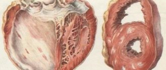
Phleboliths are a kind of evidence of the development of undetected thrombosis in the past. They are somewhat reminiscent of pulmonary calcification due to tuberculosis infection. General – the duration of the process. Many months ago, an asymptomatic thrombosis occurred in the patient’s body. Immune forces made efforts to limit the spread of the pathological process, encapsulated the thrombus with a shell of calcium, while maintaining the lumen, and therefore the patency of the vein. Therefore, such calcified blood clots are not dangerous, do not require special therapy, and have a favorable prognosis.
Prevention of pathology depends on the stage at which the phlebolitis was detected. If we are talking about the very beginning of mineralization of blood clots, the root cause of the disease or provoking factors are known, then treatment begins immediately and no trace remains of the stones. In the absence of precise knowledge of the trigger of the disease, the patient will have to undergo a long examination before prescribing adequate, that is, effective, therapy.
Patients with phleboliths need to understand that their vessels are prone to thrombosis. If blood clots are detected in the pelvis, the patient’s management tactics are thoroughly known and do not pose a threat to life. But do not forget that about 2% of mineralized blood clots are localized in the brain, for example, and this is already dangerous due to the development of a stroke. Proper nutrition is considered to prevent calcification of blood clots:
- do not eat after 18-00;
- control your weight;
- give preference to fruits and vegetables;
- exclude atherogenic foods (fatty, sweet foods);
- drink enough water;
- to refuse from bad habits;
- move more, walk;
- visit a doctor once a year (medical examination or medical examination).
The main thing is motivation, and this means maintaining the quality of life until old age, active longevity.
general information
Phleboliths (venous stones) are pathological inclusions in venous vessels, calcified blood clots. Essentially an asymptomatic pathology. A similar finding is diagnosed by chance, when examining a patient for another disease. But sometimes blood clots saturated with calcium cause blood flow disturbances, lead to stagnation, and provoke complications and pain.
They are found in half of the cases in the pelvis, but can be located everywhere. With age, the frequency of such inclusions increases, which is explained by involutive changes in the blood coagulation system and structural degeneration of the vascular wall. Phleboliths of the pelvis are more often formed in women; in other cases there are no gender differences.
Stones in veins - what are they?
Phleboliths in women and men are specific deposits in the lumens of small vessels, which are calcareous in nature. If you search for information about phleboliths in the pelvis and what it is, you may also come across names such as calcifications or venous stones.
You can feel stones that are especially large in diameter yourself - they feel like small compactions. However, a more reliable research method, of course, is an x-ray.
Manifestations of this pathology are most often observed in the following areas:
- veins of the pelvic organs in both women and men,
- vessels of the legs affected by varicose veins,
- veins of the gastrointestinal tract and spleen,
- tumors of the vascular walls.
Causes
There are many root causes of phlebolithiasis; calcified blood clots in the pelvis deserve special attention. Pathology triggers are:
- Physiological or genetic features of the blood coagulation system: antiphospholipid syndrome, hereditary thrombophilias, life-threatening without timely medical intervention.
- Inflammatory changes in the pelvic organs, especially common in women of reproductive age: salpingitis, salpingoophoritis, endometritis, metroendometritis, which destroy the vascular wall and trigger thrombus formation.
- Vascular anomalies: malformations, hemangiomas, venolymphatic malformations with multiple stones.
- A sedentary lifestyle (the work of programmers, accountants, secretaries, drivers, cashiers) is an obstacle to the venous outflow of the legs and pelvic organs, which contributes to the thickness of the blood.
- Long-term varicose veins of the lower extremities and pelvis.
- Pregnancy is a trigger for hypercoagulation.
- Taking oral contraceptives, when estrogen triggers a cascade of abnormal blood clotting, thrombus formation in the lungs, brain, and liver.
- Lack of sexual activity and anorgasmia are the causes of venous stagnation, and the occurrence of calcifications is the process of development of intravascular blood clots against the background of slowing venous blood flow and damage to the endothelium.
- Thrombophlebitis and postthrombophlebitis syndrome, when mineralization of blood clots becomes the result of chronic inflammation of the venous wall, occurring under conditions of local hypercoagulation.
- Increased pressure inside the pelvic area: straining during bowel movements, severe coughing, heavy lifting, during childbirth with damage to the endothelium and the development of stagnation.
- Liver diseases with portal hypertension, stasis in the portal vein system, varicose deformation of the vessels of the stomach wall, the formation of calcified blood clots.
- Benign and malignant neoplasms.
- Vein injuries.
- Defects of the valve apparatus.
- Physical inactivity, passive lifestyle, excess weight.
- Intestinal diverticulosis.
Phleboliths of the pelvis
After suffering venous thrombosis, various dangerous changes occur in the vessel. Phleboliths in the pelvis are calcareous neoplasms that are located on the venous wall. Most vein stones occur in women than in men. In this case, they appear as pain in the lower abdomen, disruption of the monthly cycle, and vaginal discharge. If such symptoms appear, you must go to a specialist and undergo all the necessary diagnostic tests.
What are the causes of the disease?
Most often, phleboliths are discovered by chance, during the diagnosis of other diseases.
The following are the causes of vein stones:
- Pregnancy and labor. In such conditions, in women, the location of the venous vessels and phleboliths in them changes. As a result, they accumulate and cause pain.
- Sedentary lifestyle. Physical inactivity leads to impaired blood flow from the veins of the legs and pelvic organs. Stagnation in blood vessels leads to increased blood density and the formation of clots.
- Varicose veins of the legs and pelvis. Tortuous, dilated vessels change the flow of blood, provoking the development of venous stagnation and the formation of blood clots. Blood clots can appear in the veins of the pelvic cavity affected by varicose veins or transform from the vessels of the legs.
- Genetic predisposition or diseases in which increased blood clotting is observed and there is a high risk of blood clots.
- Inflammatory processes in the pelvic organs. In diseases of the uterus, ovaries, and pelvic peritoneum, the process of destruction of the vascular wall begins and blood clotting increases.
- Oral contraceptives. When taking COCs, blood clotting increases and blood clots form both in the pelvic organs and in others: in the kidneys, eyes, lungs, liver and brain.
- Lack of sex life and orgasm. With irregular sexual relations and anorgasmia, vasospasm and venous stagnation occur, which leads to increased blood viscosity and the appearance of blood clots.
What are the symptoms of the disease?
If a patient develops multiple or single phleboliths, the following symptoms develop:
- changes in the menstrual cycle,
- frequent vaginal discharge,
- spider veins in the thigh and buttocks area,
- in a prolonged standing position, pain appears in the legs and pelvis,
- pain in the thighs and buttocks with tension in the muscles of the vagina and abdomen,
- pain during sexual intercourse,
- exacerbation of all symptoms before menstruation.
Subtleties of diagnosis
If a patient has a single phlebolitis, it is necessary to undergo a thorough examination by a specialist. The doctor will collect a medical history and examine the patient. Then the doctor will conduct a differential diagnosis with other pathologies of the pelvic organs and refer you to undergo special informative diagnostic techniques. These include:
- general blood and urine tests,
- biochemical blood test,
- plasma clotting tests,
- eye examination (fundus),
- tests for hormone levels,
- X-ray examination (shadows of phleboliths are revealed in the projection of the small pelvis),
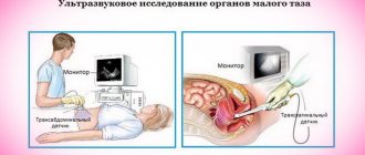
- Ultrasound,
- CT,
- MRI.
Treatment for the presence of phleboliths in the pelvis
If symptoms appear that indicate the presence of phleboliths, you should go to the doctor. Only a good specialist can conduct a thorough diagnosis and choose the right treatment. To remove all unpleasant symptoms and cure the patient, the doctor will conduct an examination, make a diagnosis, prescribe medications, diet and physical therapy. Each patient must also wear compression garments. Surgical intervention is prescribed only in cases where vein stones reach large sizes and interfere with the functioning of organs.
Drug therapy
If a patient has multiple phleboliths, he is prescribed the following drugs:
| Pharmacological groups | Medicines |
| Antiplatelet agents | "Ticlopidine" |
| "Iloprost" | |
| Antispasmodics | "No-shpa" |
| "Drotaverine" |
Physiotherapy
When stagnation appears in the veins of the pelvic organs and in the legs, as well as when phleboliths form, each patient is prescribed exercise therapy. The specialist selects a set of exercises that improves blood flow in the venous vessels and prevents the development of blood clots. At the same time, all symptoms go away in patients and their quality of life improves. This complex includes squats, the “birch” exercise and all movements that affect the group of gluteal muscles and legs. This also includes leg swings, heel-to-toe rolls, and exercises with a special gymnastic ball.
Diet food
Patients with phleboliths also need to adhere to a diet. It assumes:
- Elimination of “bad” cholesterol (fried and fatty foods).
- Avoid foods that contain processed vegetable fat.
- Limit sweet and salty foods.
- Drinking more than 2 liters of water.
- Adding a lot of vegetables, fruits and dairy products to your diet.
What's the forecast?
With timely access to specialists, a favorable outcome is observed. Also, the prognosis of the disease will be positive if the patient stops smoking and drinking alcoholic beverages, improves sleep and adheres to proper nutrition, improves sex life and begins to lead an active lifestyle. It is also recommended to abandon uncontrolled use of oral contraceptives.
Pathogenesis
The mechanism of calcium impregnation of blood clots is not completely clear. Phleboliths are the result of a slowdown in local blood flow, lymphatic drainage, and vascular dilatation. The onset of pathology is marginal intravascular thrombosis with endothelial defects, impaired coagulation and fibrinolysis. Then, fibrosis, mineralization as natural healing.
Passive precipitation of calcium crystals forms the core of stones; connective tissue fibers undergo secondary calcification. Each repetition of the pathological process provokes the growth of layers with the formation of phleboliths of the small pelvis or venous calculi. Phlebolithiasis is explained by a high concentration of phosphatase in the tissues around the bladder with deposition of the enzyme on the surface of the blood clot.
Phleboliths have an oval shape with the diameter of a vein and a multilayer membrane. According to the chemical composition, the stones contain calcium, magnesium, iron, which have high radiopacity. Therefore, shadows of phleboliths are the basis for x-ray diagnosis of the disease.
Prevention
To avoid the appearance of disturbances in the functioning of the blood flow, it is important to regularly carry out a number of preventive measures.
In particular, you should avoid wearing tight, constricting or simply uncomfortable shoes that interfere with normal blood circulation in the vessels. It is also worth giving the leg muscles and the whole body regular physical activity, which helps strengthen elasticity and improve the tone of the vascular membranes, and also prevents blood thickening, which can also lead to the formation of blood clots.
Proper nutrition is another factor that helps maintain healthy veins and prevent phlebolitis:
- avoidance of fatty foods rich in cholesterol;
- proper nutrition;
- limiting the consumption of flour and sweets, as well as avoiding processed foods and high-carbohydrate foods are the basic principles of a healthy diet.
Girls of childbearing age, as well as women and girls who have already given birth, need to pay special attention to the health of the pelvic organs. Inflammation of the appendages or complications after a difficult birth can negatively affect the condition of the cardiovascular system. With insufficient and improper treatment, such diseases can harm the entire body.
The appearance of freely moving phleboliths inside the vessels is a sure sign of the presence of the above diseases in the body, often occurring in a chronic form.
Classification
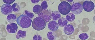
Phlebolith, large photo
There is no generally accepted classification of phleboliths. Calcified thrombi are distinguished by location, number, size, and degree of contact with the endothelium. There are stones:
- Pelvic and suprapelvic: paraprostatic, perivesical in men, perivaginal in women. In other places, phleboliths are much less common.
- Fixed and free, capable of migrating into the inferior vena cava, creating a risk of embolism.
- Single and multiple.
- Small (1–5 mm), medium (5 to 10 mm), large (more than 1 cm).
- By chemical composition: radiopaque or radiotransparent.
Types
Depending on the nature of the petrification of the blood clot, layered and homogeneous phleboliths are distinguished:
- Layered phleboliths can form when the processes of gluing the walls of the vessel and deposition on the blood clot in the vessels of salt formations occur alternately.
- In the event that these processes are observed simultaneously, we are talking about homogeneous phleboliths.
Externally, phlebolith looks like a small bead, sometimes hollow inside. The size of the phlebolith ranges from seven to ten millimeters. Sometimes you can observe an accumulation of phleboliths in a vessel - from 100-150 particles or more.
Single phlebolith
Symptoms of pathology
Phleboliths are practically asymptomatic. Few clinical symptoms manifest differently according to the localization zone. In the pelvic area - symptoms of gynecological or urological pathologies:
- spontaneous appearance of a vascular network on the buttocks and thighs;
- pain during sexual intercourse;
- discomfort in the lower abdomen when standing for a long time;
- Irregular menstruation, heavy discharge.
More dangerous is the development of symptoms of varicose veins of the lower extremities, provoked by stones as obstacles to regional blood flow:
- heaviness in the legs, chronic fatigue syndrome;
- swelling;
- cramps of the calf muscles;
- pain syndrome;
- aesthetic discomfort in the form of swollen veins;
- prodrome syndrome with fever and malaise – rare.
But the most dangerous is infertility due to pelvic varicose veins.
Briefly about diagnosis and treatment
Having recognized phleboliths, you should not immediately rush to search for them, even if there is a confirmed diagnosis of varicose veins of the legs or pelvis. These formations do not cause harm, and therefore do not require special diagnostic measures. Typically, phleboliths are discovered by chance and the diagnosis is limited to only stating the fact of their existence.
Among doctors of various specialties, radiologists most often encounter phleboliths. According to some authors, on a quarter of all pelvic radiographs taken for various reasons, shadows of phleboliths are found. In addition, it has been noted that the older the person, the greater the number of stones that can be identified and the larger they are. About 2/3 of all inhabitants of the earth after 50 years have phleboliths of one location or another.
Phleboliths in the pelvic area require additional examinations, because it is necessary to exclude the presence of stones in the ureter and other parts of the urinary tract. For differential diagnosis in such cases, an X-ray contrast study of the urinary tract is performed.
Multiple phleboliths can be detected in vascular tumors (hemangiomas) by radiography, in the veins of the legs by ultrasound or x-ray.
Of course, having learned about the presence of phleboliths, any patient will be interested in the treatment of this condition. Since vein stones are not a disease, they do not require treatment.
On the other hand, it is worth thinking about the prevention and fight against varicose veins , which is a much more important problem, so people with phleboliths will benefit from recommendations regarding an active lifestyle, physical exercise, etc. If necessary, the issue of prescribing venotonics and wearing compression garments is considered .
Diagnostics
Since phleboliths do not have classical symptoms, their diagnosis is based on instrumental examination methods. The algorithm includes:
- History taking, physical examination.
- UAC, OAM, biochemistry.
- Analysis of plasma coagulability, prothrombin index. coagulogram.
- Fundus examination.
- Blood for hormones.
- Ultrasound scanning of veins, which makes it possible to assess the anatomical and hemodynamic features of the affected segments: phleboliths are defined as echogenic oval foci with a flickering artifact.
- Radiography reveals phleboliths in the projection of the small pelvis in the form of rounded shadows with many layers. An important diagnostic sign is the presence of radiotransparent).
- Computed tomography with contrast visualizes intravascular calcifications with multiple voids and branches.
- MRI of soft tissues visualizes the condition of nearby tissues and the degree of vascular damage (the “void flow” phenomenon).
- Biopsy with histological analysis of stones.
- Excretory urography.
- Sialography is a radiopaque or radionuclide study of the major salivary glands.
In order not to confuse phleboliths with stones of the kidneys, bladder, ovaries, prostate, you need to focus on their distinctive features:
- after filling the bladder, phleboliths do not change position;
- not associated with contrast enhancement of the kidneys or ureters;
- close to the pubis.
Predisposing factors
Today, the issue of the formation of phleboliths does not cause controversy among specialists. Vein stone is the result of calcification of a blood clot, which means that the causes of its occurrence are similar to those of thrombosis.
Predisposing factors to the appearance of phleboliths are considered:
- Varicose veins with slow blood flow, venous regurgitation, and a tendency to thrombosis;
- Thrombophlebitis and thrombosis, when blood clots become a matrix for lime deposits;
- Heredity;
- Heavy lifting and sedentary lifestyle;
- Childbirth.
As you can see, the formation of stones in the veins is also caused by reasons that contribute to venous stagnation and varicose veins.
The most common localization of phleboliths are the vessels of the lower extremities, pelvic cavity, spleen , much less often these formations occur in the veins of the arms, lungs, and liver. Phleboliths can also become a peculiar finding in vascular tumors (hemangiomas).
Features of therapy
Phleboliths in the pelvis and stones in other locations do not require active treatment. Therapeutic correction is necessary when symptoms appear. For this purpose, the following methods are used in clinical phlebology:
- Sclerotherapy is the exclusion from the general blood flow of deformed veins with branches and malformations, with which phleboliths are in direct contact, by introducing into the vein a special sclerosant substance that glues the walls of the veins with subsequent death and desolation. Classic sclerotherapy and precision sclerotherapy are used under the control of visualization tools.
- Endovascular laser coagulation as monotherapy or in combination with vein sclerosis, surgery (for multiple stones).
- Phlebectomy in the classic version or after preoperative sclerotherapy to reduce the risk of uncontrolled bleeding.
- Selective removal of phleboliths - removal of stones from open access under the control of ultrasound, microscope or palpation. The technique alleviates symptoms, but, as a rule, is not performed independently: only in combination with other methods of correction.
In essence, conservative therapy is the correction of symptoms caused by phleboliths in the pelvis or other regions. In women, the symptoms of gynecological diseases predominate, so the entire arsenal of anti-inflammatory, absorbable, antibacterial therapy is used, which is correlated with the main gynecological pathology. In men, urological problems predominate, so treatment is focused on urethritis, prostatitis of various etiologies, and tumor processes.
Conservative treatment of phleboliths themselves involves taking the following medications:
- Anticoagulants (heparin-based) or antiplatelet agents: Ticlopidine, Iloprost.
- Non-steroidal anti-inflammatory drugs (NSAIDs, NSAIDs) to relieve pain and inflammation: Nise, Ibuklin, Voltaren, Nurofen, Ketorol.
- Antispasmodics: No-Shpa, Drotaverine, Spazmalgon.
To reduce congestion in the pelvis, an individual complex of exercise therapy and the use of a contrast shower are recommended. A special diet is required, excluding “heavy” foods that cause constipation, dyspepsia and filling the daily menu with fresh vegetables and fruits. The drinking regime should also be calculated - at least 1.5 liters of water per day.
If pathological inclusions are found in the inferior vena cava, surgery to implant a vena cava filter may be required.
In addition, varicose veins caused by phleboliths require the use of compression hosiery or elastic bandages. Underwear is selected according to size together with the attending physician. These can be special stockings, socks, tights.
Compression knitwear is of high quality and can normalize pressure in the vessels of the legs and pelvic veins. When using it, blood flows to the lower parts of the body evenly and measuredly. The density of knitwear is determined individually. The main thing is the absence of discomfort, since the patient will have to wear such underwear throughout the working day, during heavy physical activity: lifting weights, sports exercises, pregnancy.
What are phleboliths and are they safe?
These calcifications are common, especially in the pelvic area. They are usually not a cause for concern and rarely cause pain. However, if they cause pain, then treatment is available.
In this article we will look at the causes, symptoms, diagnosis and treatment of phleboliths.
What is phlebolitis?
Phleboliths are round accumulations of calcium that form in the walls of the vein. They can vary in size, but are usually around 5 millimeters in diameter. They most often appear in the veins surrounding the pelvis.
Phleboliths are a type of vascular calcification. They are considered a sign of an abnormality of the venous system.
Phleboliths usually do not move and are harmless, but they can signal the presence of another condition. Many adults have phleboliths and are not affected by them.
These tumors are more common in adults over 40 years of age and are equally common in men and women. Their frequency increases with age.
Causes
Researchers are not sure why phleboliths form. They are usually present from birth or congenital.
Non-genetic causes of phlebolitis include:
- abnormalities in the veins or vein malformations that slow blood flow and cause calcium buildup.
- damage to the vein walls.
- varicose veins
- poor blood flow
- constipation and deformation, which can damage the pelvic vein.
Symptoms
Phleboliths often occur without symptoms. You can find out about them only after an x-ray or ultrasound examination that is not related to them.
However, depending on their size and location, phleboliths can cause the following signs and symptoms:
- slight pain in the area.
- Varicose veins or blood clots caused by decreased blood flow.
- dilated veins and swelling in the area.
- constipation when phleboliths are in the pelvic area.
Because symptoms are similar to those of other diseases, phleboliths may be missed or misdiagnosed.
diagnosing
These tumors are usually detected on plain x-rays and are often found with other vein abnormalities.
On an x-ray, phleboliths appear white or pale. The doctor may need additional scans, such as an ultrasound or MRI, to determine the exact location and size of the phlebolith.
Depending on where the phleboliths are located, they may be misdiagnosed as kidney stones. If a person experiences severe abdominal pain, they may have kidney stones rather than phleboliths.
Treatment
Most often, phleboliths do not require treatment and do not indicate a risk of disease. If phlebolitis causes severe pain, your doctor may suggest the following treatment options:
- Sclerotherapy. In this procedure, the vein containing the phlebolith is shortened using an injection.
- Endovascular laser therapy. This involves using laser fiber to isolate the veins and is a common treatment for varicose veins.
- Operation. Depending on the severity of the vein problem, the surgeon may need to remove the vein and surrounding tissue. It is performed under general anesthetic.
- Anti-inflammatory drugs. Nonsteroidal anti-inflammatory drugs such as naproxen can reduce the overall pain caused by phleboliths.
Perspective
Phleboliths are small accumulations of calcium in veins. They usually appear from birth and may occur with vein problems that require treatment.
In most cases, phleboliths do not indicate the presence of a serious condition. If these tumors are identified, the person may be referred to a specialist to discuss treatment options.