Thrombosis of abdominal vessels
Blockage of blood vessels or thrombosis of the abdominal cavity occurs as a result of excessive blood viscosity or mechanical damage to the vessel wall. Minor ischemia leads to functional disorders of the peritoneal organs. Acute circulatory disorders cause symptoms that are life-threatening to the patient.
Thromboembolism can be triggered by smoking or eating foods rich in cholesterol.
Course of the disease
The disease progresses very quickly. The heart attack stage occurs within 6-12 hours and can last a day. Patients at this time feel relief, but there is nothing good about this - the patient stops feeling pain, since pain receptors die as a result of the death of the intestine. After another 12 hours, new symptoms appear: high leukocytosis, increased heart rate, dry tongue and increased pain. The prognosis at this stage for the patient is extremely unfavorable and in almost all cases ends in death.
Reasons for development
Vascular thrombosis in the abdominal cavity occurs as a result of excessive activity of the blood coagulation system. This is ensured due to its excessive viscosity and mechanical damage to the walls of arteries or veins. As a result, a blood clot forms on the inner endothelial wall, which breaks off and clogs the vessel, preventing normal blood flow. Excessive stickiness of red blood cells may also contribute to this.
The occurrence of thrombosis of abdominal vessels can be provoked by the influence of the following predisposing factors on the human body:
- atherosclerosis;
- smoking;
- alcohol consumption;
- poor nutrition;
- the presence of excess amounts of bad cholesterol in food;
- sedentary lifestyle;
- blood stagnation in the pelvis;
- Varicose veins;
- hormonal imbalances;
- intestinal infections.
Return to contents
Rehabilitation
After all surgical procedures, the patient usually spends some more time in the hospital. Over the next two weeks, he is contraindicated for any physical activity. Otherwise, you can provoke a hernia.
During rehabilitation, doctors recommend bed rest and, if necessary, massage the abdomen yourself, lightly stroking it clockwise.
It is very important to follow all recommendations from doctors. After all, only in this way can you forget forever about such a problem as intestinal thrombosis.
After surgery, it is equally important to adhere to a dairy-vegetable diet. The diet should consist of rice porridge, fruits, lean boiled meat/fish, and dairy products. All canned and smoked foods, alcoholic beverages, garlic and onions are prohibited. It is not recommended to drink whole milk in the first month after surgery, so as not to provoke digestive upset.
In conclusion, it should be noted that timely treatment of this disease almost always results in complete recovery. You should not delay visiting a doctor and subsequent therapy.
Main symptoms
The degree of clinical manifestations of vascular blockage and the development of ischemia depends on the level of circulatory failure. Symptoms also vary depending on which artery is affected. With minor and chronic occlusion, severe pain occurs in the abdominal cavity after a heavy meal. The patient is bothered by a constant feeling of intestinal fullness, dyspepsia, nausea, and vomiting. Diarrhea may alternate with constipation, which leads to dystrophy and weight loss. With a long course of the pathology, peptic ulcer of the stomach and duodenum occurs, a constant feeling of fatigue appears and overall performance decreases. If the vascular bed supplying the kidneys suffers, then the patient develops arterial hypertension, which develops as a result of excessive release of angiotensin.
If thrombosis and impaired blood flow are acute, they can lead to the development of the following dangerous consequences in the patient:
- necrosis of part of the intestine;
- kidney infarction;
- liver damage;
- peritonitis;
- sepsis;
- fatal outcome.
With the development of acute thrombosis of abdominal vessels, the patient develops the following clinical symptoms:
If the pathology develops in an acute form, then a person’s heart rate may increase against the background of a decrease in blood pressure.
- sharp and severe pain in the abdomen;
- lack of protective muscle tension in the abdominal cavity;
- frequent urge to defecate and loose stools;
- drop in blood pressure and increase in heart rate;
- possible improvement in well-being after 6 hours;
- deterioration of the condition with the development of peritonitis and paralytic intestinal obstruction.
Return to contents
Risk factors
In a normal state, blood flows unhindered through the venous and arterial beds, carrying oxygen and nutrients to functioning internal organs and tissues, as well as washing away various waste products. Vascular thrombosis can develop if:
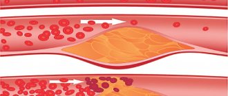
- the speed of blood flow decreases in some places - with vascular atherosclerosis, heart failure, varicose veins;
- blood density increases due to an imbalance of various biological substances - the level of low-density lipoproteins increases during atherosclerosis, the protein composition of blood serum changes during inflammatory processes;
- Atherosclerotic lesions and inflammation appear inside the walls of blood vessels, narrowing the vessel - endocarditis, various forms of arteritis, aneurysm of blood vessels and heart muscle;
- blood clotting increases due to the absence or insufficiency of coagulation factors;
- agglutination (clumping) of platelets is observed - exacerbation of myocardial infarction, sepsis, intoxication, extensive burns and injuries.
Read also: Vein thrombosis with hemorrhoids
The disease can affect veins and arteries.
Signs of thrombosis are directly dependent on the location of the thrombus that blocks the blood vessel. Pathologies such as stroke and myocardial infarction develop as a result of thrombosis of the cerebral and coronary vessels. Retinal thrombosis can lead to loss of visual function.
A thrombus localized in the abdominal aorta is most often located in the bifurcation (branching area). At the same time, it clogs not only the abdominal aorta, but also the iliac arteries. Given the gradual development of the disease, blood circulation can be compensated for a long time thanks to auxiliary blood vessels.
Patients may complain of pain and weakness in the lower extremities and impaired erection for several years, and upon examination the doctor diagnoses thrombosis. If the disease is not treated, then after 8-10 years you may encounter gangrene.
Thrombosis of the mesenteric vessels provokes significant discomfort in the abdominal cavity, less often in the lumbar and navel areas.
With prolonged insufficient blood supply to the intestines, the following symptoms develop:
- acute pain in the abdomen after eating, and it can radiate to the lower back, neck, chest and back of the head;
- disruption of the intestines, accompanied by constipation, diarrhea, vomiting and nausea;
- weight loss associated with decreased appetite;
- depressive disorders;
- weakness and decreased performance.
In more severe cases, a significant deterioration in the patient's condition may occur, including loss of consciousness.
Diagnostic procedures
Thrombosis of the abdominal vessels can be suspected based on the presence of characteristic clinical symptoms in the patient. To confirm the diagnosis, various instrumental and laboratory methods are used. It is important to take a general and biochemical blood test, conduct a coagulogram and test a urine sample. Signs of hypercoagulation may be observed with significant thickening of the blood and an increase in the number of formed elements relative to plasma. Ultrasound with Doppler ultrasound of the abdominal vessels is also performed. This helps to visualize the thrombus and eliminate the cause of ischemia in a timely manner.
Magnetic resonance imaging and angiography are used as additional methods.
Diagnostic measures
If the patient experiences unpleasant symptoms, it is better to consult a doctor as soon as possible. He will listen to the patient’s complaints and prescribe an examination, which includes:
- external examination and history taking. This will help make an approximate diagnosis and identify the cause;
- donating blood for general analysis. The results will show the erythrocyte sedimentation rate and the number of leukocytes;
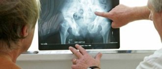
- radiography. Determines the degree of difficulty in the passage of the intestinal canal;
- laparoscopy. A small incision is made in the abdominal wall. An optical tube with a camera is inserted through it, with the help of which the internal organs are visible;
- laparotomy. It is carried out if it is impossible to perform laparoscopy;
- computed tomography;
- angiography. An iodine-containing substance is injected into the vessel, and then an x-ray is taken.
In some cases, the patient is prescribed a colonoscopy or endoscopy.
Treatment for abdominal thrombosis
Therapy for pathology should be comprehensive and aimed at eliminating the main cause of circulatory disorders. It is important to remove or dissolve the blood clot. For this purpose, fibrinolytics, antiplatelet agents and antithromboxants are prescribed. If this cannot be done conservatively, then surgical intervention is resorted to. An endoscopic procedure is performed. A special catheter is directed through the vessels to the embolus. After this, a blood-thinning substance is injected through it to the destination, it dissolves the blood clot, after which blood circulation is restored. To prevent the recurrence of abdominal vascular thrombosis, it is important to lead a healthy lifestyle, engage in active sports and eat right. It is necessary to carry out timely treatment for atherosclerosis and varicose veins, if these diseases are present.
Surgery
If mesenteric intestinal thrombosis progresses, or the disease cannot be overcome with medication, the doctor prescribes surgery, and drug therapy acts as an additional treatment.
In the case of intestinal ischemia, the disease rarely goes away on its own, however, as a preventive measure, antibiotics are recommended to remove toxins from the body.
The operation involves the removal of damaged organ segments and subsequent stitching of healthy tissues together. In some cases, bypass surgery is additionally required. During this procedure, the specialist creates a “bypass” around the blocked vessel so that the blood can move further.
If thrombosis of intestinal vessels occurs in an acute form, surgical intervention is also prescribed. The doctor independently determines what exactly needs to be done (remove a blood clot, perform angioplasty, bypass surgery, etc.). These manipulations help stop the development of the disease, subsequently tissue necrosis does not appear.
Etiology of the disease
All arteries and veins in the human body are susceptible to the formation of blood clots. In the abdominal area, this problem develops for several reasons:
- Damage to the walls of blood vessels. Occurs under the influence of an infectious, allergic or mechanical factor. Violation of the integrity of the inner surface of the vessel leads to the accumulation of blood cells in this area. As a result, a blood clot forms.
- Increased blood clotting. The problem appears against the background of a hereditary predisposition, under the influence of negative external factors. Blood counts change significantly in people with bad habits or after illnesses.
- Slow blood circulation. The presence of pathologies of the cardiovascular system and a sedentary lifestyle increase the risk of thrombosis. In the presence of such problems, the blood is not able to move at an optimal speed and stagnates in the veins.
The combination of these reasons is called Virchow's triad. They are necessarily taken into account in the process of diagnosing pathology.
Factors that increase the risk of blood clots
Blood clots rarely form in the superior mesenteric artery and inferior vena cava (IVC) due to the structural features of the abdominal vessels. Usually blood clots get there as a result of migration. Initially, they form in the vessels of the lower extremities or pelvis. The following factors contribute to the negative process:
- injuries of the limbs, abdominal cavity;
- tumor malignant process of any localization;
- taking hormonal contraceptives;
- varicose veins;
- surgical intervention in the lower extremities, pelvis, abdominal cavity;
- allergies, various autoimmune diseases.
Women with complicated pregnancy and childbirth, people with endocrine pathologies or hormonal imbalances are susceptible to the development of blood clots. Problems are possible in the presence of infections that are transmitted through the circulatory system. Very often, thrombosis is detected in patients who are forced to adhere to bed rest for a long time. Elderly people, people with low physical activity or excess weight are also at risk.
Causes
- Atherosclerosis. With atherosclerosis, fats settle in the vessel wall, which accumulate and form plaques. Over time, the plaques increase in size and fibrin and platelets settle on them, forming a blood clot.
- Myocardial infarction and other heart pathologies. Against the background of pathologies of the heart and blood vessels, blood circulation is disrupted, it stagnates in the intestinal vessels, which contributes to the formation of clots.
- Hypertension and symptomatic hypertension. Constantly increased pressure leads to pathological changes in the wall of blood vessels, which is a factor contributing to the development of atherosclerosis. At the moment, some authors consider hypertension and atherosclerosis as a single pathology, a frequent complication of which is thrombosis of the great vessels.
- Thrombophlebitis of the lower extremities. Thrombophlebitis is an inflammation of the veins, which most often affects the vessels of the legs. Against the background of the inflammatory process, blood clots form in the vessel, which can break off, migrate through the circulatory system and enter other organs, including the intestines.
- Postpartum complications. Childbirth is a big burden for a woman’s body, so disturbances in the hemostatic system are often observed in the postpartum period. This leads to increased blood clotting and increased thrombus formation.
Clinical picture
When a blood clot is located directly in a vein, protrusion of blood vessels in the abdomen and chest is observed.
Swelling of the limbs and pain in the lower body also occur. Such a clinical picture develops very rarely, since a blood clot usually penetrates into the vessels of the abdominal cavity from other parts of the circulatory system. Classification of IVC segments that are susceptible to acute thrombosis:
- infrarenal;
- hepatic;
- renal;
- suprarenal.
A floating thrombus of the inferior vena cava is characterized by mild symptoms. In this case, the blood clot is localized near the wall of the vessel.
Symptoms of a thrombus in the infernal segment of the IVC are intense lower back pain, cyanosis of the skin, swelling of the extremities. Veins are clearly visible in the lower abdomen.
With thrombosis of blood vessels in the renal and suprarenal segment, symptoms arise that are characteristic of a kidney tumor. No blood flow disturbances are observed. Signs:
- severe pain in the lumbar region;
- hematuria;
- decrease in urine volume or its complete disappearance;
- development of intoxication;
- nausea and vomiting.
When blood flow is restored, the symptoms disappear completely or significantly reduce their manifestation.
Due to a thrombus in the liver segment, a large volume of blood accumulates in the organ, which leads to stretching of its fibrous membrane. This provokes the following symptoms:
- bursting pain in the right side that radiates to the scapula;
- ascites;
- development of jaundice;
- increase in the size of the spleen;
- cyanosis of the anterior abdominal wall.
On palpation, the smoothness of the liver and a change in its contour are felt. Additionally, doctors observe dilation of the veins in the upper abdomen.
The main symptom of mesenteric thrombosis is intense pain in the right side of the abdomen. Nausea develops, turning into vomiting, and loose stools. With intestinal necrosis due to circulatory disorders, a significant increase in body temperature occurs. Additionally, a complete absence of peristalsis and fecal retention are observed.
What should you know about the disease?
Blood, as you know, has the property of clotting. In medicine, this process is called coagulation. This is a very important function, without which any person, after receiving wounds, would lose all his blood and, accordingly, die. On the other hand, coagulation promotes the formation of clots over time, otherwise known as thrombi. According to experts, they can form in absolutely any part of the body. For example, when they enter an intestinal artery, blood clots consistently clog its lumen, thereby preventing normal nutrition of a certain area of the organ. As a result, tissue necrosis in the intestine is observed. This disease is called thrombosis (mesenteric) of the intestine. In particularly serious cases, it can be fatal.
Mesenteric intestinal thrombosis is a disease that occurs due to obstruction of the superior, celiac or inferior mesenteric artery. This pathology often causes changes in blood circulation in the digestive tract. According to experts, this disease is equally common among women and men, but it is especially common in older people.
Diagnostics
The developing clinical picture does not always allow the doctor to accurately determine the patient’s thrombosis in the abdominal cavity. The following tests are used to make a diagnosis:
- blood test for D-dimer;
- radionuclide diagnostics using labeled fibrinogen;
- duplex ultrasound;
- iliocavography.
When performing diagnostics, the doctor gives preference to minimally invasive methods. However, the information content and economic feasibility of research are no less important.
Diagnostic methods
An initial surgical examination performed by an experienced specialist can quickly suggest the presence of an acute abdomen. In addition to conducting the necessary palpation tests, the doctor will order the following examinations:
- determination of the number of leukocytes in a general clinical blood test;
- assessment of blood coagulation using a coagulogram;
- ultrasound scanning of internal organs;
- plain x-ray of the abdomen;
- CT scan;
- angiographic examination to determine the location of the blockage;
- diagnostic laparoscopy.
Depending on the symptoms and severity, the examination tactics are individual. All diagnostic measures must be carried out quickly in order to prevent deterioration of the condition and progression of the disease: thrombosis of the mesenteric vessels in the compensation stage can be cured without dangerous consequences, and against the background of peritonitis, the risk of death increases to 90%.
Possible complications and prognosis
The most dangerous consequence of the pathology is pulmonary embolism. If medical care is not provided in a timely manner, the prognosis is disappointing. This condition usually ends in the death of the patient.
When the lumen of the vessel is incompletely blocked, a circulatory disorder is observed. The heart does not receive enough oxygen, which leads to the death of its tissues. In this case, with timely treatment, the prognosis is favorable. But given the blurred clinical picture of the pathology, medical assistance is provided with a delay. This leads to the patient’s loss of ability to work and his disability.
Conservative therapy
When a patient enters the hospital, the doctor first assesses at what stage of development the intestinal thrombosis is. Treatment through conservative methods is usually used if the disease has not begun to progress. Here are used:
- A parenteral method of administering anticoagulants, the main purpose of which is to thin the blood. The most commonly used drug is Heparin and some of its analogues.
- Injections of thrombolytics and antiplatelet agents (medicines “Trental”, “Reopoliglyukin”, “Hemodez”).
Despite the fairly high mortality rate from this disease, if treated in a timely manner, there is a good chance of a complete recovery for the patient.
Preventive measures
To prevent thrombosis, you must adhere to a healthy lifestyle. It is important to control your weight, maintain optimal physical activity, and eat right.
You need to take care of your own health. At least once a year it is necessary to check the blood for sugar and clotting ability, and undergo preventive examinations of internal organs. If there is a tendency to form blood clots, the use of oral contraceptives is prohibited.
Features of postoperative rehabilitation
During the postoperative period, the patient remains in the surgical department for 7-10 days. He is then discharged for outpatient treatment. During this period, you must adhere to the following rules:
- Avoid strenuous physical activity to avoid causing rupture of intestinal or intravascular sutures.
- Normalize the activity of the gastrointestinal tract, avoid constipation. It is recommended to consume dairy products, vegetables, and fruits containing large amounts of fiber.
- In the first days, try not to cough too much or laugh, so as not to cause an increase in intra-abdominal pressure.
- Showering rather than bathing should be used as hygiene procedures.
- Constantly take prescribed anticoagulants and antiplatelet agents.
After surgery, it is recommended to take a shower instead of a bath.
Following all the recommendations of the attending physician will allow you to restore your ability to work and prevent the development of postoperative complications.
Symptoms
With thrombosis of the abdominal aorta, the symptoms depend on the degree of occlusion, as well as the localization of the pathological process - in the area of bifurcation, mesenteric artery, etc.
Acute course
Thrombosis of the abdominal cavity is accompanied by a sharp disruption in the functioning of vital organs. This may be myocardial infarction, kidney, liver or spleen, necrosis of the intestinal walls.
Symptoms are characterized by an abrupt onset and rapid progression of the clinical picture. First signs:
- unbearable abdominal pain;
- rapid deterioration of the patient’s condition (in the absence of high tension in the abdominal muscles);
- diarrhea, possible involuntary bowel movements;
- a sharp decrease in blood pressure due to blockage of blood flow;
- improvement of the patient’s well-being and complete disappearance of all symptoms when patency is restored (occurs no earlier than after 6-8 hours).
When the blood supply to the intestines is disrupted, ischemia first occurs, characterized by a lack of oxygen supply to the tissues. In the absence of timely treatment, intestinal infarction occurs, complicated by peritonitis.
If the spleen is damaged, the pain is localized in the left hypochondrium. Intensifies with movements of the chest (cough, deep breathing).
Chronic course
When a small thrombus forms, closing the lumen of the artery by no more than 10-20%, collateral blood flow is activated. Most of the blood passes through other arteries, so internal organs are not damaged. However, the formation of a clot is accompanied by a spasm of blood vessels, which aggravates the situation. After some time, symptoms appear indicating an insufficient supply of oxygen and nutrients to the internal organs. The kidneys, intestines and lower extremities are most affected.
The main signs of clots in the abdominal cavity:
- Nagging abdominal pain appears after eating. The pain spreads to the lower back, the back of the head, and the neck. They go away on their own within a few hours.
- Disorders of the digestive tract. The main manifestations are nausea, vomiting, and bowel dysfunction.
- Weight loss and asthenia. Associated with a deterioration in well-being after eating, as a result of which a person refuses to eat. There is a feeling of constant fatigue, drowsiness, and decreased performance.
If the kidneys are damaged, blood pressure may increase, which is extremely difficult to normalize.
If the occlusion is localized in the bifurcation zone of the abdominal aorta, signs of damage to the extremities appear. Intermittent claudication develops, pain occurs, a feeling of crawling and numbness of the legs, trophic disorders, thrombophlebitis.
Acute course
This course of thrombosis of the abdominal aorta occurs when it is completely blocked, and, depending on the localization of the process, the following may occur: intestinal necrosis, infarction of the kidneys, spleen, and liver. Most often, thrombosis occurs at the level of the renal and superior mesenteric arteries, which supply blood to the gastrointestinal tract.
Read also: Where is varicocele surgery performed?
When intestinal damage occurs, three successive stages are distinguished: ischemia, infarction and general peritonitis. If assistance is provided in the first 2 stages, then the prognosis for the patient is satisfactory. If the diagnostic search is delayed, then at the stage of peritonitis the process is irreversible with a possible fatal outcome.
The main symptoms to suspect acute occlusion:
- severe abdominal pain that occurs unexpectedly;
- inconsistency with the severity of the condition and lack of tension in the abdominal muscles;
- frequent, loose stools, involuntary bowel movements;
- a sharp improvement in well-being after 6 hours;
- drop in blood pressure, increased heart rate;
- sudden deterioration of the condition with the development of paralytic intestinal obstruction.
With a splenic infarction, a sharp pain occurs in the left hypochondrium, which intensifies with coughing and deep inhalation. The palm detects a rustling friction noise of the peritoneum. The blood test is dominated by low white blood cells and high platelets.
If the kidneys are damaged, intense back pain and transient arterial hypertension occur. Acute kidney failure develops quickly with a sharp decrease in the amount of urine and the appearance of blood in it in the normal state of the urinary tract.
In the absence of blood supply to the lower extremities, sensory disturbances and immobility occur. There is a decrease in temperature and pale skin. There is no pulse below the knee or in the ankle area. Against this background, gangrene of the legs quickly develops.
The formation of a blood clot in the abdominal aorta can lead to life-threatening conditions. Without timely surgical assistance, death or organ loss is possible. Difficulties in establishing a diagnosis are due to the fact that thrombosis can occur under the guise of an “acute abdomen” or other diseases - cholecystitis, pancreatitis, hypertension. Therefore, the vigilance of the doctor and the patient suffering from atherosclerosis is very important.
Thrombosis of abdominal vessels
Blockage of blood vessels or thrombosis of the abdominal cavity occurs as a result of excessive blood viscosity or mechanical damage to the vessel wall. Minor ischemia leads to functional disorders of the peritoneal organs. Acute circulatory disorders cause symptoms that are life-threatening to the patient.
Thromboembolism can be triggered by smoking or eating foods rich in cholesterol.
Treatment of thrombosis
It is worth understanding and adequately assessing the current situation. This condition will never go away on its own; it cannot be treated at home. Treatment for gastric thrombosis takes place only in a hospital. When choosing a treatment method, the volume of the lesion, the severity of the patient’s condition, and allergies to medications are taken into account. The decision on the choice of therapy belongs only to the attending physician.
Possibility of conservative treatment
This type of therapy includes:
- Parenteral administration of antispasmodics to avoid vasospasm.
- Thrombolysis (drugs that dissolve blood clots). All the pros and cons are taken into account, because the method has many contraindications.
Return to contents
Surgical method for eliminating gastric thrombosis
The main method is surgery, in which the damaged part of the organ is removed along with nearby vessels. During the operation, the surgeon examines not only the stomach, but also the intestines. The volume and complexity of the operation depends on the advanced stage of the disease. If tissue necrosis has not yet begun, then only the vessels containing the blood clot are removed.
Reasons for development
Vascular thrombosis in the abdominal cavity occurs as a result of excessive activity of the blood coagulation system. This is ensured due to its excessive viscosity and mechanical damage to the walls of arteries or veins. As a result, a blood clot forms on the inner endothelial wall, which breaks off and clogs the vessel, preventing normal blood flow. Excessive stickiness of red blood cells may also contribute to this.
The occurrence of thrombosis of abdominal vessels can be provoked by the influence of the following predisposing factors on the human body:
- atherosclerosis,
- smoking,
- alcohol consumption,
- unhealthy diet
- the presence of excess amounts of bad cholesterol in food,
- sedentary lifestyle,
- stagnation of blood in the pelvis,
- Varicose veins,
- hormonal imbalances,
- intestinal infections.
Symptomatic picture
The acute type of thrombosis occurs when a large thrombus suddenly enters the vessel. This process leads to the appearance of severe pain. In this case, the localization can be different: on the right in the appendix area, in the umbilical area or on the left in the lower abdomen.
After some time, diarrhea occurs. Blood streaks are observed in the impurities. Against the background of dehydration and low blood pressure, some symptoms arise in the form of:
- weakness;
- confusion and loss of consciousness;
- sudden blanching of the skin.
Over the course of 6-12 hours, the pain gradually subsides. But at the same time the patient feels worse and worse.
Then the last stage is observed, which is accompanied by:
- nausea and repeated vomiting;
- increase in temperature values;
- bloating, constipation, lack of gas discharge.
In chronic conditions, the whole process occurs differently.
The first stage is observed first. The patient is not worried about anything. Thrombosis can only be seen with a contrast x-ray.
Then the second stage is formed. After eating, the pain intensifies even more. Against this background, there is a loss of appetite. The intestines and stomach are gradually freed. At this time, the patient feels relief.
At the third stage, the pain is constant. The skin becomes dry. Regular diarrhea and bloating occur.
The fourth stage is considered the most severe. Symptoms progress rapidly further. Gases stop passing, the pain becomes stronger, and the body temperature rises sharply.
Main symptoms
The degree of clinical manifestations of vascular blockage and the development of ischemia depends on the level of circulatory failure. Symptoms also vary depending on which artery is affected. With minor and chronic occlusion, severe pain occurs in the abdominal cavity after a heavy meal. The patient is bothered by a constant feeling of intestinal fullness, dyspepsia, nausea, and vomiting. Diarrhea may alternate with constipation, which leads to dystrophy and weight loss. With a long course of the pathology, peptic ulcer of the stomach and duodenum occurs, a constant feeling of fatigue appears and overall performance decreases. If the vascular bed supplying the kidneys suffers, then the patient develops arterial hypertension, which develops as a result of excessive release of angiotensin.
If thrombosis and impaired blood flow are acute, they can lead to the development of the following dangerous consequences in the patient:
- necrosis of part of the intestine,
- kidney infarction,
- liver damage,
- peritonitis,
- sepsis,
- fatal outcome.
With the development of acute thrombosis of abdominal vessels, the patient develops the following clinical symptoms:
- sharp and severe pain in the abdomen,
- lack of protective muscle tension in the abdominal cavity,
- frequent urge to defecate and loose stools,
- drop in blood pressure and increase in heart rate,
- possible improvement in well-being after 6 hours,
- deterioration of the condition with the development of peritonitis and paralytic intestinal obstruction.
What is intestinal thrombosis
Intestinal thrombosis is a rare and more dangerous disease
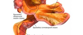
All parts of the human intestine are located in the abdominal cavity and are attached to its posterior wall using a special structure, the mesentery. This structure contains arteries, veins, nerve plexuses and lymph nodes. It is thanks to the mesentery that all parts of the intestine constantly receive blood and are ensured by the timely outflow of venous blood. Thus, intestinal thrombosis is a blockage of the mesenteric vessel that feeds the organ.
Intestinal thrombosis can be caused by blockage of an arterial or venous link, but most often the blood flow is disrupted in the arteries. In this case, a blockage may occur in one of the three main mesenteric (mesenteric) arteries, which will almost immediately lead to ischemia of the intestinal tissue. Ischemia is understood as an acute insufficiency of blood supply to tissues and organs, the long-term development of which can lead to cell destruction.
The intestines can also be damaged if the blood supply is not compensated for.
Intestinal thrombosis is an extremely dangerous condition, which is the preceding stage of ischemia and infarction of the organ. Delayed diagnosis often causes death in patients with this pathology.
Types of ischemia and pathophysiology
Insufficient blood supply to the small and large intestines can be the result of embolism or thrombotic occlusion of a vessel. Such conditions are associated with a coagulation disorder, in which the lumen of the vessel is blocked by a blood clot. In rare cases, intestinal ischemia is also associated with vasospasm (narrowing) or low cardiac output. The embolic pattern of ischemia is observed in 50% of cases, with arterial thrombosis accounting for up to 25%.
The severity of intestinal tissue damage is inversely proportional to mesenteric blood flow and depends on the number of vessels involved, mean blood pressure, duration of ischemia, and collateral circulation. Collateral circulation is understood as the occurrence of compensatory blood supply to tissues through additional (usually less involved) vessels.
Pathology most often occurs in old age
Types of ischemia:
- Acute mesenteric arterial thrombosis is a common complication of pre-existing visceral thrombosis. Symptoms usually do not develop until two of the three main arteries are blocked. Due to the rather long progression of atherosclerotic stenosis into acute occlusion (blockage), the opportunity arises for the development of collateral circulation. Most patients with this type of ischemia have atherosclerotic damage to other vessels, including the coronary and cerebral arteries. A drop in cardiac output caused by heart failure can also trigger the condition.
- Nonocclusive mesenteric ischemia is a severe reduction in the mesenteric blood supply with secondary arterial spasm that occurs due to heart failure, septic shock, or hypovolemia (insufficient blood volume). This is a less dangerous condition.
- Mesenteric venous thrombosis is the occlusion of large venous areas of the mesentery. This is a fairly common complication of surgical interventions on the abdominal organs. The condition can also occur with sickle cell anemia, coagulation disorders and malignant neoplasms. The mechanism of ischemia in this case is due to a massive influx of fluid into the intestinal walls and its lumen, which leads to systemic hypovolemia. Severe swelling of the intestines and impaired blood flow prevent sufficient blood supply to the organ. Venous thrombosis occurs more often in young patients.
The degree of intestinal damage varies from reversible ischemia to transmural infarction with necrosis and rupture of the organ wall (perforation). Arterial insufficiency causes acute tissue hypoxia, leading to spasm of the intestinal walls. As a result, the intestinal contents are evacuated through vomiting or diarrhea. Damage to the intestinal mucosa can cause bleeding in the gastrointestinal tract.
Diagnostic procedures
Thrombosis of the abdominal vessels can be suspected based on the presence of characteristic clinical symptoms in the patient. To confirm the diagnosis, various instrumental and laboratory methods are used. It is important to take a general and biochemical blood test, conduct a coagulogram and test a urine sample. Signs of hypercoagulation may be observed with significant thickening of the blood and an increase in the number of formed elements relative to plasma. Ultrasound with Doppler ultrasound of the abdominal vessels is also performed. This helps to visualize the thrombus and eliminate the cause of ischemia in a timely manner.
Magnetic resonance imaging and angiography are used as additional methods.
Therapeutic measures for thrombosis
If mesothrombosis is detected in a timely manner, the outcome of the disease will be favorable. It is almost impossible to cure the disease on your own. This will only make the condition worse.
Treatment of intestinal thrombosis depends on the stage of the disease.
- In the first stage, intestinal function is completely restored.
- At the second stage, an operation is performed to remove the damaged areas.
- The third stage is the most difficult. Intestinal function cannot be restored.
If thrombosis of a small intestinal vessel is diagnosed in a timely manner, then treatment is carried out with the help of drugs.
The treatment regimen is as follows.
- Medicines that reduce blood clotting are administered through a vein. Droppers are given every 6 hours. Treatment lasts 2-3 days.
- Preparations for restoring blood flow in blood vessels.
- Medicines to prevent blood clots.
The mesenteric thrombus can be completely removed using laparoscopy or laparotomy. In more complicated cases, thrombophlebitis is treated by removing the affected tissue. But this is only possible if the intestinal function is partially working.
The main task is to prevent the occurrence of peritonitis. In approximately 25% of cases everything is successful. And after the operation, the body recovers quickly. Treatment lasts about two weeks, and the recovery period takes up to 3 months.
After surgery, you need to think about following a strict diet. In the first week, it is better to give preference to liquid meals. Once the function of the digestive system is restored, you can diversify your diet.
Fatty and fried foods, fast foods, semi-finished products, raw vegetables and fruits with peels are prohibited. You need to eat a little, but often. You cannot drink alcohol or smoke. Carrying heavy objects should be avoided. Walking is prohibited for two weeks. After this, it is recommended to walk daily at a calm pace for 1-2 hours.
Intestinal thrombosis is often detected in older people. But this does not mean that the disease can occur in young patients. To prevent the development of complications, you need to listen to your body. And when the first signs appear, consult a doctor in a timely manner.
Treatment for abdominal thrombosis
Therapy for pathology should be comprehensive and aimed at eliminating the main cause of circulatory disorders. It is important to remove or dissolve the blood clot. For this purpose, fibrinolytics, antiplatelet agents and antithromboxants are prescribed. If this cannot be done conservatively, then surgical intervention is resorted to. An endoscopic procedure is performed. A special catheter is directed through the vessels to the embolus. After this, a blood-thinning substance is injected through it to the destination, it dissolves the blood clot, after which blood circulation is restored. To prevent the recurrence of abdominal vascular thrombosis, it is important to lead a healthy lifestyle, engage in active sports and eat right. It is necessary to carry out timely treatment for atherosclerosis and varicose veins, if these diseases are present.
Features of the disease
The intestine is supplied with blood by 2 large vessels, which drain from the aorta. They are called the superior and inferior mesenteric (mesenteric) arteries. In this case, the outflow of blood is carried out through the superior and inferior mesenteric veins. The superior mesenteric artery supplies all parts of the small intestine, as well as some of the large intestine. The inferior mesenteric artery supplies the sigmoid and rectum. The small branches of the arteries are connected in a special way: the upper vessel in a critical situation helps to nourish the lower part of the large intestine, but the lower mesenteric artery cannot replace “foreign” functions. Therefore, with thrombosis of the intestinal arteries, the blood supply to the small intestine is disrupted.
The venous outflow from the intestinal arteries is organized in such a way that when the superior mesenteric vein is blocked, the small intestine is also left without nutrition, and therefore the processes of tissue hypoxia and death will be observed in it. If a mesothrombus, or thrombus in the intestine, clogs one of the vessels, one or another part of the small intestine will undergo necrosis. This condition is called “acute mesenteric thrombosis,” or intestinal infarction. Intestinal thrombosis, therefore, can be arterial or venous, and in especially advanced cases it can form a mixed form.
In 90% of situations, thrombosis affects the superior mesenteric artery, which supplies the largest part of the intestine, so this type of pathology will have the most severe consequences. It is believed that after thrombosis of this artery develops, acute mesenteric arterial insufficiency, or AMA, develops. Since replacement (collateral) blood circulation does not occur in this disorder, there will be no compensation of blood flow with all the ensuing consequences. Only with segmental thrombosis or embolism of the inferior mesenteric vein can the blood circulation be compensated, and only the ileum is affected.
OUR READERS RECOMMEND! The most effective remedy for getting rid of varicose veins, according to our reader Ksenia Strizhenko, is. Varius is considered an excellent remedy for the treatment and prevention of varicose veins. For you, it has become that “lifeline” that you should use first!
Thrombosis of mesenteric vessels is practically not recorded in patients under 50 years of age. Age-related changes, namely, atherosclerotic decrease in the lumen of blood vessels, which occurs in the second half of life, is the main “provocateur” of this pathology. Mortality in this disease is very high, even after surgery to remove the blood clot due to the rapid progression of necrotic processes.
Classification and types of thrombosis
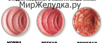
In medicine, there are three main types of intestinal thrombosis. Their division depends on the severity of the disease and the state of the blood flow.
- Thrombosis is compensated. Small vessels become clogged. Blood flow is restored over time and does not affect the function of the rectum and intestines.
- Thrombosis is subcompensated. There are blood clots and blood flow is not fully restored.
- Thrombosis is decompensated. Thrombosis in the vessel completely blocks blood circulation, and intestinal infarction may subsequently occur. This type of thrombosis often leads to the death of the patient.
Mesenteric intestinal thrombosis has three stages:
- Intestinal ischemia. The vessels were slightly damaged. If you consult a doctor for help in time, you can prevent the disease from progressing. At this stage, the patient begins to vomit bile, complains of pain in the intestinal area and loose stools. But before the first symptoms appear, the patient will have a hypertensive attack (blood pressure rises sharply).
- Intestinal infarction. The lumen of the vessel becomes clogged and blocked, which leads to destruction of the walls of the intestinal mucosa. Leads to intoxication of the body. The patient finds it difficult to go to the toilet, often suffers from constipation, and blood stains can be seen in the stool. The patient complains of pain in the intestinal area, everything swells near the navel (Mondor's symptom). The pain is severe, which is impossible to endure; there are times when painkillers do not help.
- Inflammation of the abdominal cavity (peritonitis). Intoxication passes throughout the body, while the circulatory system is disrupted. The patient is in serious condition, which is accompanied by vomiting, bloating, and when pressing on it, the patient experiences sharp acute pain. If medical assistance is not provided, the intestines become paralyzed and blood pressure drops sharply. Possible death.
Diagnosis of thrombosis
To make an accurate diagnosis, specialists must conduct many laboratory tests.
- The first step is to study the history of all congenital and acquired diseases. The doctor also conducts an external examination.
- A blood test to determine the level of erythrocyte sedimentation rate and the number of leukocytes. If a person has thrombosis, then the indicators are several times higher than normal.
- X-ray allows you to notice how difficult the passage in the intestines is.
- Laparoscopy. A small incision is made in the peritoneum, and an optical tube with a camera is inserted into it. The camera shows all the patient's internal organs.
- Laparotomy. If for some reason it is impossible to perform laparoscopy, then this type of medical examination is performed. If the doctor finds affected areas of the intestine, they are immediately removed through surgery.
- Computed tomography accurately determines the condition of all internal organs.
- Angiography. An iodine-containing substance is injected into the mesenteric vessel and then an x-ray of the peritoneum is taken. This type of examination will determine where and how severely the mesenteric vessel is blocked.
- Colonoscopy. A colonoscope with a camera, which is inserted through the rectum, will help determine the general condition of the intestines and its walls.
- Endoscopy. Similar to a colonoscopy, but the camera is inserted through the mouth.
Thrombosis of intestinal mesenteric vessels
The symptoms of the disease depend on the blockage of the arterial lumen and how many vessels are blocked.
- Often at the first stage of the disease, the patient experiences pain attacks in the abdominal area. Over time, the pain becomes permanent. Due to unbearable pain, the patient often spends time lying down. To get rid of the pain a little, the patient should lie on his side and press his legs to his stomach.
- Vomiting blood.
- Frequent loose or pasty stools with blood.
- At the initial level of development of the disease, blood pressure increases, and over time it becomes below normal levels. The normal blood pressure for a healthy person is 110/70.
- The visible mucous membrane and skin turn pale, this is the first sign that blood does not pass through the vessels in full.
- Increased body temperature above 37.5.
- Facial features become sharper.
- The patient notices the elasticity of the abdomen and its bloating.
- If you press on your stomach and then suddenly remove your hands, the pain becomes even stronger.