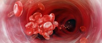
The blood supply to the brain is a separate functional system of blood vessels, through which nutrients are supplied to the cells of the central nervous system and the products of their metabolism are removed. Due to the fact that neurons are extremely sensitive to a lack of microelements, even a minor disruption in the organization of this process negatively affects a person’s well-being and health.
Today, acute cerebrovascular accident or stroke is the most common cause of human death, the origins of which are in damage to the blood vessels of the brain. The cause of the pathology can be clots, blood clots, aneurysms, looping, and kinks in blood vessels, so it is extremely important to conduct a timely examination and begin treatment.
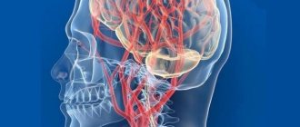
Brain blood supply device
As you know, in order for the brain to work and all its cells to function correctly, a continuous supply of a certain amount of oxygen and nutrients to its structures is required, regardless of the physiological state of a person (sleep - wakefulness). Scientists have calculated that the needs of the central nervous system consume about 20% of the oxygen consumed, while its mass in relation to the rest of the body is only 2%.
The brain is nourished through the blood supply to the organs of the head and neck through the arteries that form the arteries of the circle of Willis on the brain and penetrate through it. Structurally, this organ has the most extensive network of arterioles in the body - its length in 1 mm3 of the cerebral cortex is approximately 100 cm, in a similar volume of white matter about 22 cm.
The largest amount is located in the gray matter of the hypothalamus. And this is not surprising, because it is responsible for maintaining the constancy of the internal environment of the body through coordinated reactions, or in other words, it is the internal “steering wheel” of all vital systems.
The internal structure of the blood supply to arterial vessels in the white and gray matter of the brain is also different. For example, the arterioles of the gray matter have thinner walls and are elongated compared to similar structures of the white matter. This allows for the most efficient gas exchange between blood components and brain cells; for this reason, insufficient blood supply primarily affects its performance.
Anatomically, the blood supply system of the large arteries of the head and neck is not closed, and its components are interconnected through anastomoses - special connections that allow blood vessels to communicate without forming a network of arterioles. In the human body, the largest number of anastomoses is formed by the main artery of the brain - the internal carotid. This organization of blood supply allows you to maintain constant movement of blood through the circulatory system of the brain.
Structurally, the arteries of the neck and head are different from the arteries in other parts of the body. First of all, they do not have an external elastic shell and longitudinal fibers. This feature increases their stability during surges in blood pressure and reduces the force of blood pulsation shocks.
The human brain works in such a way that at the level of physiological processes it regulates the intensity of blood supply to the structures of the nervous system. In this way, the body’s defense mechanism is triggered - protecting the brain from surges in blood pressure and oxygen starvation. The main role in this is played by the sinocartoid zone, the aortic depressor and the cardiovascular center, which is associated with the hypothalamic-mesancephalic and vasomotor centers.
Anatomically, the largest vessels bringing blood to the brain are the following arteries of the head and neck:
- Carotid artery. It is a paired blood vessel that originates in the chest from the brachiocephalic trunk and the aortic arch, respectively. At the level of the thyroid gland, it, in turn, is divided into an internal and external artery: the first delivers blood to the medulla, and the other leads to the facial organs. The main branches of the internal carotid artery form the carotid basin. The physiological significance of the carotid artery lies in supplying microelements to the brain; about 70-85% of the total blood flow to the organ flows through it.
- Vertebral arteries. The vertebrobasilar basin is formed in the cranium, which provides blood supply to the posterior sections. They begin in the chest and follow the bony canal of the spinal part of the central nervous system to the brain, where they unite into the basilar artery. According to estimates, the blood supply to the organ through the vertebral arteries supplies about 15-20% of the blood.
The supply of microelements to the nervous tissue is ensured by the blood vessels of the Circle of Willis, which is formed from the branches of the main blood arteries in the lower part of the skull:
- two forebrains;
- two midbrain;
- pairs of hindbrain;
- anterior connecting;
- pairs of rear connecting ones.
The main function of the circle of Willis is to ensure stable blood supply during blockage of the leading vessels of the brain.
Also in the circulatory system of the head, experts identify the Zakharchenko circle. Anatomically, it is located on the periphery of the medulla oblongata and is formed by the union of the collateral branches of the vertebral and spinal arteries.
The presence of separate closed systems of blood vessels, which include the Circle of Willis and the Zakharchenko Circle, makes it possible to maintain the supply of the optimal amount of microelements to the brain tissue when blood flow in the main channel is disrupted.
The intensity of blood supply to the brain of the head is controlled using reflex mechanisms, the functioning of which is controlled by nerve pressoreceptors located in the main nodes of the circulatory system. For example, at the site of the branch of the carotid artery, there are receptors that, when excited, can give a signal to the body that it needs to slow down the heart rate, relax the walls of the arteries and lower blood pressure.
Blood circulation in various classes of animals
Depending on the class to which a particular type of living organism belongs, the circulatory system differs, which is due to evolutionary development.
Blood circulation of annelids
More information: Annelids
In most species of annelids, blood circulation occurs in a closed circuit; it is based on the dorsal and abdominal vessels, connected by annular vessels that resemble arteries and veins. There is no heart; its role is played by sections of the spinal and circular vessels containing contractile elements. Depending on the type of respiratory pigments, some annelids have red blood, while others have colorless or green blood. Respiration is cutaneous, in marine species - with the help of gills on parapodia.
Blood circulation of arthropods
More information: Arthropods
Representatives of arthropods have an open circulatory system. The vessels open into the body cavity and mix with the cavity fluid, forming hemolymph.
Blood circulation of primitive chordates
Lancelet blood circulation diagram: 1. Carotid arteries.
2. Efferent branchial arteries. 3. Roots of the dorsal aorta. 4. Cuvier's ducts. 5. Dorsal aorta. 6. Anterior cardinal veins. 7. Afferent branchial arteries. 8. Abdominal aorta. 9. Portal system of the hepatic outgrowth. 10. Venous sinus. 11. Hepatic vein. 12. Posterior cardinal veins. 13. Subintestinal vein. 14. Tail vein Additional information: Lancelets
The blood circulation (circulatory system) is represented by a closed circuit and is delimited from surrounding organs and tissues by the walls of blood vessels; the heart is absent. The arterial part of the circulatory system of lancelets is represented by a system of vessels and valves. Below the pharynx is the abdominal aorta (aorta ventralis) - a large vessel whose walls constantly pulsate and circulate blood, thus replacing the heart. The pulsation occurs through a slow, uncoordinated contraction of the myoepithelial layer of adjacent coelomic cavities[3]. Through the abdominal aorta, venous blood moves to the head end of the body. Through the thin covers of hundreds of gill arteries (efferent), extending according to the number of interbranchial septa from the abdominal aorta, oxygen dissolved in water is absorbed by the blood [4]. The bases of the gill arteries—the bulbs—also have the ability to pulsate[5]. The branchial arteries flow into the paired (right and left) roots of the dorsal aorta (aorta dorsalis), which is located at the posterior edge of the pharynx and extends under the notochord to the end of the tail. The anterior end of the body is supplied with blood by two short branches of the paired roots of the dorsal aorta (aorta dorsalis) - the carotid arteries. Arteries branching from the dorsal aorta carry blood to all parts of the body.
Having passed through the capillary system, venous blood from the intestinal walls is collected in the azygos intestinal vein, which runs in the form of the hepatic vein to the hepatic outgrowth. In it, the blood again crumbles into capillaries - the portal system of the liver is formed. The capillaries of the hepatic process again merge into a short hepatic vein, which flows into a small extension - the venous sinus (lancelet). From both ends of the body, blood collects in paired anterior and posterior cardinal veins. On each side they merge and form the right and left ducts of Cuvier (common cardinal veins), which flow into the sinus venosus, which is the beginning of the abdominal aorta. It follows from this that lancelets have one circle of blood circulation. Their blood is colorless and does not contain respiratory pigments. The oxygen saturation of blood in arteries and veins is similar - the small size of animals and single-layer skin make it possible to saturate the blood with oxygen not only through the gill arteries, but also through all the superficial vessels of the body.
Blood circulation of fish
Two-chambered heart of fish
Additional information: Two-chambered heart and fish
According to evolutionary teaching, for the first time the heart as a full-fledged organ is observed in fish: the heart here is two-chambered, a valve apparatus and a cardiac sac appear. The fish heart, consisting of one ventricle and atrium (two-chambered heart), pumps only venous blood. In fish, the circulatory system is represented by only one closed circuit (a single circle of blood circulation), through which the blood circulates through the capillaries of the gills, then collects in vessels and is again divided into capillaries of the body tissues. After which it again collects into the hepatic and cardiac veins, which flow into the venous sinus of the heart. Thus, the heart of a fish is represented by only one pump, consisting of two main chambers: the atrium and the ventricle.
The circulatory system of primitive fish can be roughly represented as a sequentially located “four-chambered” heart, completely different from the four-chambered heart of birds and mammals:
- the “first chamber” is represented by the venous sinus, which receives non-oxygenated (oxygen-poor) blood from the fish tissues (from the hepatic and cardinal veins);
- “second chamber” - the atrium itself, equipped with valves;
- “third chamber” - the ventricle itself;
- The “fourth chamber” is the aortic cone, which contains several valves and transmits blood to the abdominal aorta.
The abdominal aorta of fish carries blood to the gills, where oxygenation
(oxygen saturation) and the dorsal aorta delivers blood to the rest of the fish’s body[6].
In higher fish, the four chambers are not arranged in a straight line, but form an S-shaped formation with the last two chambers lying below the first two. This relatively simple picture is observed in cartilaginous fishes and lobe-finned fishes. In bony fishes, the cone arteriosus is very small and may be more accurately defined as part of the aorta rather than the heart.
Blood circulation of amphibians and reptiles
More information: Amphibians, reptiles, and the three-chambered heart
Unlike fish, amphibians ( amphibians)
) and reptiles (
reptiles
or
reptiles
) already have two circles of blood circulation and their heart is three-chambered (an interatrial septum appears). The only modern reptiles that have, although an inferior (the interatrial septum does not completely separate the atria, which is most likely due to the transition of their ancestors to a semi-aquatic lifestyle and decreased activity), but already a four-chambered heart, are crocodiles. It is believed that the first four-chambered heart appeared in primitive archosaurs and advanced synapsids. Subsequently, this heart structure was inherited by the direct descendants of dinosaurs - birds and the descendants of primitive mammals - modern mammals.
So, the circulatory system of amphibians is more complex than that of fish: amphibians have 2 circles of blood circulation connected in a sequential closed circuit and a 3-chambered heart consisting of 2 atria and 1 ventricle in which arterial and venous blood are mixed. However, complete separation into two independent circulation circles does not occur, since venous and arterial blood are mixed in the ventricle of the heart, common to both circulation circles.
Like amphibians, most reptiles have a three-chambered heart, consisting of a ventricle and two atria. The ventricle is divided by an incomplete septum into two halves: upper and lower.
With this design of the heart, a gradient (difference) in the amount of blood oxygen is established in the slit-like space around the incomplete ventricular septum. After the atria contract, arterial blood from the left atrium enters the upper half of the ventricle and displaces venous blood flowing from the right part of the ventricle into the lower half. Mixed blood appears in the right side of the ventricle. When the ventricle contracts, each portion of blood rushes to the nearest hole: arterial blood from the upper half - into the right aortic arch, venous blood from the lower half - into the pulmonary artery, and mixed blood from the right part of the ventricle - into the left aortic arch. Since it is the right aortic arch that carries blood to the brain, the brain receives the most oxygen-rich blood. In crocodiles, the septum completely divides the ventricle into two halves: the right - venous and the left - arterial, thus forming a four-chambered heart, almost like in mammals and birds.
In contrast to the common truncus arteriosus of amphibians, reptiles have three independent vessels: the pulmonary artery and the right and left aortic arches. Each aortic arch bends back around the esophagus, and when they meet each other, they merge into the azygos dorsal aorta. The dorsal aorta stretches back, sending arteries to all organs along the way. From the right arch of the aorta, which extends from the left arterial ventricle, the right and left carotid arteries branch off with a common trunk; from the right arch, both subclavian arteries branch off, carrying blood to the forelimbs.
A complete division into two independent circles of blood circulation in reptiles (including crocodiles) does not occur, since venous and arterial blood mix in the dorsal aorta.
Like fish and amphibians, all modern reptiles are cold-blooded animals.
Blood circulation of birds and animals
Blood circulation in birds and mammals (or animals) is represented by completely separated two circles of blood circulation, connected in a sequential closed circuit: a small one, in which gas exchange occurs, and a large one, through which blood enriched with oxygen and nutrients is sent to the systems of organs and tissues and returns to the four-chambered heart , carrying away carbon dioxide and other metabolic products. The blood circulation diagram can be represented as follows: from one or two anterior (upper) and posterior (lower) vena cava, blood enters the right atrium, then into the right ventricle, then through the pulmonary circulation the blood passes through the lungs, where it is enriched with oxygen (oxygenated), enters the left atrium, then the left ventricle and, further, the main artery of the body - the aorta (birds have a right aortic arch, mammals have a left one). The heart of birds and animals (mammals) is four-chambered. They are distinguished (anatomically): right atrium, right ventricle, left atrium and left ventricle. Between the atria and ventricles there are fibromuscular valves - on the right is the tricuspid (or tricuspid)
), left bicuspid (or
mitral
). At the exit from the ventricles there are connective tissue valves (pulmonary on the right and aortic on the left). Thus, a four-chamber heart can be represented as two completely independent pumps connected in series and in a loop.
Human blood circulation
Circulation of blood through the heart.
The pulmonary circulation passes through the right atrium, right ventricle, pulmonary artery, pulmonary vessels, and pulmonary veins. The great circle passes through the left atrium and ventricle, the aorta, organ vessels, and the superior and inferior vena cava. The direction of blood flow is controlled by the heart valves. Main article: Human circulation
Blood circulation occurs along two main paths, called circles, connected in a sequential chain: the small and large circle of blood circulation.
In a small circle, blood circulates through the lungs. The movement of blood in this circle begins with the contraction of the right atrium, after which the blood enters the right ventricle of the heart, the contraction of which pushes the blood into the pulmonary trunk. Blood circulation in this direction is regulated by the atrioventricular septum and two valves: the tricuspid valve (between the right atrium and the right ventricle), which prevents blood from returning to the atrium, and the pulmonary valve, which prevents blood from returning from the pulmonary trunk to the right ventricle. The pulmonary trunk branches into a network of pulmonary capillaries, where the blood is saturated with oxygen due to ventilation of the lungs. The blood then returns from the lungs through the pulmonary veins to the left atrium.
The systemic circulation supplies organs and tissues with oxygenated blood. The left atrium contracts simultaneously with the right and pushes blood into the left ventricle. From the left ventricle, blood enters the aorta. The aorta branches into arteries and arterioles that go to various parts of the body and end with a capillary network in organs and tissues. Blood circulation in this direction is regulated by the atrioventricular septum, bicuspid (mitral) valve and aortic valve.
Thus, blood moves through the systemic circulation from the left ventricle to the right atrium, and then through the pulmonary circulation from the right ventricle to the left atrium.
Venous system
Along with arteries, the veins of the head and neck participate in the blood supply to the brain. The task of these vessels is to remove metabolic products from nervous tissue and control blood pressure. The venous system of the brain is much longer than the arterial system, which is why its second name is capacitive.
In anatomy, all veins of the brain are divided into superficial and deep. It is assumed that the first type of vessels serves as a drainage of the decay products of the white and gray matter of the terminal section, and the second type removes metabolic products from the structures of the trunk.
A cluster of superficial veins is located not only in the meninges, but also extends into the thickness of the white matter up to the ventricles, where it unites with the deep veins of the basal ganglia. Moreover, the latter entangle not only the nerve ganglia of the trunk - they are also sent to the white matter of the brain, where they interact with external vessels through anastomoses. Thus, it turns out that the venous system of the brain is not closed.
The superficial ascending veins include the following blood vessels:
- The frontal veins receive blood from the upper part of the terminal section and send it to the longitudinal sinus.
- Veins of the central sulci. They are located on the periphery of the Rolandic gyri and run parallel to them. Their functional purpose is to collect blood from the middle and anterior cerebral arteries.
- Veins of the parieto-occipital region. They differ in branching in relation to similar structures of the brain and are formed from a large number of branches. They supply the blood to the posterior part of the terminal section.
The veins draining blood in a descending direction will unite into the transverse sinus, the superior petrosal sinus and the vein of Galen. This group of vessels includes the temporal vein and the posterior temporal vein - they send blood from the same parts of the cortex.
In this case, blood from the lower occipital zones of the terminal section enters the inferior occipital vein, which then flows into the vein of Galen. From the lower part of the frontal lobe, the veins run to the inferior longitudinal or cavernous sinus.
Also, the middle cerebral vein, which is neither an ascending nor a descending blood vessel, plays an important role in collecting blood from brain structures. Physiologically, its course is parallel to the line of the Sylvian fissure. At the same time, it forms a large number of anastomoses with branches of the ascending and descending veins.
Internal connection through anastomosis of deep and external veins allows the removal of cell metabolic products in a roundabout way when one of the leading vessels is insufficiently functioning, that is, in a different way. For example, venous blood from the superior Rolandic fissures in a healthy person drains into the superior longitudinal sinus, and from the lower part of the same convolutions into the middle cerebral vein.
The outflow of venous blood from the subcortical structures of the brain goes through the large vein of Galen; in addition, venous blood from the corpus callosum and cerebellum collects in it. The blood vessels then carry it to the sinuses. They are a kind of collectors located between the structures of the dura mater. Through them it is directed to the internal jugular (jugular) veins and through the reserve venous outlets to the surface of the skull.
Despite the fact that the sinuses are a continuation of the veins, they differ from them in their anatomical structure: their walls are formed from a thick layer of connective tissue with a small amount of elastic fibers, which is why the lumen remains inelastic. This structural feature of the blood supply to the brain promotes the free movement of blood between the meninges.
Circle of Willis
The cephalic arterial ring, consisting of vessels that merge at the base of the brain, is called the circle of Willis. It combines the vascular elements of the carotid and vertebrobasilar basins. If blood circulation in the head worsens due to impaired blood flow in any cerebral artery, it is compensated due to the redistribution of blood from other feeding vessels.
The circle of Willis forms bypass, lateral blood flow paths, which allows maintaining normal blood circulation in the brain in cases of vascular damage and the development of other pathological processes. The classic (normal) type of structure of the circle of Willis network is detected in 30-50% of people.
In other cases, cerebral arteries differ from the norm in location and anatomical structure (shape, presence and number of branches). Anomalies in the formation of the circle of Willis structure occur with a frequency of 75% of cases. Common development options:
- Trifurcation (splitting into 3 branches) of the internal carotid (25% of cases).
- The posterior medulla retreats from the bed of the internal carotid (25% of cases).
- The forebrain recedes from the bed of the internal carotid (16% of cases).
The collateral network is represented by a system of arteries through which the brain is supplied with blood, bypassing the main routes of movement. Extracranial collateral networks are groups of anastomoses (the junction of arteries) that unite the subclavian-vertebral and carotid:
- Branches of the occipital and vertebral.
- Occipital and 2 extending from the subclavian - cervicothyroid and cervicocostal.
- The superior thyroid (a branch of the external carotid) and the inferior thyroid (a branch of the subclavian).
The circle of Willis forms the basis of the intracranial collateral network. Other intracranial networks of bypass blood flow are represented by anastomoses - ocular, corpus callosum and leptomeningeal (located in the area of the meninges). The ophthalmic anastomosis is formed by combining the arteries - distal, terminal branches (supratrochlear, supraorbital) of the ophthalmic and branches of the external carotid.
Impaired blood supply
The arteries and veins of the head and neck have a special structure that allows the body to control blood supply and ensure its consistency in the structures of the brain. Anatomically, they are designed so that in a healthy person, with an increase in physical activity and, accordingly, an increase in blood movement, the pressure inside the vessels of the brain remains unchanged.
The process of redistribution of blood supply between the structures of the central nervous system is carried out by the middle section. For example, with increased physical activity, blood supply to motor centers increases, while in others it decreases.
Due to the fact that neurons are sensitive to a lack of nutrients, and especially oxygen, disruption of blood flow to the brain leads to a malfunction of certain parts of the brain and, accordingly, a deterioration in a person’s well-being.
For most people, a decrease in the intensity of blood supply causes the following signs and manifestations of hypoxia: headache, dizziness, cardiac arrhythmia, decreased mental and physical activity, drowsiness and sometimes even depression.
Disruption of cerebral blood supply can be chronic and acute:
- A chronic condition is characterized by insufficient provision of brain cells with nutrients for a certain amount of time, with a smooth course of the underlying disease. For example, this pathology may be a consequence of hypertension or vascular atherosclerosis. This may subsequently cause gradual destruction of gray matter or ischemia.
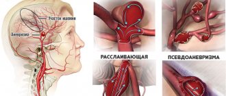
- An acute circulatory disorder or stroke, unlike the previous type of pathology, occurs suddenly with sharp manifestations of symptoms of poor blood supply to the brain. Usually this condition lasts no more than a day. This pathology is a consequence of hemorrhagic or ischemic damage to the brain substance.
Arterial blood supply
A feature of cerebral circulation is the high intensity and almost constant volume of blood, which within the cranium is about 75 ml. More than 20% of the blood during a heartbeat enters the brain tissue. The anatomy of the arteries running in the brain is represented by the elements:
- Carotid system. The pool is formed by the carotid arteries running in parallel. The carotid artery divides into branches. Carotid blood flow is fast and direct.
- Vertebro-basilar system. The pool is created by the vertebral and basilar arteries. Vertebro-basilar blood flow is slow.
- Circle of Willis. Provides blood saturation of areas of the cerebrum. The circle is formed by the branches of the carotid artery, which feed the frontal, parietal, and temporal lobes.
- Circle of Zakharchenko. It lies within the inner surface of the medulla oblongata. The contour is formed by two pairs of arteries: vertebral and anterior spinal.
The walls of the small arteries running in the head and neck are three-layered. They consist of layers - internal (endothelium), middle (smooth muscle) and external (connective tissue). A complete circle of blood circulation is completed in 4-5 seconds.
Diseases due to circulatory disorders
In a healthy person, the middle part of the brain regulates blood supply to the brain. It also controls human breathing and the endocrine system. If he stops receiving nutrients, then the fact that a person’s blood circulation to the brain is impaired can be identified by the following symptoms:
- frequent attacks of headache;
- dizziness;
- difficulty concentrating, memory impairment;
- the appearance of pain when moving the eyes;
- the appearance of tinnitus;
- absence or delayed reaction of the body to external stimuli.
To avoid the development of an acute condition, experts recommend paying attention to the organization of the arteries of the head and neck of certain categories of people who, hypothetically, may suffer from a lack of blood supply to the brain:
- Children who were born by cesarean section and experienced hypoxia during intrauterine development or during labor.
- Teenagers are going through puberty, as their body undergoes some changes at this time.
- People engaged in increased mental work.
- Adults who have diseases accompanied by depletion of peripheral blood flow, for example, atherosclerosis, thrombophilia, cervical osteochondrosis.
- The elderly, since their vessel walls are prone to accumulation of deposits in the form of cholesterol plaques. Also, due to age-related changes, the structure of the circulatory system loses its elasticity.
To restore and reduce the risk of developing serious complications from subsequently impaired cerebral blood supply, experts prescribe medications aimed at improving blood flow, stabilizing blood pressure and increasing the flexibility of vascular walls.
Despite the positive effect of drug therapy, these medications should not be taken independently, but only with a prescription, since side effects and overdose threaten to worsen the patient’s condition.
Diagnosis of vascular disorders
The classification of cerebrovascular disorders can be based on the nature of the pathological processes. Possible:
- Acute stage. In this case, the patient most often experiences a stroke. It occurs suddenly, is characterized by a long course and the development of negative consequences (impaired vision, speech, etc.);
- Chronic cerebrovascular accident. Most often it appears as a result of atherosclerosis or persistent arterial hypertension.
Vascular genesis of the acute type of brain is divided into two main groups:
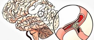
- ischemic stroke, which is characterized by the formation of blood clots in the vessels of the brain, as a result of which a sufficient amount of blood does not flow to it. There is an acute lack of oxygen and the death of some areas of neurons;
- hemorrhagic stroke, which is accompanied by the rupture of a blood vessel and the release of a blood clot.
Chronic cerebrovascular accidents develop gradually and do not always have severe symptoms. There are three main stages, which are accompanied by various symptoms.
When the first signs appear, you must immediately consult a doctor who will conduct a comprehensive diagnosis and determine the cause of this condition. Among the main studies are:
- MRI of cerebral vessels;
- ultrasound examination;
- consultation with a neurologist.
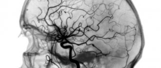
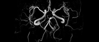
The first method is most often used; it allows you to most reliably determine the location of the blood circulation disorder. Magnetic resonance angiography is considered a more modern technique.
It is not carried out in all clinics and requires special equipment and highly qualified specialists. Using this type of study, it is possible to determine how well the cerebral circulation is functioning and to identify possible pathologies.
Today, the method of electroencephalography remains popular. It is performed for epileptic seizures, speech problems or brain injuries. Thanks to fluctuations in electronic potentials, the doctor can identify possible disorders.
The difficulty of diagnosis lies in the absence of characteristic signs of the disease. The symptoms are very similar to other pathologies, so doctors need to conduct several studies simultaneously to obtain reliable data.
How to improve blood circulation in the brain at home
Poor blood circulation to the brain can significantly impair a person’s quality of life and cause more serious diseases. Therefore, you should not ignore the main symptoms of the pathology and at the first manifestations of a blood supply disorder, you should contact a specialist who will prescribe competent treatment.
Along with the use of medications, he can also offer additional measures to restore the organization of blood circulation throughout the body. These include:
- daily morning exercises;
- simple physical exercises aimed at restoring muscle tone, for example, when sitting for long periods of time and in a hunched position;
- a diet aimed at cleansing the blood;
- use of medicinal plants in the form of infusions and decoctions.
Despite the fact that the content of useful substances in plants is negligible compared to drugs, they should not be underestimated. And if a sick person uses them independently as a preventive measure, then you should definitely tell the specialist about this at the appointment.
Folk remedies for improving cerebral blood supply and normalizing blood pressure
I. The most common plants that have a beneficial effect on the functioning of the circulatory system are the leaves of periwinkle and hawthorn. To prepare a decoction of them, 1 tsp is required. pour a glass of boiling water over the mixture and bring to a boil. Afterwards it is left to infuse for 2 hours, after which half a glass is consumed 30 minutes before meals.
II. A mixture of honey and citrus fruits is also used for the first symptoms of poor blood supply to the brain. To do this, grind them into a pasty state, add 2 tbsp. l. honey and leave in a cool place for 24 hours. For good results, you need to take this drug 3 times a day, 2 tbsp. l.
III. A mixture of garlic, horseradish and lemon is no less effective for vascular atherosclerosis. In this case, the proportions of mixing the ingredients may change. Take it 0.5 tsp. an hour before meals.
IV. Another surefire remedy for improving poor blood supply is an infusion of mulberry leaves. It is prepared as follows: 10 leaves are poured into 500 ml. boiling water and let it brew in a dark place. The resulting infusion is consumed instead of tea every day for 2 weeks.
V. For cervical osteochondrosis, rubbing the cervical spine and head can be done as an addition to the prescribed therapy. These measures increase blood flow in the vessels and accordingly increase blood supply to brain structures.
Gymnastics is also useful, including exercises for moving the head: bending to the side, circular movements and holding the breath.
Venous drainage
Blood channels that collect blood, which is enriched with carbon dioxide, from nerve tissue are presented in the form of jugular veins and sinuses of the dura mater. From the cortex and white matter, movement through the vessels occurs towards the inferior, medial and superolateral surfaces of the hemispheres. An anastomotic venous network is formed in this area. Then it runs through the superficial vessels to the hard shell. A network of deep vessels opens into a large vein. They collect blood from the brain base and internal parts of the hemispheres, including the thalamus, hypothalamus, choroid plexuses of the ventricles, and basal ganglia. The outflow from the venous sinuses is carried out through the jugular canals. They are located on the neck. The superior vena cava is the last link.
Drugs to improve blood circulation
Poor blood supply to the brain of the head is a consequence of serious pathologies of the body. Typically, treatment tactics depend on the disease that causes difficulty in blood movement. Most often, blood clots, atherosclerosis, poisoning, infectious diseases, hypertension, stress, osteochondrosis, vascular stenosis and defects interfere with the proper functioning of the brain.
In some cases, to improve blood circulation in the brain, drugs are used that act to relieve the main manifestations of the pathology: headache, dizziness, excessive fatigue and forgetfulness. In this case, the drug is selected so that it has a comprehensive effect on brain cells, activates intracellular metabolism, and restores brain activity.
When treating poor blood supply, the following groups of drugs are used to normalize and improve the organization of the cerebral vascular system:
- Vasodilators. Their action is aimed at eliminating spasm, which leads to an increase in the lumen of blood vessels and, accordingly, a rush of blood to the brain tissue.
- Anticoagulants, antiplatelet agents. They have an anti-aggregation effect on blood cells, that is, they prevent the formation of blood clots and make it more fluid. This effect helps to increase the permeability of the walls of blood vessels and, accordingly, improves the quality of supply of nutrients to the nervous tissue.
- Nootropics. They are aimed at activating the functioning of the brain due to increased cellular metabolism, while those taking such drugs experience a surge of vitality, the quality of functioning of the central nervous system improves, and interneuronal connections are restored.
Taking oral medications in people with minor disorders of the organization of the circulatory system of the brain helps to stabilize and even improve their physical condition, while patients with severe circulatory disorders and pronounced changes in the organization of the brain can be brought to a stable state.
The choice of dosage form of medications is influenced by a large number of factors. So, in patients with severe manifestations of brain pathology, to improve blood supply, preference is given to intramuscular and intravenous injections, that is, using injections and droppers. At the same time, to consolidate the result, prevent and treat borderline conditions, medications are taken orally.
On the modern pharmacological market, the bulk of drugs to improve cerebral circulation are sold in the form of tablets. These are the following medications:
- Vasodilators:
Vasodilators. Their effect is to relax the walls of blood vessels, that is, relieve spasm, which leads to an increase in their lumen.
Correctors of cerebral circulation. These substances block the absorption and removal of calcium and sodium ions from cells. This approach prevents the work of spasmodic vascular receptors, which subsequently relax. Drugs with this effect include: Vinpocetine, Cavinton, Telektol, Vinpoton.
Combined cerebral circulation correctors. They consist of a set of substances that normalize blood supply by enhancing blood microcirculation and activating intracellular metabolism. They are the following drugs: Vasobral, Pentoxifylline, Instenon.
- Calcium channel blockers:
Verapamil, Nifedipine, Cinnarizine, Nimodipine. Focused on blocking the flow of calcium ions to the tissues of the heart muscle and their penetration into the walls of blood vessels. In practice, this helps to reduce the tone and relaxation of arterioles and capillaries in the peripheral parts of the vascular system of the body and the brain.
- Nootropics:
Drugs that activate metabolism in nerve cells and improve thought processes. Piracetam, Phenotropil, Pramiracetam, Cortexin, Cerebrolysin, Epsilon, Pantocalcin, Glycine, Actebral, Inotropil, Thiocetam.
- Anticoagulants and antiplatelet agents:
Medicines intended to thin the blood. Dipyridamole, Plavix, Aspirin, Heparin, Clexane, Urokinase, Streptokinase, Warfarin.
A frequent culprit of “hunger” of brain structures is atherosclerosis. This disease is characterized by the appearance of cholesterol plaques on the walls of blood vessels, which leads to a decrease in their diameter and permeability. Subsequently, they become weak and lose their elasticity.
Therefore, the use of restorative and cleansing drugs is recommended as the main treatment. These drugs include the following types of drugs:
- statins prevent the body from producing cholesterol;
- fatty acid sequestrants, blocking the absorption of fatty acids, while they force the liver to spend reserves on the absorption of food;
- Vitamin PP - dilates blood vessels, improves blood supply to the brain.
In addition, it is recommended to give up bad habits, fatty, salty and spicy foods.
How to improve cerebral circulation
The normal functioning of the circulatory system is facilitated by a reduction in stressful situations. When a person is under stress, the hormone adrenaline is actively produced, which affects the activity of the central nervous system. Under its influence, disorders appear - tachycardia, hypertension, arrhythmia. Insulin production decreases, which leads to increased blood glucose levels.
Hyperglycemia (increased glucose levels) causes an increase in oxidative stress, tissue acidosis, endothelial dysfunction, and increased permeability of the blood-brain barrier. To improve blood supply to areas of the brain, methods are used:
- Medications – vasodilators, antispasmodics, drugs that prevent the formation of blood clots, improve rheological blood parameters, and normalize intracellular metabolic processes.
- Balanced healthy diet. Reducing table salt, the proportion of animal fats, and foods that contain cholesterol in the diet.
- Sports activities. Physical activity stimulates the activity of the cardiovascular system, accelerates blood flow, improves the flow of oxygen to tissues, and trains the vascular walls, increasing their flexibility and elasticity.
To improve the functioning of the blood supply system, weight correction is carried out if indicated. Breathing exercises will reduce the negative effects of stress. Tinctures and decoctions prepared according to traditional medicine recipes will help improve the condition of the vascular walls.
Prevention
As a complement to the main treatment, preventing the underlying disease will help improve blood supply to the brain.
For example, if the pathology was caused by increased blood coagulation, then establishing a drinking regime will help improve well-being and improve the quality of the therapy. To achieve a positive effect, an adult needs to consume 1.5 to 2 liters of fluid daily.
If poor blood supply to brain tissue was caused by congestion in the head and neck area, then in this case, performing basic physical exercises to improve blood circulation will help improve your well-being.
All the steps below must be done carefully, without unnecessary movements or jerks.
- In a sitting position, place your hands on your knees and keep your back straight. Straightening your neck, tilt your head to both sides at an angle of 45%.
- This is followed by rotation of the head to the left, and then in the opposite direction.
- Tilt your head forward and back so that your chin first touches your chest and then looks up.
Gymnastics will allow the muscles of the head and neck to relax, while the blood in the brain stem begins to move more intensely through the vertebral arteries, which provokes an increase in its flow to the structures of the head.
You can also stabilize blood circulation by performing a head and neck massage with improvised means. So, you can use a comb as a handy “simulator”.
Eating foods rich in organic acids can also improve blood circulation in the brain. Such products include:
- Fish and seafood;
- oats;
- nuts;
- garlic;
- greenery;
- grape;
- bitter chocolate.
A healthy lifestyle plays an important role in recovery and improvement of well-being. Therefore, you should not get carried away with eating fried, highly salted, smoked foods, and you should completely stop drinking alcohol and smoking. It is important to remember that only an integrated approach will help improve blood circulation and improve brain activity.
Clinical picture
If the blood supply to the brain undergoes changes, subjective sensations may be observed that are not accompanied by objective neurological symptoms. These include, in particular:
- Paresthesia.
- Headache.
- Organic microsymptoms without pronounced signs of central nervous system dysfunction.
- Dizziness.
- Disorders of the higher functions of the cortex of a focal nature (aphasia, agraphia and others).
- Disorders of the sensory organs.
Focal symptoms include:
- Movement disorders (impaired coordination, paralysis and paresis, extrapyramidal changes, decreased sensitivity, pain).
- Epileptic seizures.
- Changes in memory, emotional-volitional sphere, intelligence.
Blood circulation disorders, by their nature, are divided into initial, acute (intrathecal hemorrhages, transient disorders, strokes) and chronic, slowly progressive manifestations (encephalopathy, discirculatory myelopathy).
How to detect damage to cerebral arteries?
Early detection of cerebrovascular diseases is of great importance. This may require examination by an experienced neurologist, therapist, vascular surgeon, osteopath, who will quickly determine the main cause of pathological changes in the cerebral arteries and the actual blood supply to the brain using a thorough clinical examination and highly informative instrumental research methods. Usually, ultrasound scanning of extracranial vessels is performed, as well as magnetic resonance imaging with an angioprogram (study of intracranial vessels), as the most informative method of visualizing intracranial cerebral arteries. A thorough examination of the heart and peripheral vessels, endocrine system, kidneys, adrenal glands and other organs is also necessary.