How to take blood from a vein with a vacuum tube
An objective assessment of the patient's condition, based on laboratory data, provides more accurate results than subjective data obtained from the patient.
The results of laboratory tests allow not only to make a timely accurate diagnosis, but also to evaluate the quality of the therapy. That is why medical personnel need to ensure a high degree of reliability of the results. Several factors can influence the degree of reliability:
- preliminary preparation of a person for blood collection;
- time of day at which the material was taken for laboratory research;
- the instruments used for sampling and the technique used to obtain the material;
- compliance with the sampling algorithm.
Why is it important to use vacuum systems?
Laboratory diagnostics is carried out in three stages:
- Preanalytical.
- Analytical.
- Post-analytical.
The duration of the stages and the degree of their influence on the reliability of the data are different.
The longest is the first stage, occupying two-thirds of the duration of any study. Errors made at the preanalytical stage lead not only to an increase in time spent on making a diagnosis, but also to unnecessary waste of budget funds due to the appointment of a repeat procedure. They affect the entire process of correct diagnosis and evaluation of therapy.
The degree of reliability of the data obtained depends on a huge number of variables:
- personal characteristics of a person (gender, age, race, etc.);
- features of eating behavior before submitting laboratory material (fasting, abuse of a certain type of food, etc.);
- intensity of physical and emotional stress;
- natural changes in hormonal levels (phases of the menstrual cycle, pregnancy, menopause, etc.);
- weather and climatic conditions;
- medicines taken by humans;
- position of the patient at the time of material collection.
In addition to the above, the accuracy and correctness of the results depends on the technique of taking blood from a vein, the instruments used for this, the conditions of transportation and storage of the collected material.
When collecting blood from a vein using needles or syringes, it is impossible to standardize the technology for collecting the material. Using needles to collect venous blood can result in the collected material and pathogens of bloodborne infections getting into the hands of medical staff. This creates the danger of further transfer of pathogens to other patients. Taking biomaterial with a syringe practically eliminates this possibility, but when transferring it from a syringe to a test tube, hemolysis of red blood cells caused by mechanical action is possible.
Thus, vacuum systems have become the optimal tool for collecting venous blood.
What you need to know about the needle butterfly?
A butterfly needle is a standard medical needle that has a catheter attached to it. In practice, only two types of needles are used - hollow and surgical. "Butterfly" belongs to the first category. It can be used to perform injections, collect/transfer samples and biological samples. Most often, the needle is used to collect venous blood in children and patients with thin veins. The tubes are made of austenitic stainless steel of one of the types specified in the relevant state documentation.
The production of medical instruments is strictly regulated by the state to ensure maximum safety and quality. The needles themselves are used in pediatrics, some intensive care units, veterinary medicine and gerontology (the science of aging of all living organisms).
A catheter is a medical instrument in the form of a tube, with the help of which the vessels are connected to the external environment for subsequent emptying. A soft catheter is attached to the butterfly needle. It is made of plastic materials such as plasticized polyvinyl chloride or rubber. For the “butterfly”, vascular catheters with a venous cannula are used. A cannula is a hollow soft tube that is inserted into the internal cavity of the human body. The catheter allows not only blood sampling, but also the introduction of drugs into the bloodstream or detoxification.
Absolutely all catheters require additional fixation. They are attached to the skin with patches, suture material or special fasteners. The cannula is equipped with a flexible retainer (made of lightly colored polyethylene), which visually resembles the wings of a butterfly. Because of this similarity, the instrument got its name. The color of the retainer is not chosen for ethical reasons, but to indicate the size of the needle. Size range of needles: 18, 19, 21, 22, 23, 25, 27. Each number is assigned a specific shade of the palette, which helps doctors and nurses quickly navigate the instruments. To avoid confusion, the needle size is marked on the wings of the fixator and is additionally indicated on the sterile individual packaging.
Specifications
| Size(G) | Fixative shade | Inner diameter (mm) | Flow rate(ml/mm) |
| 18 | Pink | 1,2 | 60 |
| 19 | Yellow | 1,0 | 42 |
| 21 | Green | 0,8 | 21 |
| 22 | Black | 0,7 | 11 |
| 23 | Blue | 0,6 | 5 |
| 25 | Orange | 0,5 | 3,2 |
| 27 | Grey | 0,4 | 2,5 |
Benefits of using negative pressure systems
All the benefits of negative pressure systems come from their design. Their use allows:
- completely eliminate contact of medical personnel with the patient’s blood during sampling;
- standardize the process of blood collection and sample preparation, create a simple algorithm of actions;
- reduce the number of operations spent on preparing a sample for testing in the laboratory;
- Primary tubes included in negative pressure systems can be used directly in many automated analyzers. This saves money on purchasing secondary plastic tubes and time on transferring samples into them;
- make the transportation and centrifugation of biomaterials safer, since the tubes are sealed and made of unbreakable materials;
- facilitate the identification and labeling of samples by type of examination, thanks to color coding of the caps of negative pressure systems;
- reduce laboratory material costs for the purchase and processing of additional secondary tubes;
- simplify staff training methods;
- reduce the occupational risk of infection;
- reduce the time spent collecting venous blood using the method discussed in the article.
Sequence of obtaining venous blood using vacuum systems
The process of collecting venous blood consists of three stages:
- preparation for the procedure;
- performing a fence;
- end of material collection.
At the preparation stage in the procedure for taking biomaterial from a vein, medical personnel need to:
- Treat your hands using the scheme provided by WHO.
- When working with blood, each person is considered a potential carrier of a blood-contact infection. Therefore, before starting the blood collection procedure, it is necessary to change into protective clothing.
- Fill out a referral for a blood test in the registration journal. This is necessary for labeling instruments and filling out documents related to one person. The referral contains the patient’s passport details, the date and time of the blood draw, the registration data of the analysis in the laboratory, and the details of the doctor who ordered the analysis.
- Compare the information in the referral with the data of a specific patient.
- Check whether the patient has given informed consent to the procedure, explain in detail the purpose and sequence of its implementation.
- Clarify the patient’s compliance with the rules of food restrictions adopted before taking the tests.
- Conveniently accommodate the patient.
- Prepare the workplace: arrange all the devices necessary for drawing blood, having first ensured their integrity and suitability for use (safety of sterility seals, expiration date, etc.). Select test tubes with the desired color coding and the required volume. Take a needle of the appropriate size.
- Wear a mask, safety glasses, and rubber gloves.
After completing all the steps in the first stage, you can proceed to blood sampling.
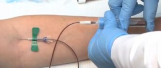
Standard “Venous blood collection using vacuum”.
Vacuum blood collection system is a safe collection system,
Transportation and qualitative analysis of blood samples.
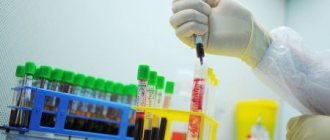
A vacutainer is a fully enclosed vacuum plastic disposable system for drawing blood from a vein.
Consists of three components:
Double-sided needle with safety valve.
Sterile tubes with a certain volume of vacuum.
Advantages:
safety, efficiency, guarantee of sample integrity and research reproducibility, minimum hemolysis, minimum microclots, the time between sample collection and its contact with the additive is constant, accuracy of the blood/additive volume ratio, minimal tourniquet effect, sample sterility.
Indication:
patient examination.
Complications:
hematoma, thrombophlebitis, sepsis.
Place of blood collection:
veins of the elbow, forearm, hand.
Prepare:
sterile: double-ended needle with safety valve, disposable holder, vacuum tubes, cotton balls, gloves, tray, 70% alcohol, tourniquet (venous cuff), oilcloth pad, mask, apron, safety glasses or screen, container for transporting tubes, KBU.
Explain to the patient the purpose and course of the procedure, obtain consent. Check to see if the patient has had breakfast.
Write a referral to the laboratory.
Assist the patient into a comfortable position.
Decontaminate your hands at a hygienic level and treat them with a skin antiseptic.
Wear a mask and gloves, safety glasses, and an apron.
Take the blood collection system.
Check the tightness of the packaging and the expiration date of the main components of the blood collection.
Take the needle, remove the protective cap (when using a 2-sided needle, remove the gray protective cap).
Insert the needle into the needle holder and screw it in until it stops.
Sit or lay the patient down, freeing the upper limb from clothing, placing a pillow under the elbow and lowering the arm down.
Place a venous cuff (tourniquet) on the middle third of the shoulder on underwear or a napkin.
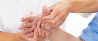
Feel the pulse on the radial artery (the pulse should be preserved).
Explore the vein. Find the most engorged vein.
Ask the patient to clench and unclench the fist several times to fill the vein, then close it.
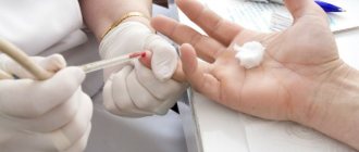
Treat the area of the elbow bend, the injection site sequentially with two cotton balls soaked in alcohol, and throw them into the KBU. Hold the third cotton ball in your left hand between the IV and V fingers.
Hold the system in your right hand.
Secure the vein below the intended puncture site with your left thumb.
Hold the needle and needle holder at an angle of 15 0 relative to the patient's hand. Insert the needle, moving it along the blood flow 1 - 1.5 cm.
Place the needle holder in your left hand, and with your right hand, insert the test tube into the needle holder until it stops and ensure blood flow.
When blood enters the sterile tube, remove the tourniquet.
Remove the tube containing the blood sample from the holder.
Mix the contents of the test tube by inverting it 8 to 10 times, but do not shake.
Insert the next test tube into the holder if you need to draw blood for other studies.
Place the tube in the blood transport container.
Apply a sterile cotton ball to the venipuncture site, remove the needle from the vein and remove it from the needle holder.
Press the sterile ball onto the venipuncture site and ask the patient to bend the arm at the elbow and hold it for 3 to 5 minutes.
Record the data in the blood collection log for testing and send the blood into a vacuum tube in a container along with a referral to the laboratory.
Remove the protective equipment and place it in the CBU.
Wash and dry your hands.
Note:
inserting the test tube into the needle holder all the way ensures the flow of blood, as the needle pierces the rubber membrane and the cap of the test tube. The blood flows into the test tube until it compensates for the created vacuum. No blood flow is an indicator that the vein has been punctured through; in these cases, it is necessary to pull the needle (without removing it) until blood enters the test tube.
When the test tube is removed from the needle holder, the rubber membrane returns to its original position and blocks the blood flow through the needle. If necessary, use different tubes depending on the tests prescribed by the doctor.
The article was written based on materials from the sites: nursehelp.ru, corway.ru, studfiles.net.
Algorithm for collecting biomaterial using a vacuum system
The second stage of the procedure is performed step by step:
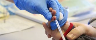
- Examine the proposed venipuncture sites, select a point for the procedure, and palpate the vein. The ulnar veins are most often used, but if necessary, blood can be taken from the veins of the wrist, the back of the hand, above the thumb, etc.
- Secure the tourniquet 10 centimeters above the venipuncture site. When applying a tourniquet, women should not use the hand on the side of the mastectomy. Prolonged compression of tissues and blood vessels (more than two minutes) can lead to shifts in coagulogram parameters and the concentration of certain substances.
- Take the needle and remove the protective cap from it.
- Connect the needle to the holder.
- Ask the patient to clench his hand into a fist. You should not make sudden movements, as this can lead to changes in blood counts. If the vein is poorly visible, you can apply a warm napkin to your arm, or massage your arm from the hand to the elbow. If there are no vessels suitable for venipuncture on one arm, the other should be checked.
- Treat the puncture site with a disinfectant using circular movements from the center to the edge.
- Wait for the antiseptic to evaporate, or remove excess with a sterile dry cloth.
- Remove the protective colored cap from the vacuum system.
- Secure the vein by clasping your forearm. Place your thumb 3˗5 centimeters below the injection site. Stretch the skin.
- Insert the needle with the holder into the vein at an angle of 15°. When inserted correctly, blood will appear in the indicator chamber of the holder.
- Secure the test tube in the holder with the cap facing up. Under the influence of negative pressure, blood will begin to flow into the test tube.
- As soon as blood begins to accumulate in the test tube, loosen the tourniquet or remove it.
- Tell the patient to relax his arm and unclench his fist.
- When the flow of blood into the test tube stops, remove it from the holder.
- Mix the biomaterial with the preservative. Do not shake! The test tube can only be gently inverted.
- If several samples are taken from the patient, the holder with the needle is left in the vein and steps 11-15 are repeated sequentially.
Butterfly catheter connected to luer adapter and needle protection mechanism
The wings of the Improvacuter butterfly needle provide effective fixation and ease of insertion into the vein. The safety mechanism ensures single use and prevents the risk of injury to personnel. Due to the flexibility of the tube, the needle does not move during manipulation, which minimizes the risk of mechanical damage to the vessel wall. Butterfly needles are sized according to needle sizes and are also color coded. Available in lengths of 190 and 300 mm and diameters from 0.5 to 0.9 mm. (20, 21, 22,23, 24, 25 G).
Price
| Code | Needle size | Needle diameter | Needle/catheter length | Price per piece in rub. | Cap color | Packaging min/box | |
| 596121300 | 21Gx3.4″x12″ | 0.8 mm | 300 mm | 32,00 | 100/2000 | ||
| 596121190 | 21Gx3.4″x7″ | 0.8 mm | 190 mm | 32,00 | 100/2000 | ||
| 596122300 | 22Gx3.4″x12″ | 0.7 mm | 300 mm | 32,00 | 100/2000 | ||
| 596122190 | 22Gx3.4″x7″ | 0.7 mm | 190 mm | 32,00 | 100/2000 | ||
| 596123300 | 23Gx3.4″x12″ | 0.6 mm | 300 mm | 32,00 | 100/2000 | ||
| 596123190 | 23Gx3.4″x7″ | 0.6 mm | 190 mm | 32,00 | 100/2000 | ||
| 596125300 | 25Gx3.4″x12″ | 0.5 mm | 300 mm | 32,00 | 100/2000 | ||
| 596125190 | 25Gx3.4″x7″ | 0.5 mm | 190 mm | 32,00 | 100/2000 | ||
Also purchased with this product:
- Medical hemostatic tourniquets, venous
- Medical pillow, blood collection pillow
- Treatment table
- Injection adhesive plaster
Related to this product:
Butterfly catheter needle (infusion cannula) - intended for administering medications into peripheral small veins during intravenous infusions, or taking blood for analysis - especially in patients in an unstable condition (neurosis, intoxication, excitability, epilepsy, etc.), as well as in infants and young children. The butterfly catheter is very often used in veterinary medicine. Best price! Read more…
Possible errors when using vacuum systems
When using vacuum systems to collect venous blood, you may encounter the following problems.
Blood does not flow into the tube after it is connected to the holder. There may be several reasons:
- the needle did not enter the vein. In this case, without completely removing the needle from under the skin, you need to change its position;
- the tip of the needle rested against the wall of the vein. It is necessary to carefully adjust the position of the needle;
- the vein is pierced through. It is also necessary to adjust the position of the needle.
In all of the above cases, you do not need to disconnect the test tube from the holder unless you need to remove the needle from under the skin.
The test tube received less blood than required. The reasons for this: the vein collapsed due to low pressure, air got into the test tube. In the first case, you need to disconnect the test tube from the holder and wait some time, during which the vein will fill again. In the second, the system must be replaced and the entire procedure must be performed all over again.
Compliance with the sequence of actions of the algorithm for taking biomaterial using vacuum systems allows you to improve the quality of laboratory analyzes and optimize the work of personnel.
source
Possible mistakes
When a medical professional takes venous blood for analysis, he may unwittingly make some mistakes. Sometimes the biomaterial does not go into the test tube after it has been connected to the needle holder. Often the reason is that the needle did not enter the vein. You need to change its position; you don’t need to pull it out completely. Sometimes this happens because the end of the needle is stuck into the vein wall. Needs to be corrected carefully. If the puncture is made through the entire vein, then blood will also not flow into the test tube. Here, too, the situation is being carefully corrected.
In all these situations, the container is not disconnected from the adapter (holder). If less than the required amount of biomaterial enters the vacuum tube, the reason may be due to air penetration into the device. You will need to do the job a second time using a new vacutainer. Low pressure may develop in the vein, causing it to collapse. You should disconnect the tube from the holder and wait until the vein is filled with blood.
What are the features?
Vacuum system is a set consisting of sterile double-sided needles of different diameters with holders and test tubes with special reagents for taking a particular analysis. Vacuum blood sampling, in contrast to a disposable syringe, has a number of features, which include:
- obtaining accurate answers, because when conducting a study, venous blood no longer has to be transfused into different tubes,
- eliminating confusion and substitution of tests, since each test tube is provided with a number,
- absence of broken glass, because the coating of the systems is made of unbreakable plastic material, which guarantees the safety of the blood directly in the test tube,
- completing the procedure in less than 10 seconds,
- absence of contact of the biomaterial with the environment and people, because not a single drop of blood will fall on clothing, which is important in order to protect against infection for health workers and hospital technical staff,
- absence of venous injuries, since when performing 2-3 examinations at once, there is no need to remove the needle, search for and grab the vein again - just change the container.
All test tubes for taking biochemical analysis are marked according to the GOST table and have a specific cap color that matches the composition contained in the test tube.
So, if the vacuum system has a cover:
- green – contains heparin for immunochemical, biochemical studies,
- gray - it is used for taking glucose, diagnosing varicose veins,
- blue – contains sodium citrate in the form of a gel for testing serum coagulation, ESR,
- purple – the reagent is suitable for immunochemical tests,
- blue – contains ADTA acid to detect heavy metal salts in the blood,
- pink is used for testing donor blood,
- red – for biochemistry, immunochemical test, determination of the Rh factor.
The vacuum system consists of a set of sterile needles, varying in size, length and diameter to suit the patient's veins. For more convenient sampling, the needles are double-sided and are inserted with one side into the patient’s vein, and the other into the stopper of the elastic cap of the tube for collecting blood from the vena cava. It is allowed to use butterfly needles with protrusions, which are convenient for piercing thin and poorly accessible veins.
Butterfly shaped
If you need to take blood from a vein, use a special double-ended needle. Only it guarantees the tightness of the system when the blood enters the test tube without any contact with the external environment. The part of the needle that is intended for insertion into the patient’s vein is treated with silicone in order to make this process as smooth and almost painless as possible. The other part is covered with a soft rubber valve to prevent blood from flowing back.
The nurse first inserts the needle with one side into the holder, then, after applying a tourniquet to the forearm and treating the skin with an antiseptic, inserts the other end of the needle into the vein. All you have to do is wait until the tube is filled with blood - thanks to the vacuum, this will happen quickly, in just 7-8 seconds. The vacuum tube and needle cannot be reused.
For children, there is a modification of the needle with a long (30 cm) catheter, similar to a butterfly. It is needed so that the baby’s hand can move during manipulation: children get scared and cannot force themselves to sit without moving. Such needles are very thin, they do not move inside the vein and cannot damage the vascular wall. Thanks to this, “butterflies” are widely used in providing care to intensive care patients.
Article on the topic
Diagnostic rules: who needs an immunogram and what the analysis will tell you about. By the way, such needles are also called “butterflies” because on both sides of the needle there are flexible clamps - “wings”.
How does a vacutainer work?
The operating principle of vacuum systems is similar to the Westergren method. After collecting clinical blood, the container is shaken so that the biological fluid is mixed with the chemical components to begin the reaction. This is important, especially for identifying the exact characteristics of red blood cells and their sedimentation rate.
Today, vacuum containers are used in pediatrics to take blood from a child, which makes the procedure painless, unlike a regular needle puncture. Children and even newborns behave calmly, without tears.
The vacuum closed-loop blood sampling system confidently replaces traditional finger or vein sampling because it is effective, convenient and safe. The sampling is carried out quickly, and this is also important for obtaining an accurate analysis result.
Technique for collecting blood from a vein using vacuum tubes
Taking blood from a vein using a vacuum system is practically no different from using a conventional syringe with a needle. Only the procedure has become safe and comfortable for patients and doctors. When the material enters the test tube, a volumetric vacuum is created, allowing you to take exclusively sterile blood and in the required quantity. You can see how blood is drawn using a vacuum system in pictures and videos.
Today, a vacutainer is the safest way to take blood from a finger or vein. This device greatly simplifies the collection procedure; it can be purchased for personal use at a pharmacy without any prescription.
This is important primarily for laboratory technicians, since it is enough to select the right tube with the appropriate marking and perform the procedure safely, conveniently and quickly. There are no contacts with biomaterial. The main thing is to follow the algorithm of actions when working with the vacuum system.
A blood sample is taken using a vacutainer by a nurse as follows:
- in the laboratory, the vacutainer is unsealed, the cap is removed from the disposable needle,
- a tourniquet is applied just above the elbow,
- the puncture site is treated with medical alcohol,
- the needle is inserted with one side into the test tube holder, and the other into the patient’s vein,
- All you have to do is watch when the blood itself is drawn into the vacuum tube in the required quantity, which is important for carrying out this or that study,
- the container with blood is disconnected without removing the needle from the vein, which is convenient for patients, because there will be no bruises on the skin,
- The container with liquid is tightly and hermetically sealed
- The needle is carefully removed, the puncture site is treated with an antiseptic, and a cotton swab is applied.
After this, blood collection with the vacutainer can be considered complete.
Many patients are not yet familiar with vacuum systems, so doctors should conduct an explanatory conversation immediately before taking a general blood test or from a vein to explain the meaning and essence of using such a method, talk about the pros and cons of the vacuum system. It is imperative to obtain the patient’s consent to vacuum blood sampling from a vein, tell about the course of action, and perhaps give written instructions or a description of the vacutainer device to read.
Preparing for the study is of no small importance, because it guarantees a quick and painless analysis and obtaining an accurate and informative interpretation of the results.
You can learn more about the process of collecting biomaterial using a vacuum system from the video:
source
Instructions for collecting blood from a vein
To infuse a substance into a vein or draw blood, it is important to follow the instructions. For doctors, the postulate is the phrase “do no harm.”
- First you need to wash your hands and arm yourself with sanitizer, gloves and a mask;
- Then we fill the system and secure the bottle;
- We perform the usual manipulation before puncture: see if there is air in the valve;
- We carry out point 1.;
- Open the package;
- Place a cushion under the limb, apply a tourniquet (maximum 1 minute);
- Checking the pulse;
- We massage the site where the catheter is placed;
- Select the filled vein;
- We process the place;
- Then you need to connect the product to the system;
- After a drop of solution appears, the needle is placed with the cut side up and the wings are bent. The cap is no longer needed;
- Fix the vein;
- The puncture is performed at an angle of 10-15˚;
- We wait for the blood to appear;
- Afterwards the tourniquet must be relaxed;
- Open the clamp; Important! If there is no infiltration, then we do not remove the device from the vein.
- We fix the device with adhesive tape;
- We give recommendations to the patient;
We told you about the use of the butterfly needle, its features and types. We hope you found the information useful.
Vacuum systems: equipment and purpose
A standard vacuum blood collection system consists of three components:
- double-edged venipuncture needle
- holder (adapter) for fixing the needle
- vacuum tube with reagent
All components of the system are intended for one-time use only. The principle of operation of the system: under the influence of a vacuum, blood is drawn into a test tube with a reagent and immediately mixed with it. A certain amount of reagent and the volume of blood required for the study provide the exact ratio of components for the study.
The wall of the blood collection needle is ultra-thin, which increases its internal lumen. The outer and inner surface of the needle wall is coated with silicone to reduce trauma to the patient and improve free blood flow. The V-shaped laser sharpening of the cut facilitates painless, smooth entry of the needle into the vein through the skin.
On the tip side for piercing the test tube, the needle is equipped with a thread and a protective rubber membrane. The needles are sealed in two plastic cases (caps) and sealed with a label to prevent their reuse. The color coding of the caps helps in choosing the needle diameter.
Different needle designs allow you to choose the right type of needle:
- standard double-edged needle
- double-edged needle with a visual transparent camera to control entry into the vein
- butterfly needle for collecting blood in children and patients with poorly defined small veins
Color coding of needle sizes according to ISO (International Organization for Standardization)
| View | Color | Needle diameter | G (gauge size) |
| Yellow | 0,30 | 30G | |
| Red | 0,33 | 29G | |
| Azure | 0,36 | 28G | |
| Light gray | 0,40 | 27G | |
| Brown | 0,45 | 26G | |
| Orange | 0,50 | 25G | |
| Light purple | 0,55 | 24G | |
| Blue | 0,60 | 23G | |
| Black | 0,70 | 22G | |
| Dark green | 0,80 | 21G | |
| Yellow | 0,90 | 20G | |
| Cream | 1,10 | 19G | |
| Pink | 1,20 | 18G | |
| Scarlet | 1,40 | 17G | |
| White | 1,60 | 16G | |
| Gray-blue | 1,80 | 15G | |
| Pale green | 2,10 | 14G | |
| Purple | 2,40 | 13G | |
| Blue | 2,70 | 12G | |
| Yellow-green | 3,00 | 11G | |
| Olive brown | 3,40 | 10G |
Needle holders
Holders, or adapters, are designed for safe and convenient insertion of a needle into a vein and connection of the needle to the test tube. The holder is a mandatory component of the vacuum blood collection system for fixing the needle with a vacuum tube. The thread of the holder matches the thread of the blood collection needle.
Types of holders
| Medical needle holder |
| Needle holder with needle protection |
The holder with needle protection is equipped with a special shield. Using a protective shield, the needle is closed immediately after the procedure, eliminating the risk of additional trauma to both the patient and the healthcare worker.
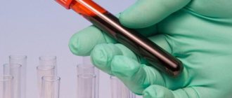
Vacuum tubes
Tubes for vacuum blood collection are made of polyvinyl chloride. They have a safe lid with a rubber stopper. Lids vary in color depending on their use for a particular study.
Medical needles
There are three types :
- Puncture needle; A needle is needed to introduce or remove fluid from organs.
- Surgical needle; The needle is designed for sewing fabrics. Used in surgery, ophthalmology, microsurgery.
- Injection needle; Everyone has seen such a needle. It is used to suck out or introduce substances into the body. For example, anesthesia is carried out in any field of medicine, so the injection needle is one of the most common.
Butterfly needle (injection needle)
Few patients pay attention to the instruments with which doctors, for example, draw blood.
A clinical blood test is necessary for every person. With its help, we learn about the level of hemoglobin in the body, how many red blood cells, leukocytes, platelets are contained in the blood, and it is easier for the doctor to make a diagnosis if the analysis shows deviations from the norm.
A butterfly needle is used to draw blood from a vein.
Varieties of needle butterflies
There are two types:
1) Blood collection needle
Structure:
- Needle;
- Catheter;
- Luer adapter (connection) is convenient because it allows you to pour the substance more than once.
- Tube – Due to the flexibility of the tube, the needle does not move during blood sampling, which eliminates injury to the vessel.
- Wings – Helps secure the needle.
Needle size – 19mm. Needle diameter – 0.6 mm. Catheter length – 190 mm.
Purpose:
When drawing blood, a butterfly needle is used in any case, but it is indispensable when the patient is “difficult.” If blood is collected slowly and it is difficult to pierce a person’s skin, then such a needle is a way out of difficult situations. In addition, the “butterfly” is used when taking blood from people in a state of alcoholic intoxication, increased excitability, or neurosis.
2) IV needle
In order for the substance to penetrate the body faster, an intravenous injection is used.
Structure:
The components of the butterfly needle for internal injections are the same.
- The needle is laser sharpened.
- Catheter;
- Luer adapter (connection);
- A tube;
- Wings;
The differences are in the length of the tube, the diameter and the length of the needle. Tube length – 30 cm. External diameter: 0.8 mm. Needle length: 19 mm.
Purpose:
Used for infusion of medications into small veins of patients and animals.
Features of needle butterflies
- Stainless steel;
- Protective cap;
- Sharpening helps to insert the needle without injuring the skin and veins;
- The needle is covered with silicone - eliminates unpleasant sensations.
- The wings are painted in accordance with GOST;
- The “butterfly” is marked – Allows you not to make a mistake in choosing a needle.
- The kit includes a flexible, thin polyvinyl chloride tube;
Advantages of a butterfly needle
The needle is universal. Suitable for both young and old people. Another advantage is the ability to manipulate newborns and children of any age. The catheter is fixed, so even the most “noisy”, frightened patients will not be injured.
Vacuum blood sampling: preparation for the procedure
The nurse before the blood sampling procedure:
- Greets the patient and introduces himself.
- Asks the patient to introduce himself and identifies him.
- Informs the patient about the procedure prescribed by the doctor and explains its progress.
- Make sure that there is voluntary informed consent for the upcoming procedure.
- Suggests and helps the patient take a comfortable position while sitting in a chair (on a chair) or lying on a couch.
- Wash hands hygienically, dry them with a towel or napkin.
- Treats hands with skin antiseptic and allows skin to dry.
Preparation of equipment and workplace
- Check that all necessary equipment is available.
- Check the expiration date and integrity of the vacuum system packaging.
- Check the expiration date and tightness of antiseptic wipes.
- Take the needle by the long colored cap in one hand, and with the other hand remove the short colored cap from the side of the rubber membrane.
- Insert the freed end of the needle with the rubber membrane into the holder and screw it until it stops.
- Place the needle and holder on the tray.
- Prepare the necessary test tubes in sufficient quantity.
- Wear a mask, safety glasses, and an oilcloth apron.
- Treat your hands with a skin antiseptic.
- Wear sterile gloves.
Vacuum blood sampling: manipulation
- Ask the patient to make room for the upcoming venipuncture (most often the elbow, possibly the forearm, the back of the hand).
- Place an oilcloth pillow under the venipuncture site.
- Apply a tourniquet 7-10 cm above the venipuncture site on top of clothing or a diaper. The tourniquet is applied so that the pulsation of the nearby artery is maintained. The tourniquet application time is no more than 1 minute.
- Ask the patient to clench his fist (do not clench or unclench, but only clench and hold in that position!)
- Inspect and palpate the vein for venipuncture.
- Treat the venipuncture site with antiseptic wipes twice - first a large area, then the venipuncture site. Use 2 or more antiseptic wipes. The skin is processed from the center to the periphery in a circular motion.
- Hold the vacuum system in one hand and remove the protective cap from the needle with the other hand.
- With your free hand, stretch the skin below the venipuncture site at a distance of 4-5 cm to secure the vein.
- Puncture the vein at an angle of 10-15º, insert the needle along the vein no more than 1/2 of the length.
- Take the test tube and insert it into the holder until it stops.
- Once the blood begins to flow into the tube, remove or loosen the tourniquet.
- Ask the patient to unclench his fist.
- Draw the required amount of blood into a test tube.
- Remove the tube from the holder when blood stops flowing into it.
- Carefully invert the tube several times to mix the blood with the filler. Do not shake the test tube!
- Place the test tube in a rack.
- If necessary, connect the next test tube to the system and repeat all steps.
- When using a two-component system, connect the needle with the holder to the syringe tube by turning it clockwise.
- After venipuncture, pull the piston towards you with a slow and smooth movement, loosen the tourniquet.
- When taking blood into several tubes, the syringe tube is disconnected from the needle by turning it counterclockwise, and the next syringe tube is connected by turning it clockwise.
- Draw the required amount of blood into all tubes.
- Apply a sterile napkin with an antiseptic to the venipuncture site and remove the needle.
- Ask the patient to press down on the tissue with his free hand.
- Place the needle in a sharps container - Class B waste.
- Place the holder in another container for collecting Class B waste.
- Replace the patient's antiseptic wipe with a dry sterile wipe, and apply a pressure bandage or adhesive bandage over it.
- Inquire about the patient’s well-being and make sure everything is okay.
- Label the tubes with the collected blood samples, indicating the patient's last name and initials and the time of blood collection.
- Place used consumables in containers for class A and B waste, in accordance with the requirements of SanPiN and internal orders.
- Make the necessary notes in medical documentation on paper and electronic media.
- Send the collected material with accompanying documents to the laboratory.
Algorithm for taking blood using a vacuum system
The method of collecting venous blood with vacuum tubes is similar to using a syringe, but provides greater safety, efficiency and convenience. The collection is carried out quickly, which is important to guarantee an accurate research result.
When collecting blood from a peripheral vein using a vacuum system, you will need:
- vacuum tubes;
- tourniquet;
- cotton wool (cotton swabs) or napkins;
- antiseptic (medicinal alcohol);
- bactericidal patch;
- sterile medical tray;
- medical clothing (gown, goggles, mask and gloves).
Before the procedure, it is necessary to fill out a patient referral, treat your hands with a special solution, and put on protective medical clothing.
Technique for collecting blood from a vein
- Prepare test tubes that correspond to the stated tests or laboratory tests required by the patient, a needle, a holder, alcohol wipes or a cotton swab, and a patch.
- Place a tourniquet on the patient's shirt or diaper 7-10 cm above the venipuncture site. Ask the patient to make a fist.
- Select a venipuncture site. The middle ulnar and saphenous veins are the most commonly used, but smaller and more plethoric veins of the dorsum of the wrist and hand can also be punctured.
- Take the needle and remove the cap from the rubber membrane side. Insert the needle into the holder and screw it in until it stops.
- Disinfect the venipuncture site with a gauze pad. You must wait until the antiseptic solution has completely dried.
- Remove the protective cap on the other side. Insert the holder-needle vacuum system into the vein in accordance with the algorithm for conventional blood collection using a syringe. Make sure that the needle is cut upward at an angle of 15º relative to the surface of the skin. Since the other end is covered with a membrane, blood does not flow through the needle. Using smooth and quick movements, the skin and vein wall are punctured. Deep immersion of the needle should be avoided.
- Insert the test tube as far as it will go into the holder. As a result, the needle pierces the membrane and the plug, and a channel is formed between the vacuum tube and the vein. The needle should not be moved when blood begins to flow. The process continues until the vacuum in the test tube is compensated.
- The tourniquet should be removed or loosened as soon as blood begins to flow into the vacutainer. Make sure the patient unclenches their fist.
- After the blood flow stops, the tube is removed from the holder. The membrane returns to its original position, the blood flow through the needle is blocked. If necessary, other tubes can be connected to the holder to collect the required volume of blood. Immediately after filling, the test tube must be carefully inverted to mix the sample with the filler: test tube without anticoagulants - 5-6 times; test tube with citrate - 3-4 times; test tube with heparin, EDTA and other additives - 8-10 times.
- After filling the last tube, disconnect it from the holder and remove the holder-needle system from the vein. To ensure safety, remove the needle from the holder and place it in a special container for disposal.
- A sterile napkin/cotton ball moistened with an antiseptic is applied to the puncture site, or a bactericidal patch is applied.
- The tubes are labeled and placed in a special container for transportation to the laboratory.