Structural features
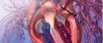
A normal umbilical cord connects the front of the abdominal wall to the placenta (an organ necessary for the bond between mother and baby). A properly formed umbilical cord has the following components:
- Two umbilical arteries. They carry blood from the fetus to the placenta.
- Umbilical vein. It works like arteries, only in the opposite direction.
- Urachus. This is the name given to the duct that connects the placenta to the baby’s bladder.
- Wharton's jelly. It is a connective matter that protects the vessels of the umbilical cord.
- Vitelline duct. Connects the intestines of the embryo with the yolk sac (responsible for hematopoiesis and the production of germ cells).
What is the pathology?
The only umbilical cord artery in the fetus is a pathology in which the structure of the connecting element changes (instead of two - one). This happens quite often: the probability is 1:20 for a multiple pregnancy and 1:200 for a singleton pregnancy. The risk of developing pathology increases if the expectant mother has diabetes.
One artery may be absent from the very beginning of pregnancy or may lose its function at any time (eg, atrophy). The structure of the umbilical cord is clearly visible on ultrasound from twenty weeks. Most often, a single umbilical cord artery is a single pathology and is not associated with any congenital anomalies (in 75% of cases). However, there is a chance that the child has developmental disabilities (25%). Most often, the pathology problem is accompanied by problems with the heart, kidneys, intestines and bones.
What is the single artery of the fetal umbilical cord?
An anomaly in the structure of the umbilical cord, when instead of two arteries one is formed, is called a single umbilical cord artery.
The absence of one artery in the umbilical cord can be initial (congenital aplasia) or develop during pregnancy (artery atrophy, which occurs as a result of the complete cessation of its functioning).
A single fetal umbilical cord artery is considered a fairly common pathology: 1 in 200 singleton pregnancies – 0.5% and 1 in 20 multiple pregnancies – 5%.
Normally, the umbilical cord contains 2 arteries and 1 vein
Diagnostics
To give birth to a healthy baby, any pregnant woman must be constantly monitored by a doctor and undergo all tests. If there is any suspicion, the unborn child must be carefully examined for various developmental defects and markers of genetic diseases. Don't be afraid of all these studies, because there is nothing more important than health.
The following methods will help identify the only umbilical cord artery during pregnancy:
- Ultrasound (ultrasound examination). The bladder must be full before passing.
- Doppler. Determines the presence and speed of blood flow.
- Examination by a gynecologist. Using special instruments, the doctor can listen to the baby’s heartbeat and, if there is any suspicion, prescribe additional examinations.
If a pathology is detected, doctors will suggest undergoing karyotyping of the fetus. This procedure involves the collection of amniotic fluid or chorionic villi, as well as detailed ultrasound and echocardiography.
The only umbilical cord artery during pregnancy can be seen on ultrasound during transverse scanning, in which the lumen of the vessels is best visible. Diagnosis of pathology is possible from the end of the first trimester. At earlier stages, the umbilical cord is not clearly visualized, so it will not be possible to make an accurate diagnosis. Difficulties may also arise if a woman has oligohydramnios, obesity, or multiple pregnancies.
The umbilical cord has 3 vessels, what is it, what is the threat of 2 vessels?
You are here: Home > Articles > Pregnancy and childbirth > Pregnancy
June 5, 2020 | views: 25,820
How many vessels should the umbilical cord have - a “conductor” connecting the fetus with the mother during pregnancy, a supplier of nutrients and oxygen. Dopplerometry of the umbilical cord vessels is among the studies required for women who are preparing for motherhood. The procedure is carried out at 21 weeks, with its help the number of arteries is determined. The study allows you to timely identify abnormalities in the development of the embryo and take the necessary measures.
If, in the results of Dopplerometry, the expectant mother discovers that the umbilical cord has 3 vessels - what does this mean? If 2 vessels are diagnosed in the umbilical cord, what threat does this pose to the embryo?
The structure of the umbilical cord is normal
To understand why the umbilical cord has 3 vessels and what this means, you need to understand its structure. This is the name given to a kind of “rope” that ensures the connection between the embryo and the mother’s body and the interaction of the circulatory systems. The formation of the connecting element is completely completed by approximately 12 weeks. The length of the formation is 40-70 cm, “used” before the birth of the child. Normally, it is adjacent to the middle part of the placenta; there are exceptions (for example, membrane attachment).
How many vessels should there be in the umbilical cord? The norm is 2 arteries and one vein – 3 vessels. The vein serves as a conductor through which oxygenated blood penetrates the baby’s circulatory system from the woman’s body. Nutrients also leak into the vein through the vein. Arteries are used to remove waste products. 3 vessels in the umbilical cord during pregnancy indicate no cause for concern.
The structure of the umbilical cord - pathology
The norm that the umbilical cord must meet is 3 vessels, what this means can be read above. However, sometimes there is a deviation from this rule - two vessels in the umbilical cord. Expectant mothers are forced to face the medical verdict “EAP”, which stands for “single artery” of the umbilical cord, in about 0.5% of cases. The risk increases when it comes to multiple pregnancies, reaching 5%. The threat increases if a woman suffers from diabetes.
2 vessels in the umbilical cord - what does this mean, if there is a reason for concern? One of the arteries may be absent immediately, or its functionality may be impaired while waiting for the baby. EAP does not belong to the category of diagnoses that are fatal to the fetus, but requires additional research.
2 vessels in the umbilical cord - danger
So, the umbilical cord has 2 vessels - what does this mean for the baby? In approximately 90% of cases, EAP pathology is an isolated abnormality and does not turn into a threat to the fetus and mother. Despite the increasing load placed on the vessel, its functioning is not impaired. A small baby is born in about 13% of cases.
The umbilical cord vessels (2 or 3) are not of significant importance at the stage of childbirth. The main thing is that the specialist and junior medical staff attending the birth are aware of the presence of pathology. Labor management tactics, selected specifically for such a case, guarantee that there is no danger for the woman in labor and the fetus.
2 vessels in the umbilical cord - what are the dangers of deviation from the recognized norm during the birth process? If it is carried out incorrectly, hypoxia of the embryo is possible (lack of oxygen, complete interruption of its supply). To avoid danger, the doctor may choose a caesarean section. It is also necessary to cut the umbilical cord as carefully as possible - there is a risk of disrupting blood flow.
The umbilical cord has 2 vessels - what does this mean according to doctors? In such situations, obstetricians-gynecologists necessarily insist on additional examination, which is prescribed to the pregnant woman.
2 vessels in the umbilical cord - examination
It’s great if there are 3 vessels in the umbilical cord. However, timely diagnosis of deviations from this norm allows one to avoid risk even with one artery. The gynecologist must prescribe an additional examination to the woman to make sure that there are no other genetic pathologies that develop against the background of the disorder.
The number of vessels in the umbilical cord, normality or pathology - these questions can be clarified already by the 20th week, when the pregnant woman can undergo Dopplerometry. Most often, the umbilical cord has 3 vessels at 20 weeks. If, as a result of the study, it is determined that the number of arteries is not normal, Doppler testing is indicated for the expectant mother until birth. The procedure will provide control of blood flow in the vessels and will allow recording deviations.
Knowing how many vessels there should be in the umbilical cord, when diagnosing a disorder, it is worth consulting with a geneticist. An examination carried out by this specialist will help to reject the assumption of chromosomal abnormalities. A pregnant woman is advised to test blood obtained from the umbilical cord (cardocentesis). An ultrasound of the embryo's heart is required to exclude heart pathology - up to 22-24 weeks. Prevention tools - ultrasound of organs (each), cardiotocography (CTG).
2 vessels in the umbilical cord - is it necessary to terminate the pregnancy?
How many vessels are in the umbilical cord is a question, the answer to which is absolutely unimportant for the future life of the child. The diagnosis of EAP only indicates the need for additional examinations. A threat arises only when other chromosomal abnormalities that can harm the fetus are attached to a single artery.
5 comments on this post
- Nnn
May 20, 2020 at 6:29My youngest son had two vessels instead of three... She carried it normally, although she often went for ultrasounds to monitor the baby’s development. Everything went well, she also gave birth without complications. The weight was 3.900. He grew and developed well... But! When he began to grow up, it was as if he had developmental delays. He only spoke when he was 4 years old. Now he is 7. He has bad memory. They tried to go to different clubs, he can’t do it, he can’t remember anything... He can’t even do simple poems, addition and subtraction don’t work either... He’s absent-minded, he always has to be reminded of everything, he even forgets his briefcase, he also constantly stumbles and falls... Not I know if this is somehow connected with his intrauterine development, but he’s the only one I have….
- Oksana
Mar 21, 2020 at 18:47I was observed by a doctor throughout my pregnancy and gave birth on my own, quickly and without complications. Weight and height are good. The child is very active, has a good memory, at the age of 6 he reads, writes, and counts; his son’s development is simply excellent. She is ahead of her peers, which I am very happy about, because I read that children with two vessels, on the contrary, lag behind... but there is one thing... vision fails -5, -7 and a whole bunch of diseases associated with the eyes. We get treatment and go for procedures, but in general everything is fine with health, I rarely get sick.
- Madina
Oct 29, 2020 at 18:14My daughter also had 2 vessels, she gave birth on her own, the birth went well, her height was normal, but her nails were too short, and today she is three years old and her nails still don’t grow, but only along with her fingers, we don’t cut them, on her toes nails are not beautiful, deformed((((
- Anna
Jun 25, 2020 at 3:57My daughter had two vessels in her umbilical cord. She is already 8 years old, an excellent student, good memory, 100% vision, generally excellent health, tall, does not lag behind in anything, but on the contrary, at school she took part in all the clubs you can, and she entered art school. The doctors told me that 2 vessels do not affect what kind of child will be born, this is a variant of the norm!
- Elena
Oct 25, 2020 at 8:16The number of vessels only affects the nutrition of the fetus. Vision, memory, and mental abilities do not depend on the number of vessels and arteries. I have a 5-year-old son, a completely healthy and active child, although there were 2 vessels. And this happens much more often than 0.5%, I only have 2 friends and everything is fine.
Leave your comment: Cancel
Causes
Problems with the vessels of the umbilical cord are not fully understood, but most often they are associated with:
- infectious diseases that the pregnant woman suffered in the first trimester;
- intoxication;
- bad habits (alcohol, smoking, drugs or potent drugs);
- diabetes mellitus;
- radiation exposure, poor ecology, hazardous production;
- multiple pregnancy;
- chromosomal disorders.
The most dangerous period of pregnancy is 6-9 weeks. At this stage, the risk of pathologies of blood vessels, placenta and blood flow increases. Artery atrophy (one of them stops functioning) can be caused by: thrombosis, tumors, hematomas or nodes (on one of the arteries). It should be remembered that such conditions occur quite rarely, but require constant monitoring.
A woman's health condition can also lead to the formation of a single umbilical cord artery during pregnancy. Causes may include: diabetes mellitus, multiple pregnancy, kidney disease, heart disease.
EAP during pregnancy: what is it, causes and consequences
Single umbilical artery syndrome ( USA ) is quite common among all fetal malformations and accounts for an average of 1.5% of all full-term pregnancies.
This pathology attracts many scientific researchers who have two opposing opinions. Some of them argue that EAP is an accidental finding that does not require any medical intervention and does not in any way affect the course of pregnancy or the outcome of childbirth.
Others, on the contrary, equate this disease to a syndrome combined with dangerous perinatal complications.
Normally, the umbilical cord has 3 vessels: 2 arteries and 1 vein . They are surrounded by connective tissue. The umbilical cord is a vascular-mesenchymal organ that exchanges blood between the tissues of the placenta and the fetus.
Moreover, 2 umbilical arteries carry out the outflow of blood saturated with carbon dioxide and metabolic products to the placenta, and the vein supplies the fetal body with oxygen and vitamins.
The intrauterine life of a child largely depends on the condition of this organ and the characteristics of its development.
Sometimes it happens that:
- 1 umbilical artery is either obliterated, that is, atrophied.
- Or it simply does not form, that is, congenital aplasia occurs.
This syndrome is called a single umbilical cord artery.
There are 4 types of syndrome:
- 1 type There is 1 artery in the umbilical cord. It arises from the right and left common iliac arteries. And 1 left umbilical vein.
- Type 2 The umbilical cord has 1 left umbilical vein and 1 artery (derivative of the vitelline duct).
- Type 3 The umbilical cord has 1 artery arising from the vitelline duct and 2 umbilical veins.
- Type 4 The umbilical cord has 1 artery and 1 persistent right umbilical vein.
This pathology is diagnosed during the first ultrasound screening at the end of the 1st trimester (from 12 weeks).
Causes of EAP
The causes of this pathology have not yet been fully studied, but attention should be paid to the main predisposing factors.
- Diseases of the mother, especially in the 1st trimester of pregnancy (ARVI, influenza, acute respiratory infections, tonsillitis, tonsillitis, pneumonia).
- The woman's age is more than 35 or less than 20 years.
- History of many births (more than 3).
- Previous artificial terminations of pregnancy.
- Excess body weight in a woman (BMI over 31).
- Male fetus.
- Structure of the umbilical cord: it is long, thin and not tortuous.
- Genetic predisposition.
- Smoking and alcohol abuse, drug addiction.
- Diabetes.
- Excessive and prolonged exposure to ultraviolet radiation (prolonged exposure to the sun, solarium).
- Harmful working conditions.
- Multiple pregnancy.
- For pathological conditions of the maternal organs : kidneys, liver, heart and blood vessels, including atherosclerosis of the arteries.
- Poisoning with toxic substances or drugs.
- The influence of unfavorable environmental conditions.
- Radioactive exposure.
- Compression of the umbilical artery by a tumor (teratoma, hematoma, hemangioma).
- Formation of nodes on the umbilical cord artery.
A decrease in the supply of nutrients and oxygen to the fetal circulatory system can lead to serious and serious consequences.
Consequences
- Delayed fetal development.
- Spontaneous termination of pregnancy, frozen pregnancy, premature birth.
- Surgical delivery is performed 2 times more often.
- The birth of a child with low body weight (less than 2500 grams), as a result of the birth of a child earlier than expected, that is, before 37 weeks.
- Congenital anomalies of fetal development , including chromosomal (pathology of the heart and blood vessels, lungs, genitourinary system, mediastinal and abdominal organs, Down syndrome, Edwards syndrome). Intestinal atresia, 2-sided renal agenesis,
- Fetoplacental insufficiency . Placental atrophy.
- Acute and chronic hypoxia (oxygen starvation). Symptoms: rapid heartbeat more than 160 beats per minute, excessive motor activity, green color of amniotic fluid due to meconium.
- Development of preeclampsia and eclampsia, starting from the 2nd trimester of pregnancy. Symptoms: swelling, headaches, increased blood pressure, protein in the urine.
- Intrauterine fetal hypotrophy.
- Placental ruptures.
- Umbilical cord prolapse.
- Placental tissue presentation.
- Anomalies of the development of the central nervous system, in particular anencephaly.
- Pathology of the musculoskeletal system, including the spine.
- Reduced Apgar scores at birth.
- Death of a newborn.
- Intrauterine fetal death.
- In the first year of life, such children often suffer from colds and are registered at a dispensary.
Recommendations
- Pregnancy must be planned . Before the intended conception, you need to give up bad habits, get examined and, if diseases are detected, treat them, take vitamins, and eat a rational and balanced diet.
- Register with the antenatal clinic as early as possible for early detection of pathology.
- Do not miss appointments with your doctor for follow-up.
- Even with minor changes in condition, observation and treatment in a hospital are recommended.
- It is necessary to observe a certain daily routine and rest .
- Eliminate any mental and emotional stress.
- Avoid heavy physical labor and lifting heavy objects.
- Walk outdoors more often.
conclusions
EAP syndrome is a fairly early developmental defect . Most often, a single umbilical cord artery does not have a negative effect on the growth and development of the fetus. But it is still worth taking into account possible risks.
It is important to follow all prescriptions and recommendations of your doctor. If you follow all the specialist’s recommendations and constantly monitor the condition of the fetus, the risks of developing negative consequences and complications are reduced to zero.
Source: https://plusiminusi.ru/eap-pri-beremennosti-chto-eto-prichiny-i-posledstviya/
Symptoms
Problems with the umbilical cord do not manifest themselves in any way in a woman. They can only be detected during ultrasound or Doppler ultrasound. But there are some symptoms that this pathology may be associated with:
- Increasing the duration of labor with a short umbilical cord. They can last more than 20 hours during the first birth, and more than 15 hours during repeated births.
- Vaginal bleeding, especially if there is a cord presentation (located near the cervix).
- Acute or chronic fetal hypoxia.
A lack of oxygen can be recognized by the following signs:
- Rapid heartbeat (more than 160 beats per minute). At a later stage, on the contrary, it can slow down to 120 beats per minute.
- Increased motor activity of the baby. Slowdown may occur over time.
- Meconium in amniotic fluid.
Accompanying illnesses
Women with a single umbilical cord artery often have a second bud, but it has simply atrophied. Atrophy of the left artery, in contrast to the right, is often combined with developmental anomalies in the baby. For example, such a pathology may be accompanied by malfunctions of the heart and genitourinary system.
Experts noted that the number of cases where one artery was present in pregnancies that ended in miscarriage due to trisomy has become more frequent. This trend was also noticed in women with dark skin and diabetes. The absence of one of the arteries increases the incidence of perinatal mortality associated with serious malformations by 4 times.
Scientists conducted a study on the relationship between problems with arteries and developmental anomalies. The result revealed that 15 fetuses (out of 30) have health problems. Moreover, 12 of them were found to have serious pathologies: heart defects, skeletal defects, and diaphragm defects. In some cases, abnormalities may appear after childbirth.
Danger to the fetus
Although it was said above that children often have concomitant pathologies with a single umbilical cord artery, there may be no consequences. In most cases, the pathology does not have a serious impact on the baby either before or after birth. Most often, one artery is able to cope with the increased load. Therefore, if the doctor sees this defect, you should not be upset right away. You will just have to undergo additional examinations and monitor your condition more carefully.
In some cases, there is a risk that the child has developmental abnormalities: chromosomal changes, serious problems with the heart, blood vessels, urinary system, abdominal organs, etc. That is why it is important to identify the presence of other anomalies as early as possible. It should be remembered that single artery syndrome without other pathologies is not considered a sign of Down syndrome or other chromosome-related disorders.
If the pathology is accompanied by other defects, there is a high risk to the baby’s health. The consequences of a single umbilical cord artery in the fetus may be as follows:
- frozen pregnancy (in long term – intrauterine death);
- developmental delays;
- chronic hypoxia;
- malnutrition.
One artery in the umbilical cord consequences
An anomaly in the structure of the umbilical cord, when instead of two arteries one is formed, is called a single umbilical cord artery.
The umbilical cord is the main highway that ensures the flow of blood with vital substances for the fetus, microelements and oxygen from the mother and the removal of waste blood.
It connects the placenta (baby place) with the anterior abdominal wall of the fetus and consists of three vessels covered with a special protective membrane (Wharton's jelly).
Through the umbilical vein, the fetus receives oxygen and all the nutrients necessary for its normal development.
Two umbilical arteries carry out the outflow of blood saturated with carbon dioxide and metabolic products back to the placenta.
Therefore, the presence of structural features or anomalies of the umbilical cord have a great impact on the viability and dynamics of fetal development, as well as on the outcome of pregnancy.
What is the single artery of the fetal umbilical cord?
A single umbilical cord artery is an anomaly in the structure of the umbilical cord, when instead of two arteries one is formed.
The absence of one umbilical artery occurs:
- congenital aplasia - the initial absence of one artery in the umbilical cord;
- arterial atrophy - pathology occurs during pregnancy, as a result of the complete cessation of the functioning of the vessel.
The only fetal umbilical cord artery is considered a fairly common pathology:
- 1 case out of 200 singletons – 0.5%;
- 1 in 20 multiple pregnancies – 5%.
The causes of this pathology
In obstetrics, this umbilical cord anomaly is defined as single umbilical artery syndrome.
The likelihood of its occurrence increases:
- for diabetes mellitus;
- during multiple pregnancy;
- for pathology of the kidneys, liver, heart and blood vessels in the expectant mother.
The reasons for the development of single umbilical cord artery syndrome are conditions and factors that negatively affect the formation of blood vessels and tissue differentiation in the early stages of intrauterine development.
The causes of umbilical cord vascular anomalies are not fully understood, but the most common predisposing and provoking factors are:
- chromosomal abnormalities (aberrations);
- severe somatic and infectious diseases of the expectant mother in the first trimester of pregnancy;
- intoxication;
- diabetes;
- bad habits (smoking, alcohol abuse, taking narcotic and other potent drugs);
- exposure to radiation, unfavorable environmental conditions, occupational hazards at work;
- multiple pregnancy.
The most dangerous period of intrauterine development with a high risk for the occurrence of congenital vascular anomalies of the placenta is considered to be 6-9 weeks of gestation - the period of formation of the future placenta.
Atrophy of the functioning umbilical cord artery can occur:
- for thrombosis;
- when it is compressed by a tumor (teratoma, hemangioma) or hematoma;
- when nodes appear in one artery.
These conditions occur extremely rarely under the influence of several pathological factors and require constant dynamic monitoring of the condition of the fetus and the expectant mother.
Symptoms and diagnosis of a single umbilical cord artery
In most cases, there are no clinical signs of the presence of this anomaly, and diagnosis is carried out by instrumental examination methods:
- screening (routine) or urgent ultrasound;
- Dopplerometry - determination of the main indicators of blood flow in the umbilical cord.
The presence of this pathology can be determined at 20-21 weeks of pregnancy by ultrasound examination of the umbilical cord in cross section.
Diagnosis is carried out with a full bladder.
If a pathology is detected, an additional examination is prescribed - Doppler ultrasound to clarify the absence of weakening of blood flow.
After identifying a vascular anomaly of the umbilical cord in the fetus, constant monitoring of the course of pregnancy is necessary until the moment of birth.
Repeated regular Doppler examinations are carried out in order to promptly diagnose changes in blood flow in the umbilical cord artery and avoid complications of the fetal condition.
Consequences and complications when identifying this anomaly
In 70% of cases, this condition does not have a negative impact on the intrauterine and postpartum condition of the child - the single artery fully copes with the increased load and diagnosing this defect in most cases is not a cause for concern.
And yet, in 25-30% of cases, this anomaly of umbilical cord development can be combined with other developmental defects and genetic disorders:
- chromosomal diseases;
- congenital pathology of the heart and blood vessels;
- defects of the urinary system;
- abnormalities of the abdominal and thoracic organs.
Therefore, when diagnosing a single umbilical cord artery, it is necessary to conduct a comprehensive examination of the fetus to exclude the presence of other anomalies.
It is important to remember that isolated single umbilical cord artery syndrome is not an indication for termination of pregnancy and is not considered a marker of Down syndrome and other chromosomal diseases.
But the combination of this pathology with other developmental defects has a high risk for the life and further development of the child.
Consequences of combined developmental defects:
- intrauterine fetal death (frozen pregnancy);
- intrauterine growth retardation (IUGR);
- chronic intrauterine fetal hypoxia;
- intrauterine hypotrophy.
Management of pregnancy in the presence of a single umbilical cord artery
The most important points in the presence of single umbilical artery syndrome are:
- Constant dynamic monitoring of the course of pregnancy by an obstetrician-gynecologist;
- Complete examination of the pregnant woman and fetus upon initial detection of pathology.
Necessary additional instrumental studies:
- additional ultrasound at 28 weeks and routine examination at 32-34 weeks of pregnancy;
- dopplerometry.
In case of any pathological changes in the condition of the expectant mother, disturbances in blood flow in the vessels of the umbilical cord, or detection of signs of intrauterine growth retardation, it is recommended that the pregnant woman be kept in a hospital under round-the-clock supervision of specialists.
The correct tactics of behavior for a pregnant woman with this pathology
Many pregnant women, after diagnosing this anomaly, are at a loss: they do not know what to do and how this pathology can affect the intrauterine development and health of the baby in the future.
First of all, you need to calm down and understand that the only artery of the umbilical cord carries an increased load, and unnecessary worries, hard work and stress have an extremely negative effect on blood flow.
Therefore it is very important:
- maintain a certain daily routine;
- completely eliminate any psycho-emotional stress;
- free yourself from heavy work and heavy lifting;
- to walk outside;
- prevent the development of constipation.
In most cases, one umbilical cord artery rarely affects the health of the child in the future, and the birth of a baby with developmental abnormalities when this anomaly is diagnosed is extremely rare.
If you follow all the recommendations of specialists and constantly monitor the condition of the fetus, the number of umbilical cord arteries does not matter for the future life of the child.
Source: https://prososudy.com/arteriya/odna-arteriya-v-pupovine-posledstviya.html
Correct behavior of a pregnant woman
Most women, after making such a diagnosis, are confused and very worried. They are concerned about intrauterine development and the future health of the baby. No medicine or remedy will add a missing artery, but you can minimize the risk of serious consequences.
Therefore, a pregnant woman should:
- stress less;
- free yourself from hard work;
- prevent constipation;
- maintain a daily routine;
- eliminate psycho-emotional stress;
- walk more and relax;
- eat more fruits and vegetables.
If you follow these simple recommendations and constantly monitor the condition of the fetus, then there will be no negative consequences. The only umbilical cord artery during pregnancy in this case may not cause any problems.
Management of pregnancy during pathology
As mentioned above, if there is a pathology, you need to monitor your well-being and the condition of the baby (especially the nature of the movements). Any changes or suspicions should be reported to the doctor managing the pregnancy. He will monitor the woman’s condition over time and prescribe a full examination if problems are detected.
Additional research will also be needed:
- Ultrasound at 28 weeks;
- screening and full examination at 32 weeks;
- weekly Doppler.
If a blood flow disorder or a delay in the child’s development is suddenly detected, the woman will be offered hospitalization. There they will provide supportive therapy until the baby’s condition stabilizes. You should be aware that a child born as a result of such a pregnancy will need careful monitoring (to identify serious abnormalities).
Research methods
A study of the structure of the umbilical cord in the early stages of pregnancy is usually not carried out, since it is not possible to study its detailed structure until the second trimester. From the 7th week of pregnancy, theoretically, you can see the umbilical cord itself, or more precisely, establish the fact of its presence, determine the place of its attachment, and see signs of pulsation in it (usually this rhythm completely corresponds to the rhythm of the baby’s heartbeat).
Later, ultrasound can establish other important details - the length of the umbilical cord, the shape of the attachment to the placenta, possible entanglement in the neck. To obtain data on the number of vessels and the speed of blood flow through them, a so-called Doppler ultrasound (USDG) is performed. Even if it was not possible to detect umbilical cord entanglement on a regular ultrasound, Dopplerometry will certainly detect it based on certain blood flow disturbances.
An ultrasound scan will determine the exact number of vessels in the umbilical cord, the vascular resistance index and other important mathematical parameters. If there are deviations in the structure of the umbilical cord: it is short - less than 30 centimeters, long - more than a meter, is not attached to the central part of the placenta, it has fewer vessels, the woman will be recommended to undergo additional examination.
Opinion of gynecologists
At the moment, there is no consensus on how dangerous it is to have a single umbilical cord artery during pregnancy. Reviews from doctors only agree that careful monitoring of the woman and baby is necessary. Experts believe that the most important research methods are ultrasound and Doppler. They are carried out at certain periods, but if there are problems, examinations are done more often.
In general, gynecologists think like this:
- There is some connection between the absence of one artery and pathologies of the baby’s organs and systems.
- If, in addition to this problem, there are other developmental defects, then the risk of chromosomal diseases is high. In this case, it is worth undergoing amnio- or cordocentesis.
- If there is nothing suspicious other than the arteries, then there is a small risk of developmental disorders. To clarify the situation, it is better to undergo a karyotyping procedure.
According to reviews, a single umbilical cord artery during pregnancy is not too scary, and is not that rare. Many women claim that everything went well and ended with the birth of a healthy baby.
Prevention of complications
Since the pathology cannot be cured, some preventive measures can be taken. This will reduce the risk of complications and various pathologies.
A woman must do the following:
- Prepare for pregnancy in advance (quit alcohol and smoking, eat right, take vitamins, improve your health).
- Register on time (up to 12 weeks).
- Regularly visit the doctor managing the pregnancy, follow all his recommendations, and undergo examinations.
- Eat rationally. You should eat more foods containing fiber (fruits, vegetables, herbs). You should also give up junk food (fried, spicy, canned, etc.).
- Undergo Doppler testing, which shows the state of blood flow and any changes in the vessels. With its help you can see the presence or absence of deviations.
- Have an ultrasound scan on time, where the doctor will immediately see the baby’s growth retardation, low weight, fetoplacental insufficiency, and hypoxia.
- Do not refuse hospitalization if there is a threat of placental abruption or increasing fetal hypoxia.
- Enough rest and sleep.
- Avoid physical activity and various experiences.
The presence of a single umbilical cord artery during pregnancy is a serious pathology that requires constant monitoring by specialists. After diagnosis, a woman should not worry; in most cases, everything ends with the birth of a full-fledged child.
What to do if only 2 vessels are found in the umbilical cord?
There are situations when, instead of three arteries, the expectant mother has two blood vessels in the umbilical cord. This syndrome does not require specific treatment. The necessary measure in this situation is a full examination, including:
- detailed anatomical ultrasound examination;
- fetal karyotype analysis with sampling of chorionic villus or amniotic fluid;
- echocardiography.
If the antenatal development of the child does not have any abnormalities, the patient successfully carries the pregnancy to term under the constant supervision of the attending physician. A woman in such a situation is recommended to avoid stress, eat well, spend as much time in the fresh air as possible, and get enough sleep.