According to world statistics, cardiovascular pathologies are the leading causes of mortality in the population. About 6 million people die from stroke every year. In addition, the pathology leads to disruption of many body functions. Often ophthalmologists and neurologists are faced with the problem of how to restore vision after a stroke. To improve visual function, complex therapy and high-quality rehabilitation measures are required.
What is a stroke
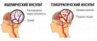
A sudden disruption of cerebral circulation that occurs due to various reasons is called a stroke. The disease belongs to the category of vascular accidents due to the fact that it leads to serious disturbances (including irreversible ones) in the functioning of many body systems. The pathological condition develops due to obstruction of the vessels supplying blood to the brain (ischemic stroke) or due to their rupture (hemorrhagic). The disease is characterized by a high level of mortality and disability.
The central part of the nervous system – the brain – is responsible for all processes occurring in the body. The exchange of information between the brain and other tissues is carried out using electrically excitable structural elements of the nervous system (neurons), which transmit electrical and chemical impulses to all cells. To maintain the constancy of all ongoing processes, brain tissue needs nutrition, which is provided by 15 percent of the total volume of blood circulating in the body and 20–25 percent of all inhaled oxygen.
Even a short-term cessation of the supply of nutrients and oxygen to brain cells entails serious consequences, which are expressed in the development of general cerebral disorders and corresponding symptoms. The absence or deterioration of blood supply to parts of the central nervous system leads to atrophic processes (death of neurons). All biological processes of the body are controlled by a certain part of the brain, and when a lesion forms in a specific area, those functions that are controlled by it are disrupted.
Non-specific events
These methods do not restore vision, but improve the quality of treatment prescribed by the doctor. When helping a sick person at home, you can carry out the following activities:
- lay out colorful carpets;
- paint the steps in different colors if a person regularly uses the stairs;
- place bright accents in the interior of the room.
These methods do not require much expense or effort, but they are good at helping the patient navigate the room and better remember obstacles and objects. In addition, a person focuses on a bright object, focuses his gaze, and transfers it to another object, thereby training the eye muscles and concentration.
Activities that work well include:
- collecting puzzles;
- drawing, it is advisable that you draw one half of the picture, and the patient completes the second, symmetrical to yours;
- educational children's games.
Daily walks have a good effect, especially if you choose picturesque places - forests, meadows, riverbanks or seashores. An excellent solution is a rehabilitation center, where the patient is 24/7 under the supervision of specialists involved in the active restoration of the patient’s brain functions.
Effect on vision
The human ability to perceive visual information is provided by the visual system. Vision is very closely integrated with the main part of the central nervous system, and it is very difficult to determine exactly which part is responsible for processing the observed images. The perception of a visual picture begins with the conversion of electromagnetic radiation emanating from surrounding objects into nerve impulses by the photoreceptors of the retina.
Light-sensitive cells transmit converted information through nerve fibers to the neural layers, where the transformation of the radiation recorded by the retina into a visual image occurs. The final picture is formed in the cerebral cortex (occipital lobe). The process of visual perception consists of numerous stages during which information is distorted, but the brain still eventually stabilizes and corrects the final image. All parts of image processing do not involve the conscious part of the brain and occur automatically.
To ensure consistency and correct visualization of observed objects, coordinated work of all elements of the visual system is necessary. The anatomical structure of the visual analyzer consists of the following formations:
- paired organ of vision (eyes);
- nervous structures (optic, oculomotor, cranial, trochlear and abducens nerves, as well as nerve tracts, pathways and chiasms);
- visual centers;
- lateral geniculate body (specific brain structure).
During a stroke, the death of neurons occurs, which leads to a deterioration in the passage of nerve impulses, a decrease in the speed of processing information received from the sensory organs and a reverse reaction to it (sensitivity). The severity of the disorders and their reversibility depends on the size of the lesion. The most negative prognosis for a large necrotic area is complete blindness. Other possible consequences of acute cerebral circulatory disorders regarding visual function include:
- Loss of peripheral vision (the area simultaneously perceived by the eyes on the sides of direct, stationary gaze) - loss of visual fields during a stroke occurs if the optic nerves or pathways are damaged. This condition leads to a decrease in the ability to navigate in space.
- Hemianopsia is a type of change in the visual fields in which loss of the same halves (homonymous) or opposite halves (heteronymous) occurs. Blinding may occur not in half the field, but in 25% (quadrant hemianopsia). In some cases, a scotoma (a blind spot of arbitrary shape) is formed.
- Abducens nerve palsy is a dysfunction of the nerve responsible for contracting the muscle that controls eye rotation. The manifestation of the pathology is esotropia (strabismus with the eye turning inward), protrusion of the eyeballs and diplopia (double vision of the observed object).
Why do eyes not open during a stroke?
One of the focal neurological symptoms of stroke, the clinical manifestations of which depend on the affected area of the brain, is ptosis (drooping) of the upper eyelid. The inability to open the eyes after an attack is due to decreased tone (paresis) or complete absence of voluntary contractions (paralysis) of the oculomotor nerve. These phenomena are observed when the formations of the central nervous system innervate the muscles that raise the upper eyelid and are located in close proximity to the brain structures.
This condition can be temporary (if the lesion is localized and its volume is small) or take a more severe form, including atrophy of the optic nerve. If measures are not taken in a timely manner to restore the functionality of the nerve fibers that conduct impulses to the levator palpebral muscle, atrophy will become irreversible. The consequence of this process is spasm of the upper eyelid, constant trembling of the eyeballs, which is the reason for the appointment of visual disability.
What is the danger of the disease?
Stroke is a complex problem with blood circulation in the head caused by blockage of a vessel . Vision will be completely lost if the vessels that supply the visual lobes with useful microelements are affected. Treatment of stroke, after damage to neurons in the visual center, is carried out similarly to other forms of the disease. Procedures are prescribed by a therapist, ophthalmologist and neurologist. The level of visual impairment during stroke is determined by the complexity of the brain damage.
When a large area is damaged, some doctors do not pay attention to a small fragment, and a blind spot appears. Sometimes double vision, oscillopsia occurs, and conjugal eye movements are lost. Changes in the visual field occur in 20% of patients, but they are difficult to diagnose due to deterioration in the level of consciousness and difficulties with communication. Changes in visual fields in active patients can cause injury.
If one side of the brain is damaged, specific areas of vision are affected. There may be problems creating a visual image.
Stroke is considered a complex disorder. Its main danger is based on the fact that, depending on the difficulties, some functions of the body will be disrupted. Vision problems after a stroke occur when the occipital region is damaged.
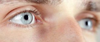
Symptoms of visual impairment
A qualified specialist can make a preliminary conclusion about the degree of damage to brain structures based on the observed clinical picture. Complete loss of vision after a stroke or inability to see laterally indicates extensive damage, the chances of reversibility of which are minimal. With paresis or paralysis of the nerve fibers innervating the extraocular muscles, the likelihood of vision restoration after a stroke increases. Signs indicating one or another visual impairment are:
- blurred visible image;
- deterioration of visual acuity;
- the appearance of floating spots before the eyes;
- severe pain in one or both eyes;
- nystagmus (oscillatory high-frequency movements of the eyeballs);
- diplopia;
- decreased ability to recognize observed objects;
- ophthalmoplegia (deterioration of eye motor ability);
- loss of sectors from the visual field while maintaining color perception;
- atrial scotoma (periodic light hallucinations - flickering, fog obscuring objects, complete darkness);
- trembling eyelids;
- presbyopia (inability to read small text or see small objects held close to the eyes);
- exophthalmos (bulging eyeballs);
- strabismus;
- lacrimation.
Medicines
How to restore vision after a stroke with drugs? Medicines are determined taking into account clinical manifestations. When disorders are caused by dysfunction of brain tissue, complex therapy is carried out aimed at stimulating blood supply and restoring brain structures.
Ophthalmic medications should not be used, but eye drops may be prescribed to help maintain eye function. When a problem with focusing vision occurs due to atrophy of nerve fibers, restorative therapy is carried out to improve blood supply and tissue function.
Homeopathic medicines are used together with traditional remedies. These medications stimulate the body's resources to fight the disease. This approach to restoring visual function is considered unique. Prescribed medications help overcome the cause of problems.
Surgery is performed in a situation where medications do not give the desired result.
How to restore vision after a stroke
Restoration of visual function after a stroke should begin as early as possible in order to prevent the aggravation of pathological processes. The forecast for the likelihood of restoration of previous visual acuity is based on the scale of the destructive changes that have occurred in the brain structures and the amount of time that has passed since the development of the disease. The longer adequate rehabilitation measures are not carried out, the longer and less effective the treatment process will be.
Temporary loss of vision during a stroke, after which the binocular optical system independently resumes, is not a reason to refuse treatment measures. If the problem has already manifested itself, it means that the structures responsible for the functioning of the visual analyzer have already undergone changes and will involute in the future. The complex of a recovery program after a stroke may include the following complementary areas:
- drug therapy;
- therapeutic oculomotor gymnastics;
- surgical intervention;
- diet (inclusion of foods high in vitamin A in the diet);
- non-specific ways to speed up rehabilitation (for example, using contrasting colors in the interior to focus the patient’s attention on surrounding objects).
The choice of method and the scope of post-stroke rehabilitation of the patient depend on the clinical manifestations of the disease and the degree of changes that have occurred. To reduce the duration of rehabilitation treatment, you can resort to homeopathy or use traditional medicine recipes. Before using one or another unconventional method, you should consult a specialist, since not all methods offered by naturopaths are equally effective and safe in every particular case.
Articles on the topic
- Stroke at a young age - symptoms of ischemic and hemorrhagic
- Exercises after a stroke at home for rehabilitation
- Speech impairment during stroke - treatment
Drugs
The main goals of drug therapy for visual function impaired due to stroke are restoration of cerebral blood flow and normalization of metabolic processes in brain tissue. Achieving these goals will ensure a reduction in the volume of necrotic tissues and acceleration of their replacement with healthy ones (when neurons die, the functions they perform are partially distributed among the remaining cells).
Depending on the location of the focus of ischemic or hemorrhagic damage, drugs of certain pharmacological groups are prescribed, aimed both at restoring lost neural connections and normalizing blood flow, and at the organs of vision themselves. The post-stroke therapy regimen may include medications from the following groups:
| Pharmacological group | Destination purpose | Drugs |
| Nootropics | Improving neural metabolism, integrative brain activity (subordination, unification and coordination of the functions of all systems). Normalizing oxygen delivery to the brain or reducing the need for it in nerve cells. | Nootropil, Encephabol, Piracetam, Glycine. |
| Antiplatelet agents | Improving the rheological properties of blood (fluidity, viscosity), restoring microcirculation, preventing blockage of blood vessels with blood clots. | Pentoxifylline, Trental, Agapurin. |
| Adenosinergic drugs | Reducing blood viscosity, stimulating the respiratory center to normalize the process of saturating the body with oxygen, increasing coronary blood flow. | Euphylline, Aminophylline, Pentoxifylline. |
| Immunosuppressants | Acceleration of recovery of brain functionality after strokes. The drugs have a specific effect on brain tissue, promoting the restoration of neurons and the normalization of nervous regulation processes. | Cortexin. |
| Drugs that improve cerebral blood flow | Stimulation of the neurotransmitter system and metabolic processes in the brain (increased oxygen uptake and absorption), antioxidant effect, reduction of pathologically increased blood viscosity. | Cavinton, Vinpocetine, Bravinton. |
| Anticoagulants | Reducing blood clotting ability, preventing thrombosis. | Heparin, Hirudin, Nadroparin. |
| Angioprotectors | Improving tissue trophism, correcting microcirculation, increasing the speed of regenerative processes, activating oxidative processes, stimulating cell proliferation (division). | Solcoseryl (solution for injection), Actovegin. |
| Vitamins and vitamin complexes | Reducing the sensitivity of brain tissue to oxygen deficiency, strengthening the vascular walls of the extraocular muscles, antioxidant effect, inhibition of degenerative disorders in the retina. | Ascorbic acid, vitamins B, A, Okuwait Forte, Complivit. |
| Local rehydrants | Replenishing the deficiency of tear fluid, moisturizing the cornea of the eye. | Optinol, Systane, Oftagel, Korneregel. |
After a stroke, it is important to restore impaired cerebral circulation, therefore stimulants of the neurotransmitter system are on the list of the most frequently prescribed drugs during the rehabilitation of patients. Cavinton belongs to this group of drugs. The main active ingredient of the drug has a multifactorial effect on the cardiovascular and nervous systems, helping to eliminate the consequences of the disease:
- characteristics: a neuroprotector that has a vasodilator and anti-aggregation effect, helps improve the rheological properties of blood and regional cerebral blood flow, the main active substance vinpocetine actively binds to tissues, improves the delivery of nutrients (glucose) to brain neurons;
- contraindications: severe coronary heart disease, acute phase of stroke, pregnancy, children (up to 18 years);
- Method of administration: orally or intravenously, take 1-2 tablets three times a day. after meals, intravenous administration is carried out by drip, the initial daily dose is 20 mg, diluted in 500 ml of saline, after 2–3 days the dosage is increased (but not more than 1 mg/kg/day), the course of treatment lasts from 10 days to 3 months .;
- side effects: headaches, drowsiness, decreased blood pressure, rapid heartbeat, dyspeptic disorders (nausea, vomiting, diarrhea, constipation);
- advantages: can be used for a long time, is not addictive;
- disadvantages: the need for constant monitoring of electrocardiogram indicators.
To regenerate damaged brain tissue during ischemia and improve the condition of healthy areas located near the lesion, stimulants of regenerative processes are prescribed. The drug Actovegin is used at all stages of post-stroke therapy in order to activate tissue energy metabolism and prevent the development of complications. This remedy is also used in ophthalmology to correct vision in case of damage to the membranes of the eye:
- characteristics: a drug of biological origin, produced from the blood of cattle, prevents the progression of encephalopathy (destruction of nerve cells), reduces the severity of neurological symptoms in strokes;
- contraindications: heart failure in the decompensation phase, allergic reaction to the components of the drug, pulmonary edema, bladder dysfunction;
- method of administration: intravenous infusion, orally or inserted into the conjunctival sac; in the acute phase of the disease, the drug is administered intravenously for a week in the form of a solution (800–2000 mg per 200–300 ml of saline), over the next 7 days the dosage is halved, after which you can switch to taking tablets - 1-2 pcs. 3 times a day before meals for 3–4 months, 1 drop of eye gel is placed into the space between the eyeball and the lower eyelid;
- side effects: focal hyperemia (redness) of the skin, rashes of allergic origin, anaphylactic shock, redness of the eye sclera;
- advantages: effective elimination of the consequences of necrosis;
- disadvantages: risk of developing a severe allergic reaction.
Operation
Restoring vision after a stroke using surgical methods is carried out only in exceptional cases. The lack of results from conservative treatment of impaired visual function is not always a reason for surgery, due to the fact that the problem often lies in the brain, and not in the organs of vision. The decision regarding the advisability of using radical methods of treating post-stroke disorders is made by an ophthalmologist in consultation with a neurologist.
Indications for surgical intervention are the presence of degenerative changes in the lens of the eye (the transparent body inside the eyeball that acts as a biological lens) or a persistent imbalance between the extraocular muscles (strabismus). The operation to replace the lens involves removing the affected area and installing in its place an artificial intraocular lens made of material biocompatible with natural tissues.
Correction of strabismus occurs by weakening overly toned eye muscles and strengthening weakened ones. Surgical manipulations involve moving the attachment points of the muscle fibers closer to the center of the eye to reduce their tone or shortening the muscle (removing part of it and suturing the remaining area to the fixation site) for toning. Not in all cases, after surgery, it is possible to completely restore visual function, but the chances of partial restoration are high.
Exercises
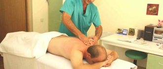
An integrated approach to post-stroke rehabilitation includes activities such as gymnastics and eye massage. Exercises performed regularly potentiate the effect of rehabilitation treatment by activating the areas affected by stroke. The load on the eye muscles helps to strengthen them and improves blood circulation in the adjacent tissues. You should not expect quick results from training - it will take several months of daily exercise to restore damaged neural connections and restore the functionality of muscle fibers.
One of the methods of vision therapy aimed at compensating for dysfunction of the areas of the brain that provide vision is exercise with prisms. This technique is indicated for use in cases of strabismus resulting from a stroke and is carried out in a laboratory setting using specialized equipment. At home, you can perform a simple gymnastic complex or use special computer programs designed to expand your visual fields.
The process of restoring vision at home involves doing exercises every day for 20–30 minutes. in the morning and in the evening. A conclusion about the effectiveness of the measures taken can only be made after 6 months. continuous classes. The training program includes the following techniques:
- Eyeball massage. Using the fingers of both hands, you should gently squeeze the bridge of your nose and, without releasing the pressure, smoothly move your fingers along the base of the superciliary arch to the temples. The exercise is performed 5–10 times until you feel warmth in the massaged area (there should be no pain).
- Rolling eyes. The essence of the exercise is to move your gaze left, right, up, down and fixate for 1-2 seconds. in the position of greatest tension. After 3–5 repetitions, you need to slowly move your eyes in a circle with the maximum possible amplitude (5 circles clockwise and 5 counterclockwise). During rotation, do not allow your head to turn following your eye movements.
- Focus shift. Extend your arms in front of you with your index fingers clasped together. The gaze is focused and fixed on the fingertips. Smoothly bring your hands closer to your face until your vision defocuses, then return to the starting position and repeat the exercise 9 more times.
- Frequent blinking. Within 10 sec. you need to blink as often as possible. After a short rest, repeat the movement 2 more times. While performing the exercise, you should try to close your eyelids as tightly as possible. It is recommended to alternate the approach of frequent blinking with a static delay of the eyelids in a closed position for 5 counts.
- Upper eyelid toning. A common problem after a stroke is drooping of the upper eyelid, which is caused by severe weakening of the muscles. To return the eyelids to their normal position, you should perform an exercise aimed at strengthening the eye muscle structures. It is necessary to place the pads of your index fingers on your eyelids, slightly pull the skin fold upward and fix it. Holding the skin with your fingers, try to close your eyelids 5–10 times.
Folk remedies
After overcoming the acute phase of a stroke attack, the patient’s rehabilitation begins, during which, in agreement with the doctor, the use of traditional medicine is allowed to speed up the recovery process. Non-traditional methods of restorative treatment are based on the use of herbal remedies made from medicinal plants.
The herbs that make up medicinal tinctures and decoctions contain biologically active substances that have a beneficial effect on the nervous and cardiovascular systems. The duration of herbal treatment is determined based on the patient’s condition and the effect of the drugs. In the absence of positive dynamics of therapy for more than 2 months. or if the patient’s well-being worsens, treatment tactics should be changed. Traditional medicine offers the following recipes for restoring visual functions after a vascular accident:
- Tincture of mountain arnica flowers. This herbaceous plant is a poisonous species, so caution is required when introducing it into medicinal potions. Arnica has a pronounced sedative effect, relieves muscle spasms, and regulates the tone of the central nervous system. To prepare the herbal medicine, add 1 tsp. flowers with 2 cups of boiling water and leave for 2 hours. Strain the finished product and take 1 tbsp. before meals. The tincture can be stored for no more than 2 days.
- Decoction of pine cones. The unique chemical composition of pine cones makes them a popular component of traditional medicine. To eliminate the consequences of acute cerebrovascular accident, the advisability of using this product is due to the tannin content in it (a tannin with a hemostatic effect). The decoction is prepared by boiling for 7 minutes. 5-6 young pine cones cut into slices, filled with 0.5 liters of water. Take 150 ml of the product up to 3 times a day before meals.
- Apple honey. If, after a stroke, diseases associated directly with the structural elements of the eyes have worsened, improvement can be achieved by instilling apple honey into the eyes. To prepare the product, remove the core from a green apple and pour 1 tsp into the hole formed. honey Make several holes on the side of the fruit (with a needle) and place the apple on an empty glass so that the resulting juice flows through the holes made into the container. Apply apple honey to your eyes every day for at least 3 months. contract.
- Motherwort decoction. The plant, known for its sedative effects, is effectively used to minimize the effects of neurological diseases. Many patients have a difficult time post-stroke, and the lack of effectiveness of rehabilitation therapy is directly related to psychological imbalance. Taking 2 tbsp. l. decoction (15 g of dry motherwort, pour a glass of boiling water and leave for 30–40 minutes) three times a day, you can normalize the functioning of the nervous system and significantly speed up the recovery process.
Compresses
To relax the extraocular muscles spasmed due to a stroke and restore blood circulation in the periocular muscle tissues, it is useful to make compresses. You can use plain water as a basis for therapeutic manipulations, wetting a towel alternately in hot and cold liquid and applying it for 5–10 minutes. to the eye area. The temperature difference helps improve the tone of the vascular walls and relax the smooth muscles.
You can enhance the effect of medical procedures using the healing properties of plants by making decoctions or mixtures for compresses from them. Effective recipes for improving vision are:
- Honey-dandelion mixture. Dandelion leaves and rhizomes must be crushed to a pasty state and combined with a tablespoon of honey. The freshly prepared mass should be wrapped in cotton cloth or gauze and applied to the eye area for 20 minutes. After completing the procedure, wipe the area where the compress was located with whey. Manipulations are carried out daily for 14 days.
- Eyebright decoction. The name of this herbaceous plant was assigned to it due to its widespread use in folk medicine for the treatment of eye diseases. Improved vision can be achieved by daily applying eyebright compresses and washing the eyes with a decoction. To prepare the herbal medicine, add 5 tbsp. dry herbs 1 liter of boiling water and leave for 3 hours.
- Honey-mint mixture. Mint in any form has a beneficial effect on the organs of vision. It can be used as a standalone remedy or combined with other components that enhance the healing effect of the plant. To improve blood flow to the tissues surrounding the eyes, you need to mix 50 g of mint leaves, agave leaf, 1 tsp. honey Pass all components through a meat grinder, add boiled water if necessary (if the mass is too thick). Wrap the resulting mixture in a cloth and apply for 20 minutes. to the eye area.
Gymnastics
The most effective method for correcting visual impairment is special eye exercises. They restore the mobility of the eye muscles, re-teaching the brain to take part in the process of vision. Sometimes this helps to completely get rid of problems, thanks to the ability of the remaining living neurons to replace the functions of dead ones.
| Exercise type | Description |
| Stretching and strengthening the eye muscles | The head remains motionless. Between each exercise, you need to blink for 10-20 seconds to relieve tension. The number of repetitions of each point is 8-12 times.
A more complicated option is to follow the movement of your finger along the same trajectory. |
| Relaxation of eye muscles | Cover your eyes with your palms as shown in the picture. Relax your shoulders, neck, and facial muscles. Eyes open. Try to look into the distance without straining. Think about something pleasant. Start with 15 seconds, gradually increasing the exercise time to a minute. |
| Eyeball stimulation |
Between all exercises, blink for 10-20 seconds to relieve tension. |
| Accommodation training | Extend both arms forward. Place your index fingers together and clench the rest into a fist. Focus your gaze on your fingertips. Slowly bring both hands closer to you and then back. Try not to lose focus. Perform 8-10 repetitions. |
Program to restore lost functions
Because the parts of the brain that control visual function are most sensitive to a lack of oxygen, they undergo changes within a minute after the onset of a stroke. This fact explains the complexity of therapy to restore vision after a stroke, not only at home, but also in a hospital during rehabilitation.
There are different approaches to restore visual functions:
- nonspecific treatment;
- drug therapy;
- therapeutic and preventive gymnastics for the eyes;
- surgical method.
The patient can combine all three approaches if the consequences are reversible and do not yet require surgical intervention. Relatives should support the patient, help and create all the conditions for full treatment.