Patients who have suffered a cerebral hemorrhage often experience a complication during the recovery period such as loss of vision. It can be complete or partial, it all depends on the degree of necrotic changes in brain tissue, the affected area and the volume of dead neurons. Doctors say that vision loss after a stroke occurs in 30% of patients; during the rehabilitation period, such patients require long-term medication and auxiliary therapy to restore lost functions at least partially.
Causes
Occlusion of the vessels of the eye rarely occurs as an independent nosology. The condition is often a manifestation or complication of another, general somatic, pathology. Diseases and conditions that provoke occlusion of retinal vessels:
- atherosclerosis;
- increased blood pressure;
- carbohydrate metabolism disorder (diabetes mellitus);
- autoimmune inflammatory vascular diseases (giant cell arteritis);
- increased blood viscosity (polycythemia, myeloma);
- thrombophilia – congenital or acquired;
- endocarditis;
- viral and bacterial vascular diseases;
- congenital vascular anomalies;
- pathologies of heart valves (rheumatism, heart defects);
- heart rhythm disturbances;
- myocardial infarction, acute coronary syndrome;
- extensive and chronic blood loss;
- acute cerebrovascular accidents (stroke, transient ischemic attack);
- hyperthyroidism;
- long-term use of diuretics, oral contraceptives, blood clotting agents;
- invasive diagnostic methods associated with the administration of contrast agents (angiography).
Local provoking conditions:
- optic disc calcifications (drusen);
- swelling of the optic disc;
- increased intraocular pressure;
- orbital tumors;
- retrobulbar hematomas.
In 20-30% of cases, the cause of an eye stroke cannot be determined.
Etiology
Ocular stroke is a rare condition, but very dangerous. The visual organs have a branched vascular pattern. Emboli and blood clots form in the bloodstream, they rupture thin walls and provoke hemorrhage. This process disrupts the nutrition of the eyes and brain, so if the pathology is not noticed in time, the consequences can be unfavorable. The following conditions can provoke the formation of blood clots:
- chronic form of hypertension;
- problems with blood clotting;
- stress;
- eye fatigue;
- avitaminosis;
- alcoholism and smoking;
- long-term use of corticosteroid drugs;
- congenital pathologies of the retina.
Typically, the tendency to such a pathology is inherited.
When studying pathology, a hereditary nature can be traced. Patients with type 2 diabetes mellitus are at risk. In this case, a microstroke of the eye is diagnosed, which can resolve on its own. Patients with this diagnosis should be registered with an ophthalmologist. With a micro-stroke against the background of a primary disease, the risk of vision loss increases by 2 times.
At-risk groups
Most patients who suffered an ocular stroke suffered from atherosclerosis and its manifestations (cardiac ischemia, angina, hypertension), and diabetes. The next position as a direct etiological factor is occupied by rheumatic pathologies, then temporal arteritis, vascular diseases.
Additional risk groups:
- people whose work activity involves working at a computer (eye strain);
- people exposed to frequent and prolonged stressful situations (doctors, lawyers, military personnel and emergency responders);
- age over 40 years;
- smokers;
- persons who abuse alcohol;
- frequently ill people (decreased immunity);
- family history of atherosclerosis, hypertension, diabetes, heart and vascular diseases.
People aged 40 years and older are more susceptible to retinal vein thrombosis. Occlusion of the retinal arteries is more common in men aged 60 to 65 years.
Arterial occlusion and retinal detachment
With retinal detachment, arterial occlusion is quite common. This is the most dangerous form of the disease, since in most cases it is asymptomatic.
Its main symptom is loss of peripheral vision. The pathology often transforms into loss of central vision. Many patients with retinal detachment and arterial occlusion are diagnosed with narrowing of the carotid artery, high blood pressure and various heart diseases. With timely treatment, the chances of complete restoration of vision are quite high and amount to 80%. However, problems with distorted image perception may still persist.
Types of eye stroke
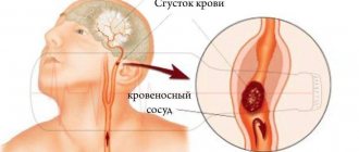
An ocular stroke can be localized in the venous or arterial bed of the retinal circulatory system. If the accident occurred in a venous vessel, they speak of retinal vein thrombosis. The blockage can occur in the central retinal vein (CRV) or in its branches (the superior temporal branch is affected in 82% of cases). A major role in retinal vein occlusion is played by disruption of the internal vascular epithelium, which predisposes to the formation of blood clots.
Obstruction of outflow leads to stagnation of blood and increased intravenous pressure. In this case, the liquid begins to seep through the walls of the vessel. Their rupture may also occur. Blood fills the eye space, swelling increases, and intraocular pressure increases even more. This is fraught with compression of small capillaries, which further limits the outflow of fluid and aggravates retinal hypoxia. This cycle of mechanisms of the pathological process is called a “vicious circle”.
Closure of the lumen of an arterial vessel is called arterial occlusion. There are occlusions of the central retinal artery (CRA), its branches and the cilioretinal artery. Often the blockage occurs in the CAS; the branches are affected with less frequency. The cause of artery blockage in most cases is cholesterol, fibrin thrombus or calcification embolism.
The embolus blocks the access of nutrition to the retina - ischemia develops. Clinical symptoms of visual impairment develop quickly. If medical assistance is provided within the first 40 minutes from the onset of symptoms, vision can still be fully restored. Delaying therapy threatens irreversible consequences.
Clinical picture
The first “danger signals” indicating the possible development of the disease may be the following symptoms:
- The field of view is noticeably narrowed. Thus, the eyes of a healthy person, with a fixed position of the head and a fixed gaze, cover a certain part of the space.
During a stroke, the “picture” transmitted to the brain becomes smaller, that is, the field of view decreases. - Floaters, stars, and sparks appear before a person’s eyes.
- Painful sensations in the eyes, in some cases vision disappears completely.
- Hemorrhages in the visual organ, minute hemorrhages.
One of the main signs that the patient has suffered an ocular stroke is the simultaneous deterioration of vision (sharp) and the appearance of white spots in front of the eyes. Upon visual examination of the visual organ, local redness and minor hemorrhages are noticeable, and the patient’s blood pressure may rise.
https://www.youtube.com/watch?v=t6JBN6Gu6Ek
Due to the fact that an ocular stroke is caused by excessive expansion (constriction) of blood vessels, this leads to “cutting off” the optic nerve from access to oxygen.
This situation certainly entails a partial or complete disruption of the main function of the eye - vision is either greatly weakened or the patient becomes completely blind.
The course of the disease is not accompanied by pain, but if not diagnosed in time, it can lead to blindness.
Symptoms
The main symptom of retinal artery occlusion is loss of visual fields. The sign is disturbing either in one or both eyes. The symptom develops suddenly. Sometimes the first signs of the disease are the appearance of glare, sparks, flickering, and mild pain in the orbit. Rapidly passing blindness is rarely a concern.
As with arterial occlusion, with venous thrombosis, symptoms develop suddenly, against the background of good health. Before the main symptom, patients are often bothered by fog before the eyes, a dark spot, image distortion, and dull pain in the depths of the orbit.
Symptoms of an eye stroke are painless and can be mistaken for a minor temporary deterioration due to fatigue or stress. This is a misconception. If the above symptoms appear, you should immediately call for medical help.
Signs
A stroke of the organ of vision often occurs without pronounced signs, and therefore often goes unnoticed. To prevent pathology, it is necessary to pay attention to the symptoms of the disease, which are characteristic of all types of stroke:
- temporary or progressive decrease in visual acuity;
- gradual deterioration of peripheral vision;
- white spots, glare, other interference in front of the eyes;
- unexpected loss of areas of the visual field;
- problems with color vision;
- hemorrhage (bleeding in the eyeball).
When an artery is blocked with subsequent retinal detachment, visibility at close range deteriorates, and the boundary of the visual angle gradually shifts inward. Sometimes there is pain in the temples and the front part of the frontal lobes, and sudden dizziness, accompanied by blurred vision, is possible.
Articles on the topic
- Lacunar stroke of the brain - signs and manifestations of the disease, drug therapy and consequences
- Stroke at a young age - symptoms of ischemic and hemorrhagic
- Speech impairment during stroke - treatment
When the retinal artery is blocked, partial or complete loss of peripheral vision is possible, which over time can transform into complete blindness if retinal detachment occurs. A stroke is accompanied by the appearance of blind spots and distorted image perception. The disease is rarely accompanied by pain, which is why patients do not pay attention to the gradual decrease in vision, and often realize when it cannot be restored.
The separation of the veins from the retina is accompanied by deterioration of peripheral vision, which eventually leads to blindness, and the signs of pathology rapidly progress. It is necessary to pay attention and urgently begin treatment if objects suddenly become unclear, glare, clouding appear, and blind spots are observed in the field of vision.
A micro-ocular stroke, known as an optic nerve stroke, is the result of a myocardial infarction or hemorrhagic stroke in the area of the brain that controls the eyes. This is a life-threatening situation that requires urgent medical attention. The following symptoms are typical for a stroke:
- sudden blindness or blurred vision in one eye combined with loss of muscle movement in an arm or leg on the opposite side of the body (hemiparesis);
- the appearance of blind spots while maintaining color perception and visual acuity;
- acute pain in the eyes;
- constriction of the pupils;
- restriction of eye mobility;
- nystagmus – involuntary oscillatory movements of the eyes of high frequency;
- doubling of objects;
- strabismus.
Treatment
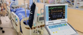
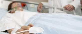
An ocular stroke is an absolute indication for emergency hospitalization and treatment under 24-hour observation.
Treatment of arterial and venous stroke does not differ significantly. The goal of treatment for these conditions is general; it is to cleanse the lumen of the vessel, strengthen the vascular wall, restore vision, eliminate intraocular hemorrhage (if any), and prevent relapse. Several groups of drugs are used for these purposes:
- Thrombolytics. Substances for resorption of blood clots. For the best effect, drugs are injected under the eye, under the conjunctiva or directly into the cavity of the eyeball.
- Drugs to dilate the lumen of the vessel. They can also reduce the sticking of red blood cells and platelets to the vessel wall, preventing blockages.
- Means for protecting and restoring the inner lining of blood vessels. They are used in treatment and as a prevention of a recurrent episode of pathology.
- Blood thinners. They help resolve blood clots and prevent relapse.
- For retinal edema, glucocorticosteroids are often added to treatment.
- Drugs to reduce intraocular pressure. If the pressure remains high, or it needs to be reduced as soon as possible, resort to puncture of the cornea.
If the blood density is high, hemodilution is performed - a method of thinning the blood by intravenous administration of solutions similar in composition to blood plasma. Surgical treatment is resorted to when conservative therapy is ineffective and in difficult cases (compression of the optic nerve head, retinal detachment, compression of a vessel from the outside).
In case of arterial occlusion, in the first 8 hours it is necessary to massage the eyeball. This helps displace the thrombus to the periphery. The head lies low. The eyelids are closed. You need to press your fingers on the eyeball over the upper eyelid for 3-5 seconds, first weakly, then harder. When pressing on the eye, intraocular pressure in the small arterioles increases, which slightly increases the blood flow to the eye.
When the pressure on the eyeball stops, the pressure drops sharply. Changes in pressure cause the clot to move, it moves, freeing up blood access. This is not a therapeutic measure, but a way to extend the time before starting therapy so as not to worsen retinal hypoxia.
For any type of stroke, treatment of the underlying disease that caused the embolism is necessary.
Types of disease and its manifestations
Blockage of an arterial vessel in combination with retinal detachment is the most dangerous and most severe type, often occurring without pain. Patients notice loss of peripheral visual fields, and sometimes partial central vision loss occurs. Sometimes accompanied by narrowing of the carotid artery.
Restorative treatment partially helps, but changes in the form of white spots and narrowing of visual fields may remain. Pain symptoms are not constant.
The main signs of occlusion are decreased visual acuity and distortion. But there are other manifestations of pathology that should concern a person and cause an immediate visit to an ophthalmologist. Ignoring them is imprudent and dangerous. These include:
- the organs of vision periodically hurt;
- from time to time double vision appears before the eyes, bright spots, flashes and lightning;
- narrowing of the central and peripheral field of vision;
- disturbance of color perception.
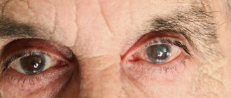
In severe cases of the disease, pinpoint hemorrhages – hemorrhages – are visible on the whites of the eyes. The vascular network is dark red in color, clearly expressed; with extensive hemorrhages and weakened vessels, the entire protein may turn red. Sometimes there is an increase in intraocular and intracranial pressure.
The pathology is classified depending on which vessel is damaged and how badly the retina is damaged. The most dangerous form of the disease is the combination of blood clot formation in the central artery with retinal detachment. The symptoms of the pathology are severe. There is usually no pain. But the following symptoms are noted:
- loss of peripheral vision;
- partial loss of the central one;
- narrowing of the carotid artery, which is the most dangerous.
Complete restoration of vision after an ocular stroke of this type is currently impossible; white spots and narrowing of visual fields will still bother you for the rest of your life.
Complications and consequences
Visual impairment can become irreversible after a stroke, even leading to blindness, if timely assistance is not provided to the patient. This is the main and most dangerous complication of the disease. An equally serious consequence is retinal detachment and macular defects, which also leads to vision loss. The transition of the pathological process to the second eye and optic nerve atrophy occur less frequently.
When the retina lacks nutrition, the body begins to come up with workarounds. This is how new blood vessels form – neovascularization. This is not a healing process - the new vessels are too brittle and tortuous, and the risk of rupture is high. There are known cases of neovascularization of the optic nerve head and neovascular glaucoma.
Symptoms of optic nerve damage due to stroke
As a result of a stroke, which occurs due to blockage of brain capillaries or insufficient oxygen supply to the brain, some neurons die. Atrophy and necrosis of large areas of tissue occurs. Those areas that are damaged cease to perform the functions for which they were responsible. If the parts responsible for visual function are affected, vision disappears and temporary blindness develops. If the damage was reversible, the foci of dysfunction are restored over time; with irreversible changes, disastrous consequences arise in the form of complete loss of vision.
Symptoms of visual impairment may indicate which parts of the brain were affected first and the extent of necrotic damage to neurons:
- loss of visual fields - the lesion had a small localization. This type of pathology is called a “blind spot” because a separate area disappears from the field of view. The syndrome is accompanied by pain in the eye sockets. Minor brain damage leads to the fact that vision is restored independently during the rehabilitation period;
- loss of peripheral vision - full visual function is provided by two brain lobes - left and right. Information from the retina of both eyes is processed by the opposite lobe (the left-sided area is responsible for the right side of the retina and vice versa). If peripheral (side) vision is lost, it means that the brain tissue has suffered extensive damage due to hemorrhage. Restoring lateral vision after a stroke is a difficult and lengthy process, requiring intensive medication and auxiliary therapy. Intact brain structures can take over some lost functions;
- paralysis of the nerve responsible for the motor function of the eyes - such an atrophic disorder that occurs in the muscle fibers responsible for the movement of the eyeballs leads to protrusion of the eyes outward. Another consequence is squint.
Successful treatment and reversibility of visual impairment depends on timely diagnosis of stroke and immediate therapeutic assistance.
Prevention
Prevention comes down to the treatment and prevention of affecting diseases. Regular visits to a therapist, vision and fundus examinations should be carried out at least once a year.
Precautionary measures:
- avoid eye strain, wear safety glasses when working with a computer and telephone;
- avoid smoking, drinking alcohol;
- dose physical and emotional stress;
- maintain a sleep-wake schedule;
- do not take any medications without first consulting your doctor;
- know and treat chronic and hereditary pathologies;
- take courses of vitamin complexes to improve immunity, especially in the spring and autumn.
Taking precautions and knowing the basic symptoms and methods of recognizing an eye stroke will help you avoid such a terrible condition. If this happens, react in time and correctly and maintain your health.
Restoring vision after a stroke - basic principles
In order to be able to restore vision in the future, if you have a stroke, you need to urgently consult a doctor and get qualified help. An ophthalmologist treats visual impairment after a stroke. All stages of recovery must be approved by a doctor. It is very important to analyze the results and adjust the rehabilitation scheme in a timely manner.
Principles of vision restoration after stroke
- moisturizing the eyes with drops and gels;
- restoration of affected visual functions with the help of medications;
- regular and correct exercise;
- diet with vitamins, especially vitamin A;
- taking nutritional supplements.
After a stroke that causes vision loss, you need to seriously engage in general rehabilitation and restoration of visual function. Without making an effort, the patient risks remaining blind for life, because medications are not able to restore vision completely. Rehabilitation may include exercises, medications, and even surgery.
Ways to improve vision after a stroke
- Gymnastics. Special exercises help stop the progression of presbyopia at the initial stage of its development. Before starting therapy, the visual organs should be prepared; it is recommended to hold your face over warm water and splash it into your eyes. There are a large number of techniques, so the choice is up to the ophthalmologist.
- Medicines. Medications for visual impairment after a stroke are prescribed when gymnastics is not effective enough. There are a large number of remedies, so the choice should also be entrusted to a doctor. It will take into account the characteristics of the disorders, concomitant pathologies and contraindications.
- Surgery. The operation is prescribed as a last resort. Patients usually require lens replacement after a stroke. In its place, a special lens is implanted, which performs all the functions of a natural lens and is combined with the structures of the eye. Surgery to remove the lens is one hundred percent prevention of cataracts.
It must be remembered that the problem of vision after a stroke is very individual. A recovery program is developed for each individual patient, taking into account the degree of damage and consequences. However, absolutely everyone is recommended to moisturize the mucous membrane with the help of drops and gels.
Methods for improving vision after a stroke
After a stroke, artificial tears, Korneregel, Taufon, Normax, Taurine are prescribed. Hydration is extremely important for patients who have been in a coma.
During the rehabilitation period, you need to reconsider your diet. After a stroke, it is recommended to eat more carrots, yellow peppers, pumpkin, egg yolks and fish. Cataract prevention is provided by grapes, blueberries and onions.
After a stroke, it is useful to massage with hot or cold compresses. This helps relax the eyes and improve blood circulation. It is enough to wet one towel in cold water and the other in warm water, and alternate them for 5-10 minutes.
The usual tossing of a ball or ball helps in restoring vision. You need to throw the object back and forth from the affected side. Focusing your gaze on an object will help to establish synchronicity between movements and vision.
Computer programs will help to rehabilitate vision after a stroke. One of these programs shows a black square; at certain intervals, a cluster of one hundred dots flashes on the side of the affected eye. Exercises on the computer take 15-20 minutes a day. The course of treatment is several months.
Comparative exercises allow you to check the extent of visual focus impairment. They make it possible to determine the required degree of rehabilitation therapy. The patient should close his eyes and direct his gaze to the injured side of the body. After determining the correct (according to the patient) direction, the eyes are opened, and the doctor determines the proximity of the gaze from the desired direction.
Compensatory vision therapy stimulates the areas of the brain that support vision. It includes scanning, visual field recognition and prism exercises. The visual field can be adapted by moving images from invisible zones.
Prisms in ophthalmology are used to correct various visual impairments. The type and placement of the prism will be determined by the symptoms. In case of double vision, the prism is placed on a glass lens, which allows you to align the direction of view. Spatial neglect requires the use of a prism on the left side of the visual field so that it reflects objects on different sides.
Restorative vision therapy aims to stimulate nerve connections in the brain. There are different techniques for all types of visual impairments.
Usually, surgical treatment of the organs of vision does not help anything after a stroke, because the problem lies in the brain. Only in some cases does surgery correct double vision by affecting the eye muscles.