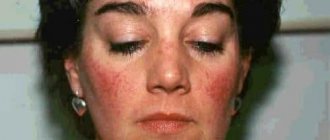
A congenital disease characterized by the appearance of wine-colored spots on the skin is called capillary angiodysplasia. The cause of the disease is the effect of a damaging factor on the body of a pregnant woman. As the child grows, the spots increase in size and turn from pink to dark red, the predominant localization is the face, neck and hands. For treatment, coagulation is performed with laser light.
What do venous changes look like in children?
As a rule, disturbances in the structure of blood vessels occur during fetal development and appear in newborns in the form of flat spots with a rich pink tint. Upon examination, they turn out to be a multilayered vascular network growing in the superficial layers of the skin. This distinguishes them from a tumor - hemangioma, which rises above the skin and has the appearance of a bruise.
Capillary angiodysplasia in young children
Capillary angiodysplasias do not resolve spontaneously, but tend to increase in size of the spots. Their color becomes more saturated over time, acquiring a characteristic red-violet hue, which gives rise to the name “wine stains.”
In addition to the visible cosmetic defect, this disease has a traumatic effect on the psychological state of the child and inhibits his social adaptation in children's and then adult groups.
We recommend reading the article about hemorrhagic vasculitis on the body. From it you will learn about the causes and forms of the disease, as well as symptoms, diagnosis and treatment.
And here is more information about the best treatment options for hemangioma.
Different forms of vascular dysplasia
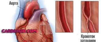
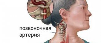
Arterial angiodysplasia as an isolated variant is quite rare; it is predominantly observed in combination with arteriovenous or venous angiodysplasia. Clinically, this form is manifested by signs of chronic ischemia of arterial vessels, slower growth of the lower limb in the affected area and impaired tissue trophism.
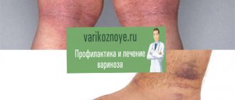
Congenital venous angiodysplasia has a clinical picture of varicose veins of the lower extremities, chronic venous insufficiency, impaired blood outflow and trophic disorders. In other cases, age spots and varicose veins may be absent, but other signs can indicate the disease:
- Reduced blood flow speed, blood stagnation, increased venous pressure;
- Lymphostasis (edema, increased volume of the limb, impaired tissue trophism);
- Increased sweating of the feet (hyperhidrosis);
- Thickening of the stratum corneum of the epidermis and its accelerated desquamation (hyperkeratosis).
An independent type of venous angiodysplasia includes Klippel-Trenaunay syndrome, which is characterized by damage exclusively to the blood vessels of the lower extremities. However, the classification of this option is also controversial - experts again disagree...
The clinical picture of the arteriovenous form (malformation) depends on the location of the vascular anomaly. Congenital, but small in size, vascular defects of the pulmonary circulation in children usually go unnoticed at first and “wait in the wings” before progression begins. And the hour of their “start”, as a rule, coincides with the period of puberty, reaching full “blooming” by the age of 20–30. And if the pathological change concerns the vessels of the small circle, the severity of the disease depends on the amount of blood discharged from the venous system to the arterial system. In such situations, the disease will quickly make itself known by a decrease in the level of oxygen in the blood - hypoxemia, which will quickly become chronic. It should be noted that the clinical picture of arteriovenous malformations is generally (in most cases) difficult:
- Increased temperature in the affected area;
- Trembling;
- Dilation of veins, which is easy to detect visually;
- Phenomena of ischemia and ulceration manifested on the skin;
- Pain and periodic bleeding.
In the absence of treatment, heart failure often develops with all the ensuing consequences.
As for arteriovenous malformations of the peripheral vessels (lower limbs, pelvis, shoulder girdle), they are mostly noticeable at the birth of the baby or in the very early years of life. Pigment spots, varicose veins, signs of partial gigantism will not hide from the “watchful eye” of parents and doctors observing the child.
Causes of capillary angiodysplasia
The disease is rarely inherited; its occurrence is associated with disruption of the formation of the vascular network during fetal development. There are known factors that can provoke capillary angiodysplasia:
use of alcohol and drugs by a pregnant woman;- smoking;
- taking medications;
- infectious diseases;
- diabetes mellitus with decompensated course;
- thyrotoxicosis;
- exposure to ionizing radiation, harmful production conditions.
When examining the tissues of the “port-wine stain,” it was found that the disease involves underdevelopment or complete absence of nerve fibers in the dilated capillaries. The vessels of the affected area cannot fully constrict due to insufficient supply of impulses to the smooth muscle fibers, so their diameter increases.
Angiodysplasia, malformation or hemangioma?
According to ICD-10, the disease is classified as a congenital defect, but experts define vascular intestinal dysplasia detected in adults as an acquired form.
It should be noted that until now scientists involved in the study of angiodysplasia have not come to a consensus regarding the use of terminology describing vascular defects; the signs of this pathology are perceived and interpreted differently, therefore in the literature you can find other names for the disease - congenital arteriovenous malformation or even hemangioma.
Angiodysplasia is the result of malformations of blood or lymphatic vessels at different stages of embryogenesis, that is, the disease originates during the stay and formation of a new organism in the womb. The localization of the pathological process can be very diverse: visible parts of the body, lungs, brain, limbs, gastrointestinal tract, etc.
The cause of vascular dysplasia is often the troubles that befell a woman during such a crucial period (gestation) or risk factors that she provoked herself:
- facial angiodysplasia
Anomalies that form at the genetic and chromosomal level (usually these are defects arising from risk factors such as the woman’s age, harmful chemicals received by the expectant mother under various circumstances in the first trimester of pregnancy);
- Viral and bacterial infections (rubella, cytomegalovirus, toxoplasmosis, herpes, tuberculosis);
- Injuries;
- Reckless use of medications (hormonal, psychotropic or antibacterial drugs);
- Use of alcohol, drugs (cocaine), smoking;
- Metabolic disorders (hormonal disorders, diabetes mellitus, endemic goiter);
- Lack of vitamins and microelements;
- Work in hazardous working conditions;
- Poisoning with carbon monoxide, salts of heavy metals (lead);
- Non-compliance with diet, work and rest by a woman expecting the birth of offspring.
All this is especially dangerous during the period of active formation of the circulatory and lymphatic systems. True, there is an opinion among the people that the pink (red, blue, purple, brown) spots with which the baby was born arise from the fact that the woman was very frightened during pregnancy and grabbed some part of her body in fear (“forbid God”, touch your face with your hands and then angiodysplasia of the child’s face is guaranteed?). Of course, this is nothing more than “women’s zabobons” (superstitions).
Signs of congenital port-wine stains
Capillary vessel pathology can occur in any area of the body, but the most common areas of port-wine stains are:
- head, especially often along the branching of the trigeminal nerve;
- neck (sides and back);
- upper limbs.
A regularity has been established - the farther capillary malformations are located from the midline, the more persistent their course and the richer their color. The size of the spots can range from 2 - 5 mm to half the surface of the body. There is a gradual penetration of the lesion into the deeper layers of the skin, which contributes to an increase in the dark shade and the formation of nodules from dilated capillaries.
"Wine Stains"
In adult patients, angiodysplasia can rise above the skin level and look like a solid spot with a bumpy surface . The skin over it becomes thinner, easily injured and bleeds, and is susceptible to infection. The disease can lead to deformation and asymmetry of facial features; some malformations transform into malignant neoplasms.
An isolated spot on the skin is not always the only sign of angiodysplasia; it can be accompanied by similar changes in blood vessels in the brain, causing a slowdown in the growth and development of the child, convulsive syndrome, and impaired movement in the limbs. About a third of patients develop glaucoma and impaired corneal transparency, various anomalies in the structure of the skull and valve apparatus.
Complex classification
Vascular dysplasia is a congenital defect, but it is not always detected at birth, since any of the systems (arterial, venous, lymphatic) can undergo pathological changes and the deformities can be deeply hidden. This does not exclude the presence of defects at the level of different vessels (hemolymphatic malformation) or the dominance of defects in only one system (arterial or venous angiodysplasia). In this regard, vascular dysplasia is classified depending on which vessels have undergone more pathological changes and, based on this, several forms are distinguished:
types of vascular malformationsArterial (rare);
- Venous (it has several forms and includes such a common variant as capillary angiodysplasia);
- Arteriovenous;
- Lymphatic.
Vascular malformation is also divided by location, for example, in the literature and in life one can more often than not come across the concepts: angiodysplasia of the face, intestines, lower extremities.
Facial angiodysplasia , as a rule, is noticeable from birth; it will be discussed in the “capillary angiodysplasia” section.
Congenital anomalies of the vessels of the lower extremities in children also manifest themselves almost immediately after birth: the formation of red spots, bruises, stretching of the limbs, an increase in their size, an increase in body temperature, and constant crying.
intestinal angiodysplasia
The situation is more complicated with the diagnosis of vascular intestinal dysplasia - it is not visible at birth, in addition, many experts are inclined to think that the pathology is acquired, however, the causes in most cases remain, as they say, “behind the scenes”. Meanwhile, given the widespread prevalence of pathological changes in intestinal vessels among the adult population, one cannot ignore the main symptoms of this process, these are:
- Bleeding from the anus, worsening after physical exertion;
- Vomiting streaked with blood;
- Blood in the stool;
- Stomach ache;
- Signs of anemia (pale skin, weakness, drowsiness, irritability).
However, without going into a detailed classification, we will focus on the main, most common forms of vascular dysplasia.
Video: angiodysplasia of the colon
Vascular diagnostics
Most often, the diagnosis can be made based on external signs; if the doctor suspects a violation of brain functions, then an additional examination is carried out:
- radiography of the skull bones;
- and MRI of the brain;
- electroencephalography.
Consultation with an ophthalmologist to measure visual acuity, pressure inside the eye, examination of the cornea, internal media, biometrics.
We recommend reading the article about benign and malignant vascular tumors. From it you will learn about the types and signs of vascular tumors, diagnostic methods and treatment options for tumors.
And here is more information about childhood congenital heart defects.
Angiodysplasia: symptoms and treatment
In this case, it is impossible to identify a general clinical picture, since everything will depend on which part of the body the pathological process is localized.
Category: Heart, blood vessels, blood 3248
Angiodysplasia is a pathological process that results in an increase in the number of subcutaneous vessels.
In the case of the gastrointestinal tract, this can lead to internal bleeding, which is extremely life-threatening. It is noted that this vascular disease can be congenital.
In newborns, capillary angiodysplasia is localized in the face, lower extremities, and less often the arms.
Only a doctor can establish an accurate diagnosis by conducting a physical examination and carrying out all the necessary diagnostic procedures.
Treatment methods in children
Before the introduction of laser coagulation into medical practice, capillary angiodysplasia was considered an incurable condition. There is no alternative to this method. In pediatric practice, pulsed radiation options are most often used, which tend to be absorbed by the dye. Hemoglobin plays the role of such a substance in the skin. Therefore, damaged vessels are the focus of action, and healthy tissues do not change their structure.
Under the influence of light in the green and yellow spectrum, dilated capillaries are subjected to intense heating, which causes the cessation of their functioning, and the surface layers of the skin remain unaffected. The coagulation technique is carried out using a point method, that is, with some intervals between places of exposure.
Within three days, the surface of the treated skin wrinkles and becomes covered with a crust. It goes away on its own after a week, revealing a red spot underneath that remains unchanged for up to 2 weeks.
Capillary angiodysplasia after treatment
A noticeable result can be seen only after the third month of treatment. To achieve a lasting effect, you need:
- undergo at least 5 sessions for medium lesions, 1 - 3 for small ones and about 10 procedures for large ones;
- after the procedure, use ointments with an antibacterial effect;
- You cannot remove the crusts, as tissue regeneration occurs underneath them;
- the start of laser therapy should be as early as possible; coagulation can be used in infants from the second month;
- For young children, treatment involves the risk of involuntary movements, so it involves the use of general anesthesia.
Adult patients are difficult to treat, so after a 2-3 month break, laser therapy is repeated.
Capillary angiodysplasia appears in newborns as pink spots on exposed parts of the body. The development of pathology is associated with a disruption in the formation of a network of nerve fibers around blood vessels. Clinical manifestations may be limited to port-wine stains only, or skin signs may be combined with brain signs. For treatment, laser coagulation is performed according to an individual scheme.
Causes
Scientists agree that vascular angiodysplasia is the result of a negative effect on the fetus during intrauterine development. Angiomas are formed in the blood and lymphatic vessels at the time of the formation of the bloodstream (from 5 to 20 weeks of pregnancy).
Pathology develops in early pregnancy in women due to a number of provoking factors:
- infection (herpes, rubella, cytomegalovirus, toxoplasmosis);
- alcoholic, drug toxicosis due to abuse of alcohol, hormonal, antibacterial and psychotropic drugs, drugs (cocaine);
- hormonal imbalances;
- deficiency of microelements, vitamins;
- diabetes;
- poisoning with salts of heavy metals, lead, carbon monoxide.
According to scientists, vascular dysplasia develops due to adverse effects on the fetus during intrauterine development. The cause may be a hereditary predisposition or chromosomal damage with the formation of an anomaly at the genetic level.
Also, if a woman abuses drugs with a teratogenic effect in the early stages of pregnancy or suffers from untreated infectious diseases.
Capillary dysplasia in newborns
Angiodysplasia (vascular malformation) is a pathological change in the vessels of the circulatory or lymphatic system resulting from a violation of the intrauterine development of the embryo.
The name angiodysplasia comes from the Greek words angio - “vessel”, and dysplasia - abnormal formation of tissues. The frequency of this congenital defect of the structure and functioning of blood vessels among all patients with vascular pathologies is about 2.6%.
Types of angiodysplasia
With all types of angiodysplasia, several types of vessels are involved to a greater or lesser extent in the process; depending on the main lesion, several types of angiodysplasia are distinguished.
- Venous - when the veins are affected, venous formations are clearly visible, the organ in which the venous malformation is localized increases in size.
- arteriovenous malformations, characterized by a direct connection of arteries with veins without the participation of capillaries.
- Veins and lymphatic vessels.
- Capillary.
- Pathologies of lymphatic vessels are rare and manifest in asymmetrical damage to the limbs. The patient's leg swells, in appearance the disease is similar to “elephantiasis”. Unlike this disease, angiodysplasia in a short time can lead to the appearance of trophic ulcers on the skin of the affected leg.
Vascular malformations are also classified according to the speed of blood flow in the lesion.
- High-speed - these include arterial dysplasia, aneurysms, arteriovenous fistulas.
- Low-speed - venous, capillary, lymphatic dysplasia.
Symptoms
Depending on the localization of the process and the nature of the malformation, a number of manifestations of angiodysplasia are possible.
Venous and lymphatic angiodysplasias
Veins and lymphatic vessels involved in the pathological process form painful lumps under the skin. Externally, such a bump resembles a mole. If the lump is formed by lymphatic vessels, it may leak lymph fluid and will require treatment with antibiotics or surgery. The lump formed by the veins may bleed.
Arteriovenous
The pathology can occur anywhere in the body and is characterized by the absence of a capillary network, which leads to direct reflux of arterial blood into the venous system.
The main symptom of arteriovenous angiodysplasia is bleeding and pain that occurs when blood moves from the arteries to the veins during heart contractions.
Arteriovenous malformations are most often localized in the bladder, intestines, uterus, and brain.
Possible complications
If left untreated, complications that threaten health and life will follow:
- hemorrhages in internal organs, causing pulmonary hemorrhage, stroke;
- extensive bleeding causing hypovolemic shock;
- thrombosis;
- extensive tissue dysplasia with malignancy into a malignant tumor, although the phenomenon is rare.
Fortunately, malformations rarely biotransform into malignant lesions. Venous dysplasia of the brain is dangerous. May result in hemorrhage and disability.
Another problem is that blood vessels are subject to pathological changes. Blood flows from the right ventricle of the heart to the left atrium, bypassing the lungs. They are not saturated with oxygen.
Patients experience shortness of breath, causeless fatigue, and cough with blood. If blood clots enter the general bloodstream through the arteries of the lungs, they can cause a stroke, cerebral edema, or thrombosis.
Who's at risk
People with a hereditary predisposition are at risk of getting venous dysplasia if close relatives have previously suffered a myocardial infarction or stroke. The risk group also includes:
- hypertensive patients;
- suffering from acute vascular diseases, cardiac ischemia, diabetes mellitus.
Provoking factors in a person’s life may include smoking, alcohol or drug abuse, and frequent stress as a powerful factor in the outbreak of vascular diseases.
Angiodysplasia of the intestines, blood vessels, brain and spinal cord is a congenital anomaly, a consequence of intrauterine disturbances in the anlage and development of the fetus. Malformations located in the upper layers of the skin are successfully treated. With angiodysplasia of internal organs (brain, lungs, intestines), the prognosis is disappointing.
The material was prepared specifically for the website venaprof.ru, edited by doctor N.A. Glushakova. Specialty: therapy, cardiology, family medicine.