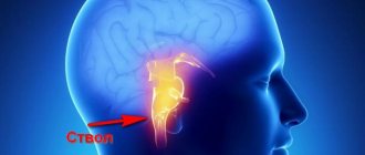
A complex disease characterized by increased blood filling of organs and tissues. Venous hyperemia is a pathology that occurs due to obstructed blood flow through the veins. This pathology can be local and widespread. When the disease occurs, patients experience a change in the color of the skin, a decrease in local temperature, tissue swelling and disruption of the functioning of the affected organ. If such symptoms appear, you need to go to the doctor to get diagnosed and choose the right treatment.
What are the causes and pathogenesis of the disease?
Most often, the development of venous hyperemia is observed in diseases of the blood system.
Pathology also occurs due to the following reasons:
- mechanical obstruction (uterus during pregnancy, hernial protrusion, postoperative scars, blood clot);
- increased blood viscosity;
- rapid blood clotting;
- varicose veins;
- heart valve dysfunction;
- hormonal imbalance;
- genetic predisposition;
- taking hormonal contraceptives.
Pathogenesis of the disease
With venous stagnation, the bloodstream is filled with carbon dioxide.
The mechanism of development of venous hyperemia begins with a violation of the outflow of venous blood from tissues or organs due to a large influx of arterial blood. Stagnation in the veins activates the release of oxygen to the tissues and the entry of carbon dioxide into the bloodstream. This is the pathogenesis of the appearance of hypoxemia and hypercapnia, in which cyanosis of the skin appears. With further progression of the disease, a disruption of oxidative processes occurs, a decrease in heat production and an increase in heat transfer, which leads to a local decrease in temperature.
Anemia (ischemia)
Anemia is the reduced blood supply to a tissue, organ, or part of the body as a result of insufficient blood flow. In tissue during ischemia, not only hypoxia develops, but also a deficiency of metabolites used by the cell in the process of glycolysis, which activates under conditions of decreased oxygen delivery, which explains the accelerated development of damage.
By prevalence, anemia can be divided into:
-general (anemia)
A disease of the hematopoietic system and is characterized by insufficient levels of red blood cells and hemoglobin.
-local
Tissue changes that occur during anemia are associated with hypoxia or anoxia (oxygen starvation). Depending on the cause of ischemia, the moment of suddenness of its occurrence, the duration of hypoxia and the degree of sensitivity of the tissue to it during ischemia, either subtle changes occur at the level of ultrastructures, or gross destructive changes, up to a heart attack.
Cell damage is also observed when blood flow is restored - reperfusion syndrome
, which includes three components:
1) calcium overload
Reperfusion of ischemic cells that have lost the ability to synthesize sufficient levels of ATP leads to loss of control over ion exchange. An intracellular increase in calcium triggers apoptosis or activates enzymes that disrupt cell membrane structures.
2) formation of reactive oxygen species
Ischemia induces the generation of reactive oxygen species such as superoxide, peroxide, and hydroxyl radical. Free radicals cause a cascade of damage to cell membranes, proteins and chromosomes.
3) development of inflammation
Also, reactive oxygen species activate the inflammatory cascade.
For acute anemia
dystrophic and necrobiotic changes occur.
In this case, glycogen disappears from the tissue, a decrease in the activity of redox enzymes and destruction of mitochondria. Acute ischemia should be considered as a pre-infarction condition. With prolonged anemia,
atrophy of parenchymal elements and sclerosis develop.
Coloring
: for diagnosis, various tetrazolium salts and potassium tellurite are used, which are reduced outside the ischemic areas and color the tissue gray or black, while the ischemic areas are not colored.
Depending on the reasons and conditions
occurrences are distinguished:
a) angiospastic anemia
Occurs as a result of artery spasm due to the action of any irritants. For example, painful stimulation can cause spasm of the arteries and anemia in certain areas of the body. Angiospastic ischemia appears both with negative emotional affects (“angiospasm of unreacted emotions”) and with exposure to low temperatures.
Angiospasm underlies the veno-arterial effect: with an increase in venous pressure, arteriolar spasm develops.
b) obstructive anemia
It develops as a result of the closure of the lumen of the artery by a thrombus (often resulting in vasospasm) or embolus, as a result of the proliferation of connective tissue in the lumen of the artery during inflammation of its wall (obliterating endarteritis), narrowing of the lumen of the artery by an atherosclerotic plaque.
c) compression anemia
Appears when an artery is compressed by a tumor, effusion, tourniquet, or ligature.
d) ischemia as a result of blood redistribution
Observed in cases of hyperemia after anemia. For example, cerebral ischemia when fluid is removed from the abdominal cavity, where a large mass of blood rushes.
Anemia due to arterial spasm is usually short-lived and does not cause any particular distress. However, with prolonged spasms, dystrophy and heart attack may develop. Acute obstructive anemia is especially dangerous, as it often leads to a heart attack. Long-term anemia sooner or later leads to atrophy and sclerosis.
What types of disease?
The following types of venous hyperemia are distinguished:
- Natural - does not cause consequences. Occurs with prolonged exposure to heat, significant emotional stress, changes in hormonal levels in women and physical overload.
- Pathological - develops against the background of exposure to negative factors (the influence of medications, cosmetics, edema and tumors, allergies, viral infections, the influence of high and low temperatures).
The following types are also distinguished:
- Working. Improves the functioning of organs and tissues.
- Reactive. Occurs with prolonged disruption of the blood supply to soft tissues and compression of organs.
There are also:
- acute;
- chronic.
Ticket 30. 1. Venous hyperemia. Causes
1. Venous hyperemia. Causes. Manifestations (changes in microcirculation and metabolism). Mechanisms of development. Stasis. Kinds. Causes. Development mechanism. Consequences
Enous blood stagnation - an increase in blood supply to an organ or tissue with a decrease in blood flowing through the vessels of the organ due to a violation of the outflow of blood into the venous system.
It occurs due to mechanical obstacles to the outflow of blood from the microcirculatory bed into the venous system. Reasons: 1) vein thrombosis; 2) increased pressure in large veins (for example, with right ventricular heart failure); 3) compression of the veins (occurs easily due to the thinness of their walls). If the pressure in the veins in front of the obstacle increases so much that it exceeds the diastolic pressure in the adductor arteries, then orthograde (normal) blood flow is observed only during systole, and during diastole, due to perversions of the pressure gradient, a retrograde (reverse) push of blood occurs. This type of blood flow is called pendular.
Symptoms of venous stagnation: 1) the temperature of superficially located organs and tissues decreases due to a decrease in the intensity of blood flow; this does not happen in the internal organs; 2) extravasation increases, resulting in tissue swelling; 3) during stagnation, blood oxygen is maximally used by tissues and most of the Hb is restored, therefore, the tissues acquire a bluish tint (dark cherry) - cyanosis.
General pathology of microcirculation itself
The main causes of violations: 1) intravascular changes; 2) changes in the vessels themselves; 3) extravascular changes.
Let's start with the characteristics of intravascular changes leading to disturbances in the microvasculature.
Hemorheology is the science of the influence of blood elements and their interaction with the walls of capillaries on blood flow. The rheological properties (fluidity) of blood as a heterogeneous liquid are of utmost importance when it moves through microvessels, the lumen of which is comparable to the size of its formed elements.
Normal blood flow through microvessels is possible under the following conditions: 1) formed elements can be easily deformed; 2) they do not stick together and do not form aggregates; 3) the concentration of blood cells is not excessive.
Otherwise, the rheological properties of blood deteriorate sharply. All of these properties are important primarily for erythrocytes (their number is 1000 times higher than the number of leukocytes), therefore, their properties have the greatest impact on the rheological properties of blood.
Impaired deformability of red blood cells. This ability of red blood cells is associated with the properties of their outer membrane, as well as the high fluidity of their contents. In the blood flow, rotational movements of the membrane occur around the contents of red blood cells, which also move. Deformability is extremely variable. It decreases with age. Red blood cell membranes become more rigid under the influence of various pathogenic factors (hyperosmolarity, loss of ATP, phospholipids, etc.). This occurs with heart disease, diabetes insipidus, cancer, stress, etc., therefore, the fluidity of blood in microvessels decreases sharply.
Violation of the structure of blood flow in microvessels. Of particular importance for the rheological properties of blood are changes in the structure of blood flow in microvessels with a diameter of 15–80 μm (arterioles). Thus, with the initial slowdown of blood flow, the longitudinal orientation of erythrocytes often changes to transverse, the velocity profile of various layers of blood in the vascular lumen becomes blunted, and the trajectory of erythrocytes becomes chaotic. This leads to changes in the rheological properties of the blood, in which the resistance increases significantly, causing an even greater slowdown in the flow of blood in the capillaries and disrupting microcirculation.
Increased intravascular aggregation of red blood cells. The ability of red blood cells to aggregate (stick together) and form “coin columns”, which then stick together, is their normal property. However, aggregation can significantly increase under the influence of various factors that change both the surface properties of erythrocytes and the environment surrounding them. As aggregation increases, the blood turns from a suspension of erythrocytes with high fluidity into a mesh suspension completely devoid of this ability.
Thus, erythrocyte aggregation disrupts the normal structure of blood flow in microvessels and is the most important factor changing the normal rheological properties of blood.
The extreme degree of aggregation of erythrocytes in modern medical literature is designated by the term sludge (thick mud, silt, swamp). Sludge phenomena disrupt the microrheological properties of blood to such an extent that blood flow in the capillaries slows down and stops completely - stasis occurs, despite the fact that the arteriovenous difference in blood pressure along these microvessels is preserved. However, initially, during blood stasis, neither hemolysis nor blood clotting occurs. For some time, the stasis is reversible: the movement of red blood cells can resume and the patency of the capillaries is restored again.
Factors causing increased erythrocyte aggregation:
1) Damage to the walls of the capillaries, as a result - increased filtration of fluid, electrolytes and albumins (low molecular weight proteins) into the surrounding tissues, which means that the concentration of high molecular weight proteins - globulins and fibrinogen - increases in the blood plasma. The absorption of these proteins on the membranes of red blood cells reduces their surface potential and promotes their aggregation.
2) Penetration of chemical damaging agents inside the capillaries and their direct effect on red blood cells, causing a change in the physicochemical properties of their membranes and promoting their aggregation.
3) The speed of blood flow in the capillaries, determined by the condition of the afferent arteries. Vasoconstriction leads to slower blood flow in the capillaries, promoting red blood cell aggregation and the development of stasis. With dilatation of the afferent arteries and acceleration of blood flow in the capillaries, erythrocyte aggregation and stasis become more difficult to develop and are much easier to eliminate.
In conclusion, we point out another factor that affects the rheological properties of blood - the concentration of red blood cells. A direct relationship has been proven between the concentration of red blood cells in the blood (hematocrit) and its relative viscosity (relative to water).
Consequences of stasis in microvessels. If no significant changes have occurred in the capillary wall and blood during the period of stasis, blood flow can be restored after the causes of stasis are eliminated. With significant disturbances of the vascular wall and red blood cells, blood stasis may be irreversible, causing necrosis of surrounding tissues. The pathogenic significance of stasis depends largely on the organ in which it originated (it is especially dangerous in the microvessels of the brain, heart and kidneys).
Pathogenetic principles of recovery
rheological properties of blood
1) Administration of low-molecular dextrans (rheopolyglucin), which leads to: a) dilution of the blood and an increase in Ronc due to the macromolecules of these hydrocarbons, leading to the transition of fluid from the intercellular substance into the vessels; b) to increase the Z-potential on erythrocytes and platelets; c) to close the damaged vascular endothelial wall.
2) The introduction of anticoagulants (heparin) increases the Z-potential on the membranes of red blood cells, platelets and, naturally, prevents the process of blood clotting.
3) Administration of thrombolytics (fibrinolysin).
4) Administration of antiplatelet agents (trental, nicotinic acid, etc.).
5) Elimination of vasospasm
2. Etiology and pathogenesis of collapse. Unfortunately, to date, in the definitions and classifications of the three main forms of ES - collapse, shock and coma, there are a number of inconsistencies or, on the contrary, repetitions, controversial points and other difficulties of various kinds. A comparison of various options for complete definitions makes it possible to identify acutely developing systemic arterial and venous hypotension as the main distinguishing signs for collapse, for coma - loss of consciousness and loss of reactions to external, including painful, stimuli, and for shock - the effect on the body of super-strong pathogenic irritants (N.I. Losev et al., 1995).
Collapse
It is known that the level of systemic blood pressure is directly dependent on several interconnected parameters: circulating blood volume (CBV), cardiac output (CO), total peripheral resistance of the vascular system (TPR) and inversely dependent on the volume of the vascular bed.
This leads to general pathogenetic mechanisms and specific etymological factors of collapse:
1) Decrease in BCC. An absolute decrease in blood volume may be due to the loss of part of the blood due to blood loss, plasmorrhagia, extensive burns, and dehydration of various origins. A relative decrease in BCC occurs with a significant increase in its deposited fraction in the low-pressure pool and capillaries. The cause of excessive blood deposition is usually a significant decrease in the tone of small vessels or right ventricular heart failure, leading to stagnation of blood in the veins of the systemic circle. An additional pathogenetic factor of collapse during blood deposition is an increase in the total volume of the vascular bed.
2) Primary decrease in cardiac output. It can occur as a result of acute heart failure due to heart attack, tamponade, certain types of arrhythmias, severe infections and intoxications, etc., leading to a weakening of the contractile function of the heart or a decrease in venous return of blood and a corresponding decrease in stroke volume.
3) A primary drop in total peripheral resistance can occur when various pathogenic factors influence the walls of resistive and capacitive vessels - this leads to a decrease in their neurogenic and myogenic tone and reactivity to pressor agents. Such pathogenic factors include infectious and non-infectious intoxications, some drugs when used incorrectly (adrenoblockers and adrenolytics), free radicals, disorders of the ionic composition (hyponatremia), excess of biologically active substances (histamine, serotonin), endocrine disorders (adrenal insufficiency, pituitary gland), hypoxia, etc. A sharp drop in OPS may also be associated with excessive irritation of depressor reflexogenic zones.
It follows from the above that three pathogenetic types of collapse are most common: cardiogenic, hypovolemic and vasodilatory. However, in addition to these types, in practice, types of collapse are often distinguished, taking into account its specific cause or group of related causes: infectious, toxic, radiation, pancreatic, orthostatic, hypocapnic, etc. Unfortunately, there is no generally accepted nomenclature of types of collapse. According to I.R. Petrov distinguishes the following types of collapse: 1) infectious-toxic; 2) hypoxemic; 3) orthostatic; 4) pancreatic (receipt of pancreatic metabolic products into the blood); 5) enterogenous (with dumping syndrome); 6) hemorrhagic.
Manifestations of collapse. It usually develops acutely and begins with severe disturbances in central hemodynamics. Mean arterial pressure drops below 70–60 mmHg. Art. Consciousness is preserved in most cases, but there is general lethargy, severe weakness, ringing in the ears, weakened vision, thirst, chilliness, decreased body temperature, pale skin, often cold sweat, tremor of the fingers, dilated pupils, sometimes nausea, vomiting, convulsions .
With severe and prolonged collapse, microcirculation disorders almost inevitably occur. Due to a decrease in perfusion pressure, blood flow in the microvasculature slows down and blood stagnates in the capillaries. The resulting circulatory hypoxia leads to an increase in the permeability of vascular membranes and the release of the liquid part of the blood into the perivascular environment. Hemoconcentration and deterioration of the rheological properties of blood can contribute to the aggregation of erythrocytes and platelets with the subsequent development of stasis and the appearance of microthrombi (DIC syndrome). As a result of severe arterial hypotension, filtration pressure in the renal glomeruli drops, oliguria or anuria occurs, leading to renal failure. In general, with an unfavorable development of the process, a picture of severe circulatory and then mixed hypoxia with an immediate threat to life is observed. This is the “average” description of the manifestations of collapse that follows from an analysis of the literature. But additional signs associated with the specific cause that caused the collapse may also be observed.
Fainting (syncope) is a sudden short loss of consciousness due to transient cerebral ischemia. It occurs reflexively. It is the mildest form of acute vascular insufficiency. The leading factor in the genesis of fainting is a decrease in blood pressure to a level at which sufficient brain perfusion is not ensured.
There are three main pathogenetic links in the development of fainting:
1) a drop in blood pressure due to a decrease in peripheral vascular resistance during systemic vasodilation (psychogenic fainting caused by hyperactivity of the parasympathetic division of the ANS, orthostatic hypotension);
2) heart rhythm disturbances (Morgagni-Edams-Stokes syndrome);
3) decrease in oxygen content in the blood.
2. Hyperfunction of the anterior pituitary gland. Causes, nature, mechanisms of disorders developing in the body.
Total hypopituitarism
1. Simmonds disease (hypothalamic-pituitary cachexia). The disease is based on diffuse damage (infection, tumor, trauma, hemorrhage) of the hypothalamic-pituitary region with loss of function of the adenohypophysis and failure of the peripheral endocrine glands. Characterized by severe exhaustion (cachexia), premature aging, and metabolic and trophic disorders. Women aged 30-40 years are most often affected.
Pathogenesis. A lack of pituitary tropic hormones leads to a sharp decrease in the function of peripheral endocrine glands. A decrease in somatotropin production causes exhaustion. Loss of gonadotropic function leads to ovarian failure, amenorrhea, atrophy of the uterus and vagina. Deficiency of thyrotropin, as a result - pituitary myxedema. A decrease in corticotropin production leads to the development of adrenal insufficiency up to Addisonian crises. Typically, this is the sequence of progression of pituitary insufficiency (loss of gonadotropic, somatotropic, thyrotropic and corticotropic functions). It is important to emphasize that the adenohypophysis has large functional reserves. Therefore, obvious symptoms of pituitary insufficiency develop only when 75–90% of the glandular tissue is destroyed. Clinically, general weakness, adynamia, emaciation, muscle atrophy, lack of appetite, drowsiness, amenorrhea, and apathy are detected. In the internal organs, changes in the form of hypofunction and atrophy are also sharply expressed (bradycardia, decreased blood pressure, suppression of secretion in the gastrointestinal tract, splanchnoptosis, etc.).
2. Sheehan's disease - postpartum hypopituitarism. The disease is usually based on significant and not timely compensated blood loss during childbirth (in combination with postpartum sepsis), accompanied by vasospasm of the anterior pituitary gland (APG). PDH hyperplasia during pregnancy is important. With prolonged vascular spasm, ischemic necrosis of the pituitary gland and a picture of pituitary cachexia develop. Unlike Simmonds' disease, it is not characterized by severe exhaustion and disorders of the gonads are relatively less pronounced.
Partial hypopituitarism
Strictly monohormonal forms of pathology are almost never found. Let us consider only the most common diseases, which are based on partial adenohypophyseal insufficiency.
Pituitary dwarfism. The main manifestation of this disease is a sharp retardation of growth associated with an absolute or relative deficiency of somatotropin. Frequency from 1:30005000 to 1:30000. In a broader sense, dwarfism is a disorder of growth and development, the occurrence of which can be caused not only by a deficiency of GH due to the pathology of the pituitary gland itself, but also by a violation of the hypothalamic regulation of its functions, and disturbances in tissue sensitivity to this hormone.
Most forms of pituitary dwarfism are genetic diseases. The most common is panhypopituitary dwarfism, which is inherited predominantly in a recessive manner. Genetic dwarfism with isolated growth hormone deficiency occurs occasionally (more common in Africa and the Middle East).
In the development of secondary dwarfism, as a symptom of any disease, chronic infections, intoxications, and poor nutrition are important.
A large group of patients with dwarfism consists of patients with various types of organic pathology of the central nervous system that arose in utero or in early childhood (underdevelopment of the pituitary gland, its cystic degeneration, atrophy due to compression by a tumor). Dwarfism can be caused by traumatic damage to the hypothalamic-pituitary region (intrauterine, birth or postnatal), which often occurs during multiple pregnancies, as well as during childbirth in the breech, leg presentation or in the transverse position with rotation on the leg (this is the mechanism of childbirth in 1/3 patients with dwarfism). Infectious and toxic damage is important (intrauterine viral infections, tuberculosis, toxoplasmosis; early childhood diseases, neonatal sepsis, meningo- and arachnoencephalitis).
Clinic. A sharp lag in growth and physical development are the main manifestations of pituitary dwarfism. Patients are born with normal weight and body length and begin to lag in growth from 2–4 years of age. Height below 130 cm for men and 120 cm for women is considered to be dwarfism. In addition to small absolute body sizes, pituitary dwarfism is also characterized by low annual dynamics of growth and physical development. The physique is proportional, but the proportions of the patients’ bodies are characteristic of childhood. The skin is pale, often with a yellowish tint, dry (due to thyroid insufficiency). The most important symptom of the disease is a delay in the timing of differentiation and ossification of the skeleton. In this regard, the dental system also suffers: there is a late change of milk teeth. The genital organs in most patients are severely underdeveloped, but malformations are rare. Sexual insufficiency is accompanied by underdevelopment of secondary sexual characteristics and decreased sexual feelings, absence of menstruation.
Thyroid insufficiency is a fairly common symptom of dwarfism. Intellect in most cases is not impaired, although some infantilism in behavior is often noted. The EEG in patients is characterized by features of immaturity, long-term preservation of a high “childish” voltage; unevenness of the alpha rhythm in amplitude and frequency; a sharp increase in the content of slow (theta and delta) rhythms.
Treatment. This is a long process. To obtain the effect, two basic principles must be observed:
1) maximum approximation of treatment-induced development to physiological conditions; 2) sparing the epiphyseal growth zones. The main type of pathogenetic therapy for pituitary dwarfism is the use of human growth hormone (human and primate somatotropin is used). For treatment with somatotropin, patients are selected with proven deficiency of endogenous growth hormone, with skeletal differentiation not exceeding the level characteristic of 13–14 years. In addition, the most important means of treating dwarfism is the use of anabolic steroids (nerabol, nerobolil), which stimulate growth by enhancing protein synthesis and increasing the level of endogenous growth hormone. In the presence of hypothyroidism, thyroid drugs are prescribed in parallel. When treating boys, the next step is the administration of human chorionic gonadotropin. Girls over 16 years of age are usually prescribed estrogens. The final stage of treatment (after closure of the growth zones) is the constant administration of therapeutic doses of sex hormones corresponding to the patient’s gender, with the aim of the full development of the genital organs.
Neuroendocrine obesity. This form of pathology includes numerous variants that differ in their pathogenetic mechanisms. Many of them are now believed to be based on insufficient biosynthesis in the adenohypophysis of the fat-mobilizing polypeptide lipotropin as a result of damage to the pituitary gland itself or the hypothalamic centers with secondary involvement of the pituitary gland. Pituitary obesity is characterized by excessive fat deposition on the abdomen, back and proximal extremities with relative “thinness” in the distal parts - forearms and legs.
Other endocrine glands are also involved in the progression of various forms of the disease. Hyperinsulinism is characteristic. The level of somatotropin decreases and the level of corticotropin increases. The gonadotropic function of the pituitary gland also decreases, resulting in hypogonadism.
Adiposogenital dystrophy. It develops more often in boys. This disease manifests itself in two main syndromes: obesity and hypogonadism. Such a pathology can be considered an independent disease only if its symptoms appeared in childhood and the cause of the disease could not be established. When establishing the nature of the process that damages the pituitary gland (inflammation, tumor, etc.), obesity and hypogonadism are considered as symptoms of the underlying disease.
The disease is based on dysfunction of the hypothalamus, which leads to a decrease in the gonadotropic function of the pituitary gland, and as a result, secondary hypogonadism. Adiposogenital dystrophy is detected more often in prepubertal age (10–12 years). The syndrome is characterized by general obesity of the “female type”: in the abdomen, pelvis, torso, face. Body proportions are eunuchoid (tall, narrow shoulders, poor muscle development, etc.). The penis and testicles are reduced in size, and cryptorchidism is often detected.
Hyperpituitarism
Overproduction of adenopituitary hormones, as a rule, is partial in nature and is expressed in the following most common forms.
3.1.monogenic diseases
2.autosomal recessive
3.mutation delF508, leading to the absence of phenylalanine at position 508 of the transmembrane regulatory protein
Signs of the disease
With this pathology, the skin on the face may acquire a blue tint.
The following signs of venous hyperemia are distinguished:
- bluish tint to the skin of the face and other parts of the body;
- decrease in temperature in the affected area;
- hyperemic mucous membranes in pathological places;
- swelling of tissues;
- soreness when touched.
How is venous hyperemia treated?
Treatment of hyperemia is aimed at eliminating the cause of its occurrence (decreased vascular tone) and normalizing blood circulation.
Treatment consists of several stages:
- Maintaining a healthy lifestyle. For example, treatment for skin hyperemia is prescribed by a dermatologist. It is necessary to wipe with special lotions, adhere to appropriate skin care requirements, and take medications to normalize blood circulation and microcirculation. When treating brain hyperemia, it is important to maintain a rest regime. The diet should contain light and non-irritating foods with a full range of vitamins and minerals. Treatment also includes body rubdown, steam baths, massages, and foot wraps. It is advisable to walk barefoot on wet grass.
- To maintain the venous wall normally , all necessary elements must be supplied to the body. Vein tone is also maintained by systematic physical activity (in moderation) and an active lifestyle. Excessive stress and prolonged immobility should be avoided. Sitting and standing positions should be alternated with rest breaks. Quitting smoking and excessive consumption of alcoholic beverages helps to prolong the youth of the veins and increase their elasticity.
- The use of medications to tone the venous wall. There are a number of drugs that are used both locally and systemically.
- Hirudotherapy (placement of leeches) copes with several functions at once: unloading the local blood flow, thanks to which the vein wall returns to normal tone; improving blood movement, microcirculation, and, accordingly, tissue nutrition; The properties of saliva deserve special praise; they ensure the normalization of cholesterol, protect blood vessels, and have an anti-inflammatory effect.
- In extreme cases, surgery .
Diagnostic features
When the first symptoms of venous hyperemia appear, the patient urgently needs to go to the hospital. The doctor will record the patient's complaints and conduct an objective examination. Then the specialist will find out what the pathophysiology of the disease is and identify the differences between the disease and other vein pathologies. After this, the specialist will refer you to special informative diagnostic measures. These include:
- general blood and urine examination;
- blood biochemistry;
- prothrombin index;
- coagulogram;
- Ultrasound;
- Dopplerography.
Diagnostics
- Doppler ultrasound. To recognize venous stasis (vein hyperemia) of internal organs, Doppler scanning is used. It measures how sound waves are reflected from moving blood particles. In this case, the data is processed using a computer and a color image is created. This diagnosis of hyperemia allows you to consider the contents of the vessel - how the blood flows and the speed of its flow.
- Ultrasonography. When using ultrasonic waves, they are reflected from the vessels to the blood elements. The computer processes these waves and produces a two-dimensional black and white image.
What treatment is needed for venous hyperemia?
To treat the pathology, you will need a medical consultation.
Venous hyperemia is a common disease that, if prolonged, leads to serious consequences. For proper treatment of the pathology, you need to contact a specialist at the clinic. The use of various therapeutic techniques leads to a worsening of the condition. The doctor will examine the patient, make a diagnosis and draw up a treatment plan. To cure such a pathology, a specialist will prescribe medications and folk remedies.
Drug therapy
If the patient has hyperemia of the skin, the medications presented in the table are prescribed:
| Pharmacological groups | Drugs |
| Phlebotonics | "Venoruton" |
| "Ginkor Fort" | |
| Anticoagulants | "Warfarin" |
| "Calciarin" | |
| Nonsteroidal anti-inflammatory drugs | "Nimid" |
| "Indomethacin" | |
| Diuretics | "Spironolactone" |
| "Furosemide" | |
| Vitamins | B1, B6, B12 |
Folk remedies
To prepare a medicinal infusion you will need rowan.
Facial hyperemia is reduced by using the following recipes:
- Infusion of horse chestnut, rowan and arnica. Take all the plants in a ratio of 5:5:1, mix, select 1 tbsp. l. and stir in 250 ml of hot water. Then the mixture is tightly closed and left for 10 minutes. Drink ½ glass 2 times a day.
- Infusion of butcher's broom. Take 6 grams of rhizome and add 750 ml of water. Then mix everything, set to boil for 5 minutes and leave to infuse. After this, add honey and consume 150 ml 3 times a day.
Treatment of skin hyperemia
When treating, you can use the following tips:
- Cosmetics must not contain alcohol;
- Using protective creams before going outside;
- The use of external agents such as petroleum jelly, zinc ointment, boric acid;
- Masks, cooling compresses, herbal medicine;
- It is forbidden to eat spicy foods, coffee, or alcohol.
Primary and secondary skin hyperemia can be successfully treated. To achieve the best results, it is important to change your lifestyle. Healthy foods and gentle physical activity will help improve blood flow through the vessels.
Consequences
Venous congestion significantly worsens the condition of the body, due to the fact that the disease is accompanied by impaired trophism, hypoxic damage to cells, tissues and organs, and compression by edema. In severe cases of pathology, there is an increase in the permeability of the vascular wall, the release of red blood cells into the tissue, resulting in the formation of small hemorrhages.
Articles on the topic
- Yeast fungi in the stool of an adult during analysis
- Diseases of the lower extremities - signs, classification, diagnosis, therapy and prevention
- Valtrex - instructions for use, indications, dosage, contraindications and reviews
In the chronic course of the disease, edematous fluid accumulates in tissues and cavities. Under conditions of oxygen deficiency, mucoid swelling of the interstitial substance occurs in the cells of parenchymal organs, atrophy and fatty degeneration develop, and organ function is lost. Such pathological changes are usually reversible if the cause of the disease is eliminated. The consequences of venous hyperemia can be as follows:
- phlebeurysm;
- stasis;
- vascular sclerosis;
- prolonged inflammatory processes;
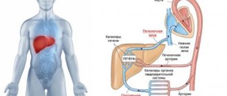
- cirrhosis.