What is cardiography
The essence of cardiography is the study of electrical currents arising during the work of the heart muscle. The advantage of this method is its relative simplicity and accessibility. Strictly speaking, a cardiogram is the result of measuring the electrical parameters of the heart, displayed in the form of a time graph.
The creation of electrocardiography in its modern form is associated with the name of the Dutch physiologist of the early 20th century, Willem Einthoven, who developed the basic ECG methods and terminology used by doctors to this day.
Thanks to the cardiogram, it is possible to obtain the following information about the heart muscle:
- Heart rate,
- Physical condition of the heart
- The presence of arrhythmias,
- The presence of acute or chronic myocardial damage,
- The presence of metabolic disorders in the heart muscle,
- Presence of electrical conductivity disturbances,
- Position of the electrical axis of the heart.
Also, a cardiac electrocardiogram can be used to obtain information about certain vascular diseases not related to the heart.
An ECG is usually performed in the following cases:
- Feeling of abnormal heartbeat;
- Attacks of shortness of breath, sudden weakness, fainting;
- Heartache;
- Heart murmurs;
- Deterioration of the condition of patients with cardiovascular diseases;
- Passing medical examinations;
- Medical examination of people over 45 years of age;
- Examination before surgery.
An electrocardiogram is also recommended for:
- Pregnancy;
- Endocrine pathologies;
- Nervous diseases;
- Changes in blood counts, especially with an increase in cholesterol;
- Over 40 years of age (once a year).
Paroxysmal ventricular tachycardia
It is the most life-threatening of all types of cardiac tachycardia. The main source of rhythm, instead of the sinus node, becomes a focus of increased impulses in the ventricles. It “imposes” its own rhythm on the ventricles, which is different from the frequency of contraction of the atria.
Causes:
- the presence of post-infarction scars in the ventricular tissue;
- chronic aneurysm of the left ventricle (protrusion of the thinned wall outward);
- inflammatory diseases of the myocardium;
- cardiomyopathies (increase in the volume and mass of the heart);
- infiltrative diseases of the myocardium (tumors, amyloidosis);
- intoxication with cardiac glycosides;
- use of antiarrhythmic drugs (quinidine, procainamide, sotalol, amiodarone);
- instrumental studies (angiography, endoscopy);
- heart surgery;
- electrolyte imbalance (decreased levels of potassium and magnesium in the blood).
Symptoms:
- a sudden attack of increased heart rate;
- dyspnea;
- dizziness;
- chest pain;
- decreased blood pressure;
- fainting;
- convulsions.
Characteristic changes on the electrocardiogram:
- non-sinus rhythm;
- wrong rhythm;
- the frequency of contraction of the atria may be normal, and that of the ventricles – 100 – 250 per minute;
- their complete separation (dissociation);
- deformation and widening of QRS complexes for more than 0.12 seconds.
Treatment
If the patient's condition is stable, you can start with the administration of the antiarrhythmic drug lidocaine. In its absence - bretylium tosylate. If there is no effect or the patient is unstable (there are “alarming” symptoms), defibrillation (electrical cardioversion) is used. The charge strength ranges from 100 to 360 Joule. Then hospitalization is carried out in the intensive care unit.
Surgical treatment is also possible, during which a cardioverter-defibrillator is implanted into the patient’s chest. This device analyzes the heart rhythm and, if it is disrupted, performs electrical cardioversion.
Implantation of a cardioverter-defibrillator (ICD) has been performed in the world for more than 30 years. The idea of its creation belonged to the French doctor F. Zacouto, although the defibrillator he invented in 1953 was still external. The first implantation of a defibrillator was performed in 1980. In Russia, this happened later, in 1990, at the Bakulev Scientific Center.
A modern ICD is a small box, up to 8 cm in size, weighing about 80 grams. It is coated with titanium alloy. Inside there is a microprocessor, battery, capacitor, voltage converter, resistors, discharge release system and database. One or more electrodes extend from the housing. The ICD is implanted under the skin in the left subclavian region, and the electrodes are implanted in the chambers of the heart.
Regularly, 2 - 4 times a year, it is necessary to check the functioning of the device and charge the battery. In addition, the ICD itself can inform its owner about a breakdown - via vibration or sound signal. Replacement of the device as a whole will be required after 5 – 8 years. In the Russian Federation, an implantable cardioverter-defibrillator will cost 900 thousand rubles.
The ICD monitors the heart rate, and if an abnormal rhythm is detected, a current discharge occurs. The patient at this moment experiences a “blow” to the chest. Not the most pleasant feeling. For this reason, installing a defibrillator is irrational for frequent paroxysms of tachycardia.
Methodology of the procedure
ECG recording is usually performed in a supine position. To take a cardiogram, a stationary or portable device is used - an electrocardiograph. Stationary devices are installed in medical institutions, and portable ones are used by emergency teams. The device receives information about electrical potentials on the surface of the skin. For this purpose, electrodes are used that are attached to the chest area and limbs.
These electrodes are called leads. There are usually 6 leads installed on the chest and limbs. The chest leads are designated V1-V6, the leads on the limbs are called basic (I, II, III) and reinforced (aVL, aVR, aVF). All leads give a slightly different picture of oscillations, but by summing up the information from all electrodes, you can find out the details of the functioning of the heart as a whole. Sometimes additional leads are used (D, A, I).
Typically, the cardiogram is displayed in the form of a graph on paper containing millimeter markings. Each electrode lead has its own schedule. The standard speed of the belt is 5 cm/s; other speeds may be used. The cardiogram displayed on the tape can also indicate the main parameters, normal indicators and a conclusion generated automatically. Data can also be recorded in memory and on electronic media.
After the procedure, the cardiogram is usually deciphered by an experienced cardiologist.
Magnetocardiography
Magnetocardiography (MCG) is a non-contact study of cardiac activity, which studies the magnetic field of the organ.
MCG and ECG have the same goals and can complement each other. As a rule, magnetocardiography is used when it is impossible to use electrodes.
Due to the weakness of the electromagnetic field of the heart, highly sensitive recording equipment is required to conduct MCG. As a rule, specialists use a toroidal coil with a large number of turns as a sensor, which is placed as close as possible to the person’s chest. The patient should be in a sitting or lying position at this time. The signals transmitted by the sensor are recorded using a recorder. The resulting magnetocardiogram is interpreted by a specialist.
The study has no contraindications, does not cause discomfort and can be carried out both in a clinic and in a hospital setting. However, patients should not have any magnetic materials (metal dentures, watches, etc.) that could cause signal distortion or failure.
Magnetocardiography device
Holter monitoring
In addition to stationary devices, there are also portable devices for daily (Holter) monitoring. They are attached to the patient's body along with electrodes and record all information received over a long period of time (usually within 24 hours). This method provides much more complete information about processes in the heart compared to a conventional cardiogram. For example, when taking a cardiogram in a hospital setting, the patient must be at rest. Meanwhile, some deviations from the norm may appear during physical activity, sleep, etc. Holter monitoring provides information about such phenomena.
Photo: kostastudio/Shutterstock.com
When is cardiography of blood vessels and heart prescribed?
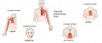
Indications for cardiography are pain, discomfort in the heart, neck, back, abdomen, chest (this is how ischemia manifests itself in some cases), shortness of breath, frequent fainting, swelling of the legs, high blood pressure, heart murmurs, rheumatism, diabetes, stroke.
A cardiogram is prescribed to patients in preparation for surgery, during preventive annual examinations, pregnancy, when completing documentation before being assigned to health institutions and sports sections, etc.
In addition, people over 40 years of age are recommended to have a heart cardiogram performed annually, despite the absence of complaints. This is the only way to detect hidden heart rhythm disturbances, ischemia, and heart attack in a timely manner.
Other types of procedures
There are several other methods for carrying out the procedure. For example, this is monitoring with physical activity. Abnormalities are usually more pronounced on the stress ECG. The most common way to provide the body with the necessary physical activity is a treadmill. This method is useful in cases where pathologies can only manifest themselves in the case of increased heart function, for example, if coronary artery disease is suspected.
During phonocardiography, not only the electrical potentials of the heart are recorded, but also the sounds that arise in the heart. The procedure is prescribed when it is necessary to clarify the occurrence of a heart murmur. This method is often used when heart defects are suspected.
Types of teeth
First we should talk a little about how the heart works. It has 4 chambers - two atria and two ventricles (left and right). The electrical impulse, due to which it contracts, is formed, as a rule, in the upper part of the myocardium - in the sinus pacemaker - the sinoatrial (sinus) node. The impulse spreads down the heart, first affecting the atria and causing them to contract, then passes through the atrioventricular nerve node and another nerve node, the bundle of His, and reaches the ventricles. The main burden of pumping blood is taken on by the ventricles, especially the left one, which is involved in the systemic circulation. This stage is called heart contraction or systole.
After contraction of all parts of the heart, the time comes for their relaxation - diastole. The cycle then repeats again and again - this process is called heartbeat.
The condition of the heart, in which there are no changes in the propagation of impulses, is reflected on the ECG in the form of a straight horizontal line, called an isoline. The deviation of the graph from the isoline is called a spike.
One heartbeat on the ECG contains six waves: P, Q, R, S, T, U. The waves can be directed both up and down. In the first case they are considered positive, in the second - negative. The Q and S waves are always positive, and the R wave is always negative.
Photo: Infmedserv.ru
The teeth reflect the different phases of heart contraction. P reflects the moment of contraction and relaxation of the atria, R – excitation of the ventricles, T – relaxation of the ventricles. Special designations are also used for segments (spaces between adjacent teeth) and intervals (sections of the graph that include segments and teeth), for example, PQ, QRST.
Correspondence between the stages of heart contraction and some elements of cardiograms:
- P – atrial contraction;
- PQ – horizontal line, the transition of the discharge from the atria through the atrioventricular node to the ventricles. The Q wave may be absent normally;
- QRS – ventricular complex, the element most often used in diagnostics;
- R – ventricular excitation;
- S – myocardial relaxation;
- T – ventricular relaxation;
- ST – horizontal line, myocardial recovery;
- U – may be absent normally. The reasons for the appearance of the prong are not clearly understood, but the prong is valuable for diagnosing certain diseases.
Below are some abnormal ECG findings and their possible explanations. This information, of course, does not negate the fact that it is more advisable to entrust the decoding to a professional cardiologist who better knows all the nuances of deviations from the norm and associated pathologies.
Main deviations from the norm and diagnosis
| Description | Diagnosis |
| The distance between the R teeth is not the same | atrial fibrillation, heart block, sinus node weakness, extrasystole |
| P wave is too tall (more than 5 mm), too wide (more than 5 mm), has two halves | atrial thickening |
| The P wave is absent in all leads except V1 | the rhythm does not come from the sinus node |
| PQ interval extended | atrioventricular block |
| QRS extension | ventricular hypertrophy, bundle branch block |
| No gaps between QRS | paroxysmal tachycardia, ventricular fibrillation |
| QRS as a flag | heart attack |
| Deep and wide Q | heart attack |
| Wide R (more than 15 mm) in leads I, V5, V6 | left ventricular hypertrophy, bundle branch block |
| Deep S in III, V1,V2 | left ventricular hypertrophy |
| ST is more than 2 mm above or below the isoline | ischemia or heart attack |
| Tall, double-humped, pointed T | cardiac overload, ischemia |
| T merging with R | acute heart attack |
Table of cardiogram parameters in adults
| Index | Value,c |
| QRS | 0,06-0,1 |
| P | 0,07-0,11 |
| Q | 0,07-0,11 |
| T | 0,12-0,28 |
| PQ | 0,12-0,2 |
Normal duration of cardiogram elements in children
| Index | Value,c |
| QRS | 0,06-0,1 |
| P | <0,1 |
| PQ | 0,2 |
| QT | <0,4 |
The norms indicated in the table may also depend on age.
Rhythm of contractions
Violation of the rhythm of contractions is called arrhythmia. The irregularity of the rhythm during arrhythmia is measured as a percentage. An irregular rhythm is indicated by a deviation in the distance between similar teeth by more than 10%. Sinus arrhythmia, that is, arrhythmia combined with sinus rhythm, may be normal for adolescents and young adults, but in most cases it indicates the onset of a pathological process.
A type of arrhythmia is extrasystole. They say it in the case when extraordinary contractions are observed. Single extrasystoles (no more than 200 per day with Holter monitoring) can also be observed in healthy people. Frequent extrasystoles that appear on the cardiogram in the amount of several pieces may indicate ischemia, myocarditis, or heart defects.
Heart rate
This option is the simplest and most understandable. It determines the number of contractions in one minute. The number of contractions may be higher than normal (tachycardia) or lower than normal (bradycardia). The normal heart rate in adults can range from 60 to 80 beats. However, the norm in this case is a relative concept, so bradycardia and tachycardia may not always be evidence of pathology. Bradycardia can occur during sleep or in trained people, and tachycardia can occur during stress, after exercise or at elevated temperatures.
Heart rate norms for children of different ages
| Age | Heart rate, beats/min |
| Newborns | 140-160 |
| 6 months | 130-135 |
| 1 year | 120-125 |
| 2 years | 110-115 |
| 3 years | 105-110 |
| 5 years | 100-105 |
| 8 years | 90-100 |
| 10 years | 80-85 |
| 12 years and older | 70-75 |
Photo: Africa Studio/Shutterstock.com
Sinus tachycardia
Sinus tachycardia is the result of a malfunction of the sinus node. It produces an electrical impulse more often than necessary (more than 90 per minute). In this case, the sequence of contraction (atria - ventricles) is not disrupted.
Such tachycardia should be considered normal in the following conditions:
- exercise stress. When working, muscles consume more energy and oxygen than at rest. Oxygen delivery is provided by blood, therefore, the faster it circulates, the greater the volume of oxygen it can transfer to the muscles. For this purpose, the body itself accelerates the work of the heart. In completely healthy people, during physical activity, the heart rate increases to 180 - 200 per minute;
- emotional stress (excitement, fear, joy, fright, anger and other emotions);
- release of adrenaline (a hormone produced by the adrenal glands) into the blood. It is produced during stress, pain, strong emotions and other conditions;
- increase in ambient body temperature; rise to height. In the mountains, the oxygen content in the inhaled air is reduced. To ensure the required level of oxygen in the blood, the body is rebuilt - breathing and heartbeat become more frequent;
- drinking alcohol, coffee or any other energy drinks;
- smoking. Substances contained in smoke stimulate the nervous system, leading to tachycardia;
- medicines. After reading the instructions, you will see that many medications have a side effect - tachycardia;
- a sharp change in body position from horizontal to vertical.
The upper limit of normal heart rate can be determined by the formula: 220 – age (in years).
After a few minutes (on average, up to 10) after exposure to the above reasons, the heart rate should return to normal. If this does not happen, or tachycardia occurs at rest, you should consult a doctor. In this case, the cause may be some disease.
The most common pathological conditions accompanied by rapid heartbeat:
- hyperthermia (increased body temperature). There is a relationship - with every 1.5°C increase in body temperature, the heart rate accelerates by 10 beats per minute;
- hypovolemia (decrease in circulating blood volume, for example, as a result of blood loss);
- anemia (decrease in hemoglobin concentration and number of red blood cells in the blood);
- infection (any bacterial or viral disease);
- malignant neoplasms;
- myocardial ischemia (poor blood supply to the heart);
- heart failure;
- pulmonary embolism (blockage with a blood clot);
- thyrotoxicosis (increased production of thyroid hormones by the thyroid gland);
- brain damage and others.
Cases of congenital familial sinus tachycardia have been described in the literature.
Manifestations of an attack of sinus tachycardia:
- feeling of heartbeat;
- weakness;
- dizziness;
- nausea;
- feeling of lack of air;
- heartache;
- sweating
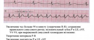
Signs on the electrocardiogram:
- sinus rhythm (positive P wave before each QRS complex in lead II. P waves of the same shape and amplitude in the same lead);
- correct (distances R - R are the same and shortened);
- heart rate from 90 to 180 per minute;
- shortening the P–Q intervals, but not less than 0.12 seconds and Q–T;
- increase in the amplitude of P waves in leads I, II, aVF;
- oblique depression of the RS – T segment, but not more than 1 millimeter below the isoline.
Treatment
Elimination of provoking factors - smoking, excessive physical activity, changing medications, etc. If necessary, you can use valocardine, corvalol or sedatives (phenozepam). Treatment of the disease that caused the tachycardia is required. Only when symptoms of myocardial ischemia appear (pain behind the sternum, lasting more than 20 minutes) is it necessary to slow down the heart rate with beta-blockers (bisoprolol, metoprolol).
The drug ivabradine has proven itself well in the treatment of tachycardia. It acts on the sinus node itself, and is able to slow down the frequency of contractions by 10 - 15 per minute. But it should not be taken instead of treating the cause, but in addition to the selected therapy.
Heart Rate Types
There are several types of heart rhythm, depending on where the nerve impulse begins to spread, causing the heart to contract:
- Sinus,
- Atrial,
- Atrioventricular,
- Ventricular.
Normally, the rhythm is always sinus. In this case, sinus rhythm can be combined with both a heart rate above normal and a heart rate below normal. All other types of rhythms are evidence of problems with the heart muscle.
Atrial rhythm
Atrial rhythm also often appears on the cardiogram. Is atrial rhythm normal or is it a type of pathology? In most cases, the atrial rhythm on the ECG is not normal. However, this is a relatively mild degree of heart rhythm disturbance. It occurs when the sinus node is suppressed or disrupted. Possible causes are ischemia, hypertension, sick sinus syndrome, endocrine disorders. However, isolated episodes of atrial contractions can also be observed in healthy people. This type of rhythm can take on both the character of bradycardia and the character of tachycardia.
Atrioventricular rhythm
Rhythm emanating from the atrioventricular node. With atrioventricular rhythm, the pulse rate usually drops to less than 60 beats per minute. Causes: weakness of the sinus node, atrioventricular block, taking certain medications. Atrioventricular rhythm, combined with tachycardia, can occur during heart surgery, rheumatism, and heart attack.
Ventricular rhythm
With ventricular rhythm, contractile impulses propagate from the ventricles. The contraction frequency drops to below 40 beats per minute. The most severe form of rhythm disturbance. Occurs in acute infarction, heart defects, cardiosclerosis, cardiac circulatory failure, and in a preagonal state.
Normal heart rate per minute in women
Palpitation is a condition in which heartbeats are felt. In the normal rhythm of life they are very difficult to track. Therefore, when such phenomena occur, a person may have concerns about his own health.
To understand in what cases it is necessary to seek help, it is important to be able to distinguish tachycardia from normal palpitations, which may occur due to a combination of certain circumstances.
To find out what the cause is, you need to count your pulse rate and pay attention to the presence of other symptoms. If the pulse rate is between 60-90 beats per minute, the blood pressure is normal and there are no other signs of deterioration in health, this situation does not require special help. If the pulse rate is within normal limits, but there are surges in blood pressure, signs of dizziness, or a state of fainting, then there is a need to seek help from specialists.
Pulse and blood pressure indicators are the main components of normal physical condition. Pressure is the force with which the blood presses on the vessels. Pulse describes the beats of the heart per minute. In adult women, fluctuations in the range of 60-100 beats are considered indicators of a normal heartbeat. Blood pressure is considered normal within 120-80.
How to determine your pulse correctly
You can easily measure the pulse from the arteries that are located as close as possible to the surface. You can feel the pulse in your neck and wrist:
- fingers must be placed on the place where the pulse can best be felt;
- the blows are counted for fifteen seconds. At this moment, the eyes should carefully monitor the movement of the arrow;
- the number obtained during the calculation must be multiplied by 4.
This method easily determines the heart rate in a given period of time for any person. To find out your maximum heart rate during physical activity, you need to subtract your age from 220. Women at the age of 20 have a maximum heart rate of 200. At age, this figure will be equal to 150. Heart contractions in this case should be 50-85% of the maximum heart rate.
There are several types of heartbeat:
- Bradycardia is a decreased heart rate. Characterized by a heart rate of up to 60 beats per minute (the state of an unhealthy person).
- Normocardia is a heartbeat that is within the normal range (60-90 beats per minute) at rest.
- Tachycardia. In this case, the heart rate is outside the normal range. Heart rate is more than 90 per minute.
Women's heart rates can change as they age and depending on their level of fitness.
During pregnancy, it is necessary to measure the fetal heartbeat. This helps to identify deviations of various types. Defects identified in advance contribute to timely treatment and elimination of the causes of the disease.
Normal blood pressure in pregnant women
During pregnancy, there are factors that influence the woman's condition. The main one is blood pressure. There are cases when a pregnant woman’s blood pressure readings often fluctuate, then there should be regular monitoring.
At the initial stage of pregnancy, a problem such as hypotension often occurs. It manifests itself as low blood pressure. There is an increased tendency to sleep and a state of dizziness. The factors that provoke such conditions are mainly hormonal changes in the body. Symptoms become more severe in the morning. Most women consider such jumps to be a normal condition, but this is not the norm. During pregnancy, low blood pressure can cause placental insufficiency in the fetus. In this case, the child will not receive enough nutrients and suffer from a small amount of oxygen.
During the second half of pregnancy, some expectant mothers complain of increased blood pressure. The reason for this phenomenon is considered to be a sharp increase in blood volume, by approximately 1 liter. At the end of pregnancy, the increase in volume can reach 1.5 liters. Symptoms that characterize high blood pressure include severe headaches, tinnitus, blurred vision, and rapid heartbeat. There are cases when changes in a woman’s body do not cause her such troubles, so it is not so easy to determine this. If, when measuring blood pressure, the readings are 140/90 or more, this is considered a reason to consult a doctor. Because high blood pressure can trigger a lot of irreversible processes in a woman’s body that will affect the life and health of the child.
Pregnant women who are prone to pressure surges should constantly pay attention to them. If a woman feels quite well, it would be a good idea to check her blood pressure readings once a week. If headaches or dizziness are present, measurements should be taken much more often, and if the indicators deviate from the norm, you should consult a doctor. For pregnant women, blood pressure is a very important indicator that should be periodically monitored.
Scientists have long proven that a rapid pulse can cause the development of many diseases, not only in the female body, but also in the male body. Therefore, monitoring your pulse is a necessary procedure. Timely diagnosed deviations from the norm will help to avoid many problems in the future.