Acute coronary syndrome (ACS) is a serious condition caused by impaired coronary circulation and myocardial ischemia. When the blood supply to the heart muscle completely stops, hypoxia occurs, which is the direct cause of heart attack and death. This deadly pathological process is popularly called a pre-infarction condition or a heart attack. ACS is a general concept that includes a number of processes that are equal in origin and similar in course, but differ in prognosis and likelihood of cure.
Diseases of the cardiovascular system, which are based on coronary insufficiency syndrome, are designated by the term ACS. This is what clinicians call heart diseases in which its blood supply deteriorates: myocardial infarction and unstable angina. These diseases have similar initial manifestations, pathophysiological mechanisms and some treatment principles. That is why the concept of “acute coronary syndrome” was introduced into medical practice. It is used to make a preliminary diagnosis, when all the characteristics of the patient’s pathological process are not fully clarified.
Acute coronary syndrome has an ICD-10 code of 124.9 and the name “Acute coronary heart disease, unspecified.” The causes of the pathology most often are: thrombosis and thromboembolism caused by rupture of an atherosclerotic plaque or erosion of the endothelium of the coronary artery. There is a special form of the syndrome that is of allergic origin. It is associated with excess production of inflammatory mediators by mast cells. Symptoms of the syndrome are: shortness of breath, irregular heart rhythm, pressing chest pain that occurs at rest or with minor physical stress. The most dangerous sign of ACS is sudden cardiac arrest. This diagnosis is usually made in the intensive care unit.
Coronary disease occurs with pronounced periods of exacerbation and remission. IHD worsens under the influence of provoking factors. In this case, its mild form develops - unstable angina or a severe form - myocardial infarction. In the first case, acute ischemia of the heart muscle does not lead to necrosis. Due to narrowing or blockage of the coronary arteries, trophic changes occur that develop gradually, slowly, in steps. After exposure to the trigger factor, an attack of ACS occurs. With myocardial infarction, irreversible cell death occurs. This pathology is much more dangerous - it is accompanied by severe chest pain, impaired breathing and consciousness, and an avalanche-like death of cardiomyocytes. Destruction of large areas is manifested by the most severe symptoms. With angina pectoris, there are no ECG signs of myocardial necrosis and specific biochemical markers in the blood. It is possible to transition from one clinical form of IHD to another.
The characteristic clinical picture of the syndrome allows you to quickly make a diagnosis and help the patient. To save his life, you should know the algorithm for providing emergency medical care and be able to carry out all the necessary measures before the arrival of qualified specialists. Emergency treatment of ACS will help avoid the development of serious complications and death. It is effective only if diagnosed early. General therapeutic measures prescribed to patients depend on clinical manifestations, the severity of pathological changes and the general well-being of the patient.
Currently, acute coronary syndrome is a pressing medical problem. This is due to the high frequency of its occurrence and the need for preventive measures. The development and implementation of a new medical strategy can save the lives of patients with ACS.
Etiology
Various provoking factors and pathological processes lead to the development of acute coronary syndrome.
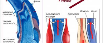
Atherosclerosis of the coronary arteries is the main cause of the disease. The fatty substances that form the plaque narrow the blood vessels and impede the flow of blood through the narrowed area. The heart stops fully pumping oxygen-rich blood. This leads to chest pain and heart attack. At early stages of the process, lipid structures are highly soluble. As the pathology progresses, they calcify and become rock-hard. You can only get rid of such formations surgically.
When the surface of an atherosclerotic plaque ruptures, a blood clot forms at the site of damage, the lumen of the vessel narrows, and its patency is impaired. These morphological elements of the disease reduce coronary blood flow and disrupt the blood supply to the myocardium. Blood stops circulating normally, and the heart rate increases to compensate for the resistance. Patients' blood pressure rises and their pulse quickens. If the blood flow completely stops due to a plaque or thrombus blocking the vascular lumen, an irreversible process develops - myocardial infarction, and generalized vascular dysfunction occurs.
Currently, there is no unified theory of the origin of acute coronary syndrome. Using statistical and experimental data, scientists were able to identify the main factors that most often lead to the development of coronary diseases.
Factors that trigger the atherosclerotic process and predispose to the development of the syndrome:
- Stress, psycho-emotional stress, nervous shock,
- Persistent spasm of blood vessels of various origins,
- Postoperative complications
- Coronary artery embolism,
- Inflammation of the vascular wall,
- Congenital anomalies of cardiac structures,
- Overweight,
- Smoking,
- Drug use
- Lack of physical activity
- Imbalance of fats in the blood,
- Alcoholism,
- Genetic predisposition to cardiovascular pathologies,
- Increased blood clotting
- High blood pressure,
- Diabetes,
- Autoimmune or infectious vasculitis,
- General hypothermia of the body,
- Taking certain medications
- Age over 55 years.
There are secondary provoking factors that lead to the development of ACS and are not associated with ischemia of the heart muscle. These are non-atherosclerotic causes of the disease that can cause myocardial infarction. These include:
- Traumatic injury
- Long-term insolation,
- Hyperfunction of the thyroid gland,
- Arteritis.
Description of the pathology, its code according to ICD-10
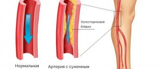
Obliterating atherosclerosis of the legs is a vascular disease in which the vascular lumen is blocked and blood circulation in the extremities is impaired. Arterial blockage occurs due to a large accumulation of lipids in them.
Cholesterol plaques form on the walls, which gradually increase in size. As a result, the vascular lumen narrows more and more, and then completely closes off. This entails a disruption in blood flow, which is observed not only in the legs, but throughout the body.
Obliterating atherosclerosis of the vessels of the lower extremities has a code according to the International Classification of Diseases (ICD-10) I70.
Pathogenesis
ACS is a deadly condition that requires emergency medical care and emergency resuscitation measures. The disease, caused by primary damage to the coronary arteries or secondary changes in them, occurs in various clinical forms that have similar diagnostic and therapeutic features. The slightest delay or incorrect first aid actions can lead to death.
Pathogenetic links of ACS:
- Impact of etiological factors,
- Coronary thrombosis,
- Isolation of biologically active substances from platelets - thromboxane, histamine,
- Arterial spasm,
- A decrease in the intensity of blood flow in the heart,
- Deterioration of blood supply to the myocardium,
- Accumulation of toxins that inhibit contractility,
- Hyperproduction of adrenaline and calcium ions,
- Persistent narrowing of the coronary vessels,
- Blocking the anticoagulant system,
- The release of enzymes into the blood that destroy cells in the necrosis zone,
- Scar formation in the heart muscle
- Impaired contractile function of the heart,
- Inability of the heart chambers to function adequately,
- A drop in oxygen saturation,
- Poor nutrition of the brain, distant organs and systems.
The degree of blocking of the heart vessels by a plaque or thrombus largely determines the mechanism of development of the syndrome:
- Partial narrowing of the lumen - periodic attacks of angina pectoris,
- Complete occlusion - the appearance of dystrophic foci in the myocardium, quickly transforming into necrosis,
- Sudden ischemic changes—ventricular fibrillation and death.
The essence of the pathology, regardless of the type and form of the process, is a malnutrition of the heart muscle caused by stenosis or blockage of the coronary arteries. The main reason that triggers a complex cascade of pathogenetic reactions is atherosclerosis, congenital or acquired defects. Acute coronary syndrome over time can lead to the death of the patient. Recovery requires urgent action.
I63 Cerebral infarction
Includes: occlusion and stenosis of the cerebral and precerebral arteries causing cerebral infarction
Excludes: complications after cerebral infarction (I69.3)
I63.0 Cerebral infarction caused by thrombosis of precerebral arteries
I63.1 Cerebral infarction caused by embolism of precerebral arteries
I63.2 Cerebral infarction caused by unspecified occlusion or stenosis of precerebral arteries
I63.3 Cerebral infarction caused by thrombosis of cerebral arteries
I63.4 Cerebral infarction caused by cerebral artery embolism
I63.5 Cerebral infarction caused by unspecified occlusion or stenosis of cerebral arteries
I63.6 Cerebral infarction caused by cerebral vein thrombosis, non-pyogenic
I63.8 Other cerebral infarction
I63.9 Cerebral infarction, unspecified
Symptoms
Chest pain or cardialgia is the main clinical sign of ACS. This symptom occurs first, is paroxysmal in nature and radiates to the shoulder or arm. With angina, the pain is squeezing, burning, pressing, short-lived, and with a heart attack it is intense, stabbing and cutting, leading to painful shock and requiring immediate hospitalization. The pain is so severe that it prevents you from moving or breathing normally. Persons with ACS cannot find a comfortable position, rush about and are afraid of dying.
Anginal pain is often associated with previous physical or emotional stress. During a heart attack, it lasts more than an hour and brings severe suffering to the patient. With angina pectoris, the attack lasts ten minutes and is repeated periodically. The pain is practically not relieved by nitroglycerin. To relieve it, narcotic analgesics are used.
Symptoms that accompany chest pain and are not mandatory:
- Cold sweat,
- Fluctuations in blood pressure,
- Euphoria and motor overexcitation,
- Worry and anxiety
- Blackout,
- Panic and fear
- Presyncope or syncope,
- Pale skin
- Cyanosis of the nasolabial triangle,
- Shortness of breath, suffocation,
- Cough,
- Nausea and vomiting,
- Heartburn,
- Abdominal pain
- Dizziness,
- Causeless weakness.
The presented points are the basis of coronary syndrome. Manifestations of the disease may not be the same in all people. Their combination allows experienced specialists to quickly and correctly make a preliminary diagnosis. Symptoms may differ depending on the gender and age of the patient, the degree of circulatory disorders and the individual characteristics of the body, as well as the presence of concomitant diseases. Such a clinical picture should alert the patient and force him to see a doctor. You should take the symptoms of ACS very seriously, as this condition is life-threatening.
Treatment regimen
Treatment of obliterating atherosclerosis is carried out using different methods. The choice of specific treatment tactics depends on the degree of damage to the arteries of the extremities. Therapy is carried out only in a comprehensive way.
Adjusting nutrition and lifestyle
Atherosclerotic plaques form when there is a large amount of harmful fats in the blood. They mostly enter the body with food. That is why patients need to follow a diet.
The diet should not contain foods high in animal fat, such as fatty meats, fast food, dairy products with a high fat content, smoked foods, processed meats, sausages, and fried foods.
It is recommended to include more plant foods in the menu: vegetables, fruits, berries, herbs. You should also eat cereals, lean meats and fish.
Patients with atherosclerosis of the arteries need to switch to a healthy lifestyle. And this applies not only to nutrition. Doctors advise moving more, performing therapeutic exercises, but avoid overstraining the limbs. You will also have to give up bad habits.
Establishing diagnosis
Patients with suspected ACS are examined, their complaints are analyzed, the heart is auscultated and percussed, and blood pressure and pulse are measured.
Electrocardiographic examination is the main diagnostic method for ACS. An ECG should be done as soon as possible after the onset of cardialgia. This technique involves recording the electrical activity of the heart using electrodes attached to the skin. First, electrical impulses in the form of waves are displayed on a monitor and then printed on paper. When the myocardium is damaged, its conductive function is impaired. The ECG shows the form of ACS - angina or heart attack.
As soon as the patient’s condition becomes satisfactory, you can move on to a full diagnostic program, including:
- 24-hour Holter monitoring – measuring blood pressure and pulse over 24 hours.
- General blood test, blood for hormones - general examination of the body.
- Clinical examination of urine to determine the functional state of the kidneys.
- BAC - detection of the level of cholesterol, glucose, as well as enzymes that, during the development of myocardial infarction, are released from damaged cardiomyocytes into the blood.
- Coagulogram is an assessment of the functional state of the blood coagulation system.
- Echocardiography is an ultrasound examination of the heart to detect lesions. Ultrasound waves are sent from the device's sensor to the heart and then returned back. The received signals are processed by a computer, and a video image is formed on the monitor screen.
- X-ray examination of the chest cavity - determining the size and shape of the heart and large blood vessels.
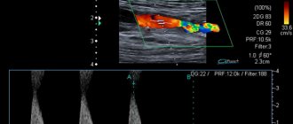
- Coronary angiography is an invasive test that identifies the location and extent of narrowing or occlusion of the coronary arteries. A long catheter is inserted into the heart vessels through the large arteries of the arms or legs. The vascular bed is filled with a liquid contrast agent and a series of x-rays are taken, in which the areas of narrowing are clearly visible. During the catheter procedure, the doctor can use tiny balloons to relieve the narrowing. They are introduced into the affected vessel and inflated. At the same time, the narrowing expands and the occlusion is eliminated. To prevent repeated spasm, a stent - a mesh tubular frame - is installed in the artery.
- Scintigraphy – detection of coronary blood flow disorders. Radioactive substances are injected into the blood, and special cameras monitor their uptake by the myocardium. Thus, where blood flow is obstructed, less radioactive material passes through. In the resulting image, such zones appear as dark spots.
- Computed tomography with contrast is performed in difficult diagnostic cases when other methods cannot determine the cause of anginal pain. Before the examination, the patient is injected with a contrast agent intravenously, and then the CT scanner takes a series of images, from which the computer creates a three-dimensional image of the heart. CT scans allow doctors to evaluate the condition of the arteries and detect narrowing or blockages in them.
- Bicycle ergometry is a stress test that is performed after acute signs of cardiac pathology have been removed. This technique allows you to determine the reaction of the heart and blood vessels to physical stress.
- Pulse oximetry - determining the level of oxygen in the blood.
ACS on ECG
All diagnostic measures are carried out under the constant supervision of a cardiologist. If the patient’s condition is serious, he is not examined comprehensively, but is limited only to visual examination data, pulse and pressure indicators, and ECG results. After stabilizing the patient’s well-being, they proceed to diagnostic measures.
Diagnostics
In an emergency, electrocardiography (ECG) is the most effective diagnostic test for angina. ECG changes that may be observed during anginal episodes include the following:
- Transitional ST segment elevations
- Dynamic T wave changes: inversions, normalizations, or hyperacute changes
- ST depressions: they can be connective, oblique or horizontal
Laboratory tests that may be helpful include the following:
- Study of levels of creatine kinase isoenzyme MB (CK-MB)
- Cardiac troponin level test
- Myoglobin level test
- General blood analysis
- Basic Metabolic Panel
Possible diagnostic methods that may be prescribed:
- Chest X-ray
- Echocardiography
- Myocardial perfusion tomography
- Cardiac angiography
- Computed tomography, including CT coronary angiography and CT analysis of the coronary artery
Healing procedures
Acute coronary syndrome is a serious pathological condition that requires emergency medical care aimed at stabilizing the patient’s condition, preserving life and preventing further progression of myocardial ischemia and necrosis.
Before the ambulance arrives, the patient needs to measure blood pressure and pulse, open the window for fresh air, sit him down and give him Nitroglycerin. Currently, there are fast-acting remedies for relieving anginal pain - Nitromint and Nitrosorbide sprays. One injection under the tongue is enough to provide relief. The patient is monitored, excluding all possible risk factors.
Inpatient treatment is considered radical. It is selected individually, depending on the severity of the pathology. Patients are prescribed strict bed rest, oxygen inhalations, and medications. Diet therapy is of great importance. Patients should avoid animal products, fatty, spicy and salty foods.
Drug treatment regimen for a patient with ACS:
- Narcotic or non-narcotic painkillers - “Morphine”, “Fentanyl”, “Promedol”,
- Beta blockers – “Atenolol”, “Propranolol”, “Metoprolol”,
- Calcium antagonists - Nifedipine, Amlodipine, Verapamil,
- Nitrates – “Nitroglycerin”, “Erinit”, “Nitromint”,
- Disaggregants – “Plavix”, “Aspirin-Cardio”, “Cardiomagnyl”,
- Statins – “Atoris”, “Simvastatin”, “Cardiostatin”,
- Fibrinolytics – “Urokinase”, “Fibrinolysin”,
- Cardioprotectors - Mildronate, Riboxin, Preductal.
If there is no effect from conservative therapy, patients undergo surgery:
- Stenting of the coronary arteries - expansion of the lumen of the vessel using a balloon and installation of a stent in the narrowed vessel,
- Coronary artery bypass grafting is the replacement of affected areas of blood vessels with special shunts, the creation of an alternative route for blood flow bypassing pathologically altered arteries, and the restoration of coronary circulation.
There are traditional medicine recipes that improve the trophism of the heart muscle. These include: decoctions of nettle or eryngium, infusion of centaury or oat grains.
Experts give patients clinical recommendations that allow the body to recover faster from illness and prevent relapse of the syndrome:
- Eliminate psycho-emotional stress,
- Limit physical activity
- Take a walk in the fresh air every day,
- Eat properly,
- Don't drink or smoke,
- Lead a healthy lifestyle,
- Normalize body weight,
- Monitor blood pressure, cholesterol and blood sugar levels.
Treatment
After confirming the diagnosis in a medical institution, the doctor selects the most suitable treatment regimen for the patient, which takes into account the characteristics of the course of the disease, the condition of the body and the stage of the disease.
Treatment can be conservative, with health measures, endovascular or surgical.
Treatment is designed to solve priority problems:
- Reduce and facilitate the passage of pain in the patient;
- Promote endurance during everyday walking;
- Stop the development of plaques in blood vessels and prevent the formation of ulcers.
With conservative treatment, medications are prescribed to restore blood supply to the legs; vitamin complexes; ointments containing antibiotics; local agents that stimulate regeneration; physiotherapy; drugs to improve blood microcirculation.
Endovascular treatment involves acting directly on damaged vessels. This is dilatation
stenting, angioplasty (its essence is in dilation of blood vessels using local anesthesia).
Surgical treatment comes to the rescue if nothing else has helped. Then doctors resort to thromboendarterectomy or bypass surgery (organizing a bypass for blood flow).
With advanced gangrene, in irreversible cases, amputation of the limb is performed.
Any treatment brings the best results with an integrated approach, including drug therapy, household health measures, and natural remedies of traditional medicine.
General lifestyle recommendations include:
- Treatment of concomitant diseases that complicate the treatment of atherosclerosis;
- Quitting smoking;
- Rationing of physical activity;
- Protect the lower extremities from hypothermia;
- Control of eating behavior in order to reduce cholesterol and lipids, adherence to dietary recommendations to reduce and normalize weight.
Forecast
The prognosis for ACS is ambiguous. It depends on the impact of provoking factors, the general condition of the body, age, existing concomitant diseases and structural and functional disorders of the cardiovascular system.
- The prognosis for unstable angina is determined by the location of the lesion: narrowing of the proximal arteries ends in death, and the distal ones are more favorable. With the development of left ventricular failure, the prognosis becomes more complicated.
- Myocardial infarction with ST segment elevation has a benign course. In the absence of this ECG sign, the area of the lesion is important - the larger it is, the more severe the patient’s condition.
Acute coronary syndrome is a dangerous pathology that, if medical instructions are not followed, can lead to serious complications: arrhythmia, persistent cardiac dysfunction, pericarditis, rupture of the dilated aorta, cardiac arrest, stroke, cardiogenic shock, and death. Even with timely and adequate treatment, there remains a high risk of complications. To avoid this, you need to monitor your health, regularly visit a cardiologist and strictly follow all his recommendations.
Signs and symptoms
Atherosclerosis is the main cause of acute coronary syndrome, and in most cases the disease occurs as a result of aggravation of a previously minor lesion. Symptoms reported by patients with ACS include the following:
- Palpitations
- Pain that typically feels like pressure, squeezing, or a burning sensation in the precordium (sternal area) and may radiate to the neck, shoulder, jaw, back, upper abdomen, or either arm
- Shortness of breath during physical activity that resolves with rest
- Diaphoresis due to stimulation of sympathetic nerves
- Nausea from vagus nerve stimulation
- Decreased tolerance to physical activity
Physical examination findings may range from normal to any of the following:
- Hypotension: indicates ventricular dysfunction due to myocardial ischemia, myocardial infarction (MI), or acute valvular regurgitation
- Hypertension: may cause angina or reflect increased catecholamine levels due to anxiety or exogenous sympathomimetic stimulation
- Increased sweating
- Pulmonary edema and other signs of left heart failure
- Extracardiac vascular diseases
- Dilatation of the jugular vein
- Cool to the touch, clammy skin and sweating in persons with cardiogenic shock
- Third heart sound (S3) and, in frequent cases, fourth heart sound (S4)
- Systolic murmur associated with dynamic left ventricular outflow tract obstruction
- Crackles on examination of the lungs (suggesting left ventricular dysfunction or mitral regurgitation)
Angina pectoris is one of the variants of clinical forms of coronary insufficiency. Angina pectoris is often called “angina pectoris” (lat. angina pectoris), because Its main symptom is pain of a different nature, localized behind the sternum in the area where the heart is located. In addition to pain, patients often characterize angina as a feeling of heaviness, squeezing, fullness, or a burning sensation in the chest. Unpleasant sensations spread to the shoulders, arms, neck, throat, lower jaw, shoulder blade and back.
Potential complications may include the following:
- Ischemia: pulmonary edema
- Myocardial infarction: rupture of the papillary muscle, left ventricular free wall and ventricular septum
Angina usually has the character of an attack that occurs in response to a sharp increase in the heart's need for oxygen, for example, during intense physical activity, strong emotional experience or stress. At rest, angina pain is almost always absent. Angina attacks can recur with varying frequencies, from several times a day to several episodes a month. Angina is always an attack of described pain in the heart area. Outside of an attack, angina has no clinical manifestations.
Based on the characteristics of the clinical course, there are three main variants of angina pectoris, called:
1. Stable angina (syn.: exertional angina). The main symptom of this angina is the occurrence of pain (unpleasant sensations) during physical activity and its rapid disappearance after stopping the exercise.
2. Unstable angina (syn.: angina at rest). It is characterized by attacks that occur spontaneously in a state of apparent rest and are not associated with physical activity. This angina is more severe in nature compared to stable angina; attacks last longer and can be provoked by minor exertion. Unstable angina is considered as a precursor to myocardial infarction, so patients with unstable angina require hospitalization and qualified therapy, unlike cases of stable angina.
3. Angina pectoris of the Printzmetal type (syn.: variant angina) is a variant of unstable angina. Attacks usually occur at night or early in the morning, have a short duration - on average 2 to 5 minutes and are effectively controlled by taking one nitroglycerin tablet under the tongue. Prinzmetal's angina develops as a result of a sharp vasospasm of the coronary vessels (hence another name for it: “vasospastic angina”). The basis for diagnosing spontaneous angina is recording an ECG during an attack. Prinzmetal's angina does not always develop against the background of coronary heart disease. This type of angina can occur with heart valve defects (eg, aortic stenosis), severe anemia, or severe myocardial hypertrophy, provoking a reflex spasm of the coronary vessels.
In the first few hours of coronary insufficiency, it is impossible to determine the type of its development in a particular patient, since clinical manifestations and ECG changes during this period are nonspecific, and biochemical markers of myocardial necrosis appear only 4-6 hours after the onset of this form of pathology. However, already in the first hours, if a heart attack is suspected, it is possible, using an electrocardiographic examination, to distinguish between two main variants of its development: a heart attack without a Q wave (synonyms: large-focal, transmural) and a heart attack with a Q wave (synonyms: small-focal, subezzocardial, “microinfarction”) .
Clinical signs, nature and frequency of complications, treatment measures and prognosis vary significantly with these types of myocardial infarction.
Evidence of the development of a Q wave infarction is the detection of pathological Q waves in two or more adjacent ECG leads. Infarctions without a Q wave usually differ from infarctions with a Q wave in being smaller in size; the cause of their development is non-occlusive thrombi of the coronary arteries or repeated short episodes of fairly severe myocardial ischemia. Acute infarctions with a Q wave are characterized by large-focal heart attacks, and infarctions without a Q wave are characterized by small-focal lesions of the heart. An ECG harbinger of the development of a heart attack with a Q wave is the rise of the ST segment (“coronary wave”), and a heart attack without a Q wave is the absence of a “coronary wave.” In the clinical aspect, it was noted that early in-hospital mortality in patients with a heart attack without a Q wave is lower, and the frequency of recurrent fatal infarctions over several months of observation is higher than in patients with a heart attack whose ECG shows pathological Q waves.
Characteristic ECG changes (primarily the appearance of a pathological Q wave) in combination with the clinical picture are sufficient to diagnose a heart attack with a Q wave. In contrast, with a heart attack without a Q wave, the QRS complex, as a rule, does not change; note (not always!) only minor changes in the ST segment and T wave. Based on such minimal and inconsistent changes in the ECG (especially in the absence of a Q wave!), it is only possible to assume the occurrence of a heart attack, and to establish a diagnosis of “infarction without a Q wave” it is necessary to determine biochemical markers of myocardial necrosis.
It has been established that the immediate cause of myocardial infarction with a Q wave is thrombotic obstruction of the coronary artery, and that of a myocardial infarction without a Q wave is incomplete occlusion of a coronary artery by a thrombus. Due to the fact that thrombus formation during a heart attack without a Q wave is of a “sparing” nature, there is the possibility of fairly rapid vascular reperfusion due to spontaneous lysis of the thrombus or weakening of the vasospasm accompanying thrombus formation. In this regard, it is believed that in case of a heart attack without a Q wave and ST segment elevation, there is no need for thrombolytic therapy, and in a heart attack with ST segment elevation and a Q wave, thrombolytic therapy is indicated as early as possible (sometimes early thrombolysis in patients with T segment elevation prevents the development of Q-wave infarction).
In addition to the above criteria for diagnosing myocardial infarction, two more must be added:
• cardiac arrhythmias are a constant companion to myocardial infarction;
• cardiac aneurysm is the most reliable radiological sign of myocardial infarction.
Sometimes coronary insufficiency is asymptomatic (latent), so special examination of the patient is necessary to recognize it. In outpatient practice, the presence of such (mainly chronic) coronary insufficiency can be detected by ECG changes during stress tests. In cardiology hospitals, pharmacological tests and coronary angiography are additionally used to diagnose chronic coronary insufficiency.
The main forms of complications of IHD are:
• heart failure;
• cardiac arrhythmias;
• sudden coronary death. Sudden death is considered to be death within one hour after the onset of the first symptoms of coronary artery disease against the background of the patient’s previous stable condition. The immediate cause of sudden death in the vast majority of cases is ventricular fibrillation.
Treatment of patients with coronary insufficiency involves influencing the underlying disease that determined it, as well as the use of antianginal, antithrombotic, and antiarrhythmic drugs. If drug therapy is ineffective and coronary insufficiency is severe, in some cases coronary artery bypass surgery and other methods of myocardial revascularization are indicated.
Why does a lack of blood supply to the brain occur?
Acute ischemic cerebrovascular accident is often a secondary pathology and occurs against the background of existing diseases:
- arterial hypertension;
- widespread atherosclerotic vascular lesions (up to 55% of cases develop due to pronounced atherosclerotic changes or thromboembolism from plaques located in the aortic arch, brachiocephalic trunk or intracranial arteries);
- previous myocardial infarction;
- endocarditis;
- heart rhythm disturbances;
- changes in the valvular apparatus of the heart;
- vasculitis and angiopathy;
- vascular aneurysms and developmental anomalies;
- blood diseases;
- diabetes mellitus
Up to 90% of patients have changes in the heart and main arteries of the neck. The combination of these reasons sharply increases the risk of ischemia.
Possible compression of the vertebral artery by the processes of the vertebrae
Transient attacks are most often caused by:
- spasm of the arterial brain stems or short-term compression of the carotid and vertebral arteries;
- embolization of small branches.
The following risk factors can provoke the disease:
- elderly and senile age;
- excess weight;
- the effect of nicotine on blood vessels (smoking);
- experienced stress.
The basis of the influencing factors is the narrowing of the lumen of the vessels through which blood flows to the brain cells. However, the consequences of such a malnutrition may vary according to:
- stamina,
- localization,
- prevalence,
- severity of vessel stenosis,
- gravity.
The combination of factors determines the form of the disease and clinical symptoms.
An ischemic stroke can be considered recurrent if it occurs in the area of the blood supply of one vessel within 28 days after the initial manifestations of the first event. A recurrent stroke is called a stroke at a later date.