What is cardiography
The essence of cardiography is the study of electrical currents arising during the work of the heart muscle.
The advantage of this method is its relative simplicity and accessibility. Strictly speaking, a cardiogram is the result of measuring the electrical parameters of the heart, displayed in the form of a time graph. The creation of electrocardiography in its modern form is associated with the name of the Dutch physiologist of the early 20th century, Willem Einthoven, who developed the basic ECG methods and terminology used by doctors to this day.
Thanks to the cardiogram, it is possible to obtain the following information about the heart muscle:
- Heart rate,
- Physical condition of the heart
- The presence of arrhythmias,
- The presence of acute or chronic myocardial damage,
- The presence of metabolic disorders in the heart muscle,
- Presence of electrical conductivity disturbances,
- Position of the electrical axis of the heart.
Also, a cardiac electrocardiogram can be used to obtain information about certain vascular diseases not related to the heart.
An ECG is usually performed in the following cases:
- Feeling of abnormal heartbeat;
- Attacks of shortness of breath, sudden weakness, fainting;
- Heartache;
- Heart murmurs;
- Deterioration of the condition of patients with cardiovascular diseases;
- Passing medical examinations;
- Medical examination of people over 45 years of age;
- Examination before surgery.
An electrocardiogram is also recommended for:
- Pregnancy;
- Endocrine pathologies;
- Nervous diseases;
- Changes in blood counts, especially with an increase in cholesterol;
- Over 40 years of age (once a year).
What to do?
First of all, every examined patient must understand that changes in the amplitude of oscillations of the waves on cardiograms are not a diagnosis at all. Any changes to the recordings of this study should only be reviewed by an experienced cardiologist.
It is also impossible not to understand that electrocardiography is not the only and final criterion for establishing any diagnosis. To detect a certain pathology in a patient, a comprehensive, full examination is necessary.
Depending on the health problems discovered after such an examination, doctors may prescribe certain medications or other treatment to patients.
Various cardiac problems can be eliminated with the help of cardioprotectors, antiarrhythmic drugs, sedatives and other therapeutic procedures. In any case, self-medication for any changes in the cardiogram is categorically unacceptable!
In conclusion, we note that any changes in the electrocardiogram should not lead to panic in the patient.
It is categorically unacceptable to independently evaluate the primary diagnostic conclusions obtained through this study, because the data obtained is always additionally checked by doctors.
Establishing a correct diagnosis is possible only after collecting an anamnesis, examining the patient, assessing his complaints and analyzing the data obtained from certain instrumental examinations.
At the same time, only a doctor and no one else can judge the health status of a particular patient with a cardiogram showing a decrease in the amplitude of indicators.
CREATE NEW MESSAGE.
But you are an unauthorized user.
If you register, you will be able to further track responses to your messages and continue the dialogue on interesting topics with other users and consultants. In addition, registration will allow you to conduct private correspondence with consultants and other users of the site.
When a cardiogram is taken, they first look at the ECG voltage as a very important indicator. What can you find out when decoding this parameter? Electrocardiography is a recording tape for subsequent decoding and analysis of the electric field indicators that are generated by the heart muscle during its activity.
Thanks to ECG studies, it is possible to identify many heart diseases at an early stage of their development and begin adequate and timely treatment. But not everyone understands the terms that are used in this type of diagnosis, including the concepts of high or low voltage electrocardiogram. Therefore, it is necessary to understand the very concept of cardiogram voltage, as well as whether it is good or bad if this indicator is reduced or increased.
A standard ECG graph reflects the dynamics of changes in the electrical field of the heart and consists of such elements as:
- 1. Teeth P, Q, R, S, T. These elements can be normal or deformed.
- 2. The U wave should normally be very smooth and barely visible on the ECG.
- 3. The QRS waves together form a separate complex or segment.
When the voltage of the electrocardiogram is pathologically low or, on the contrary, it is too high, this indicates the beginning of the development of cardiopathy, that is, heart pathology. But, in addition to the voltage indicator, you also need to look at such an indicator as the amplitude of the RS segment. For information: the norm for this parameter in the chest leads is 0.7 mV. Accordingly, when the RS amplitude decreases or, conversely, increases, they speak of emerging heart problems.
It is noted that a distinction is made between reduced voltage in the limb leads or a general decrease in ECG voltage. In this case, there is a decrease in the amplitude of those complexes on the ECG in question. Sharp fluctuations in amplitude on the cardiogram are not common. But a decrease in indicators can never be considered a variant of the individual physiological norm.
What body conditions can provoke disturbances in the amplitude of oscillations? These include fever, anemia, hyperthyroidism, and heart block.
Causes and manifestations of low voltage on the ECG
Low voltage on the ECG means a decrease in the amplitude of the waves, which can be observed in various leads (standard, chest, limbs). Such a pathological change in the electrocardiogram is characteristic of myocardial dystrophy, which is a manifestation of many diseases.
Low voltage on ECG is a sign of myocardial dystrophy
The value of QRS parameters can vary widely. Moreover, they, as a rule, have greater values in the chest leads than in standard ones.
The norm is considered to be a QRS wave amplitude value of more than 0.5 cm (in the limb lead or standard lead), as well as a value of 0.8 cm in the precordial leads.
If lower values are recorded, then they indicate a decrease in the parameters of the complex on the ECG.
Do not forget that to date, clear normal values for the amplitude of the teeth have not been determined depending on the thickness of the chest, as well as body type. Since these parameters affect the electrocardiographic voltage. It is also important to consider the age norm.
There are two types: peripheral and general decrease. If the ECG shows a decrease in waves only in the limb leads, then they speak of a peripheral change; if the amplitude is also reduced in the chest leads, then this means a general low voltage.
A decrease in voltage during an ECG can have many causes
Causes of low peripheral voltage:
- heart failure (congestive);
- emphysema;
- obesity;
- myxedema.
Overall voltage may be reduced due to pericardial and cardiac causes. Pericardial causes include:
- pericardial effusion;
- pericarditis;
- pericardial adhesions.
Cardiac reasons:
- myocardial damage of an ischemic, toxic, infectious or inflammatory nature;
- amyloidosis;
- scleroderma;
- mucopolysaccharidosis.
Dilated cardiomyopathy leads to chronic heart failure
The amplitude of the waves may be less than normal if the heart muscle is damaged (dilated cardiomyopathy). Another reason for deviation of ECG parameters from normal is treatment with cardiotoxic antimetabolites.
As a rule, in this case, pathological changes in the electrocardiogram occur acutely and are accompanied by pronounced impairments in the functionality of the myocardium.
If after heart transplantation the amplitude of the waves is reduced, then this can be regarded as its rejection.
It should be noted that pathological changes on the cardiogram, manifested by a decrease in the parameters of the amplitude of the waves, are often observed with dystrophic changes in the myocardium. The reasons leading to this are the following:
- acute and chronic infections;
- renal and liver intoxication;
- malignant tumors;
- exogenous intoxication caused by drugs, nicotine, lead, alcohol, etc.;
- diabetes;
- thyrotoxicosis;
- vitamin deficiencies;
- anemia;
- obesity;
- physical stress;
- myasthenia gravis;
- stress, etc.
Dystrophic damage to the heart muscle is observed in many heart diseases, such as inflammatory processes, coronary disease, heart defects. On the ECG, the voltage of the waves is reduced primarily by T. Some diseases may have certain features on the cardiogram. For example, with myxedema, the parameters of the QRS waves are below normal.
The goal of therapy for this electrocardiographic manifestation is to treat the disease that caused the pathological changes on the ECG. Also the use of medications that improve nutritional processes in the myocardium and help eliminate electrolyte disturbances.
The main thing is that patients with this pathology are prescribed anabolic steroids (nerobolil, retabolil) and non-steroidal drugs (inosine, riboxin). Treatment is carried out with the help of vitamins (group B, E), ATP, cocarboxylase. Prescribe drugs containing: calcium, potassium and magnesium (for example, asparkam, panangin), oral cardiac glycosides in small doses.
Based on the ECG results, the specialist will identify the problem and prescribe the necessary treatment.
For the preventive purpose of cardiac muscle dystrophy, it is recommended to promptly treat the pathological processes leading to this. It is also necessary to prevent the development of vitamin deficiencies, anemia, obesity, stressful situations, etc.
To summarize, it should be noted that such a pathological change in the electrocardiogram as a decrease in voltage is a manifestation of many cardiac as well as extracardiac diseases. This pathology is subject to urgent treatment in order to improve myocardial nutrition, as well as preventive measures to help prevent it.
Low voltage
In the winter of the early eighties, the Department of Tuberculosis and Pulmonology at our hospital decided to conduct a mass examination. It was supposed to perform a Mantoux test (test with tuberculin) on young people who had not previously had tuberculosis, with the aim of early detection of asymptomatic forms of this disease. Students from several institutes in Minsk were taken for examination. Those whose test is sharply positive or hyperergic will be invited to our dispensary for further examination and treatment.
At that time, in addition to my main job, I was managing several wards part-time in one of the tuberculosis departments of our hospital. And indeed, young men and women with suspected tuberculosis began to be admitted to the departments, and many were diagnosed with an active process. More often it was extrapulmonary tuberculosis. I remember at the conference they reported on patients with kidney tuberculosis, ovarian tuberculosis, disease of the bronchial and intra-abdominal lymph nodes. A completely exotic form was also identified - tuberculosis of the frenulum of the tongue. A mouth ulcer that had not healed for years turned out to be a manifestation of tuberculosis and disappeared after two months of proper treatment.
Sergei, a university student, tall, blond, and athletic-looking, also entered my ward. After he was diagnosed with a hyperergic Mantoux test, he was further examined and a small infiltrate with decay was found in the left apex, surrounded by several foci. During the first conversation, I confidently told the student that we would quickly cure him and he would not stay with us. As it turned out later, in vain. Sergei felt well, the tests showed no gross changes, no tuberculosis mycobacteria were found. On the ECG, my attention was drawn to the low voltage of the QRS complex and the smoothness of the T wave, but this could be due to intoxication and hyperergy. Sergei was prescribed pills and he took them carefully. He behaved calmly. He was often visited by his parents, workers of one of the rural district party committees. A girl came, also tall, beautiful blonde. He didn’t drink, he didn’t violate the regime - a typical mild patient. A month has passed. The patient's condition was not bad, but in the evenings a slight temperature began to rise - 37.3 - 37.6, weakness increased, and the pulse became more frequent. There were no positive changes on the x-ray. A week later, during his morning rounds, Sergei did not get out of bed; he lay pale and bored. -How do you feel, why don’t you get up? - Yes, over the weekend I had pain in my chest, shortness of breath when walking, and today there are some irregularities in my pulse. I examined Sergei carefully. Yes, slight swelling of the legs, tachycardia, mild shortness of breath at rest. They took an x-ray - in the picture the shadow of the heart slightly expanded in both directions. Pericarditis? Myocarditis? But when I saw single ventricular extrasystoles on the ECG, I immediately remembered the incident while working on the ambulance (Repeat call) and got on the phone to call a cardiologist from the Regional Hospital. The next day, a young, knowledgeable associate professor of the Department of Therapy at the Institute of Advanced Studies arrived. A consultation gathered - an associate professor of cardiology, our chief medical officer, an assistant at the department of phthisiology, who supervises the department, and me, the attending physician. At that time, we and the nearby hospitals did not have equipment for ultrasound or radioisotope examination of the heart, as well as computed tomography and nuclear magnetic resonance. The diagnosis of such a complex heart lesion, which we assumed, had to be decided on the basis of the anamnesis, x-ray picture, laboratory data, ECG and the personal experience of each doctor. As a result, a diagnosis was made: diffuse idiopathic Fiedler's myocarditis, possibly viral. In response to my remarks that heart disease could be associated with tuberculosis (before that I had seen two cases of cardiac tuberculosis), the associate professor said weightily: “Doctor, why do we need to think about exotic causes, when more often we encounter viral or idiopathic ones, and in a large number of cases The etiology is generally difficult to establish. They prescribed hormonal therapy, heart medications, and diuretics. There were no changes in the treatment of tuberculosis.
Where can I get a cardiogram?
If you suspect that there is something wrong with your heart, you can contact a therapist or cardiologist so that he can give you a referral for an ECG. Also, for a fee, a cardiogram can be done in any clinic or hospital.
Let's look at some terms that an ECG transcript may contain. They do not always indicate serious pathologies, but in any case they require a visit to a doctor for advice, and sometimes additional examinations.
What to do?
Everyone who undergoes an ECG must understand that low or high voltage is not a diagnosis, but only an indicator. To establish an accurate diagnosis, cardiologists refer their patients for additional heart tests.
If pathological processes are detected, the doctor will prescribe appropriate treatment. It can be based on taking medications, including dietary nutrition and physical therapy in the patient’s regimen.
Important! In this case, you cannot self-medicate, as you can only worsen the situation of the disease. Only a doctor prescribes and cancels medications or procedures.
Low voltage ECG reasons
There seem to be no complaints. The normal blood pressure is 100/60 (110/70). Cholesterol was elevated, but regulating my diet seemed to help overcome this problem. Height 165, weight 67. No sharp dynamics.
VSD. There are single extrasystoles on the holter. Intercostal neuralgia. I will be very grateful for the answer.
2) The numbers are written for the doctor to save time (so as not to count again) and have no independent meaning
3) The diagnosis is not made using any one research method, only based on the totality of data
Methodology of the procedure
ECG recording is usually performed in a supine position. To take a cardiogram, a stationary or portable device is used - an electrocardiograph. Stationary devices are installed in medical institutions, and portable ones are used by emergency teams. The device receives information about electrical potentials on the surface of the skin. For this purpose, electrodes are used that are attached to the chest area and limbs.
These electrodes are called leads. There are usually 6 leads installed on the chest and limbs. The chest leads are designated V1-V6, the leads on the limbs are called basic (I, II, III) and reinforced (aVL, aVR, aVF). All leads give a slightly different picture of oscillations, but by summing up the information from all electrodes, you can find out the details of the functioning of the heart as a whole. Sometimes additional leads are used (D, A, I).
Typically, the cardiogram is displayed in the form of a graph on paper containing millimeter markings. Each electrode lead has its own schedule. The standard speed of the belt is 5 cm/s; other speeds may be used. The cardiogram displayed on the tape can also indicate the main parameters, normal indicators and a conclusion generated automatically. Data can also be recorded in memory and on electronic media.
After the procedure, the cardiogram is usually deciphered by an experienced cardiologist.
The meaning of the teeth: a detailed examination
When analyzing a cardiogram, it is worth looking not only at the intervals and the presence of a particular wave, but also at their height and duration. A normal amplitude indicates the correct functioning of the organ, while a violation to a greater or lesser extent is a direct signal of a problem.
The waves on the ECG are normal:
- P. Width no more than 0.11 s., height depends on age, but on average no more than 2 mm. Deviation from these values indicates atrial hypertrophy.
- Q. The width is not more than 0.04 s., the height is not more than 25% of the R wave. The deepening of the wave is observed in myocardial infarction or in severe obesity.
- R. The norm is determined by ECG leads V5 and V6, where the height should not be more than 2.6 mV. When moving from V5 to V6, the amplitude should increase.
- S. There are no special standards, since the depth depends on many factors, such as body position, age of the patient, and so on. However, a tooth that is too deep is a clear signal of ventricular hypertrophy.
- T. Amplitude of at least 1/7 of the R wave.
Sometimes a U wave appears after the T wave, but it has no norms and is rarely taken into account when making a diagnosis.
Segment norm option
Along with the teeth, the spaces between them are also taken into account. If the interval or complex on the ECG deviates from the norm, this is a clear signal for additional examinations.
Complexes and intervals on an ECG should normally be as follows:
- QRS – the QRS complex should be no more than 0.07-0.11 s; widening of the complex is considered a pathology.
- PQ – interval duration is about 0.12 ms, but not more than 0.21 s.
- QT is an interval whose width depends on heart rate.
- ST segment – located directly on the isoelectric line.
It is worth remembering that prolongation of the PQ interval is provoked by AV blockade.
Variants of the ventricular complex
Important! The ST segment may be slightly above the isoelectric line in leads V1 and V2!
Correct assessment of the cardiogram helps to make the most accurate diagnosis, so you must definitely show the results to a cardiologist. Only he will correctly interpret the meaning of all the teeth and intervals. It is difficult for a person without proper education to correctly evaluate the data obtained.
To record the electrical activity of the heart, electrodes are placed on the chest, arms and legs. This arrangement records the spread of electrical impulses throughout the body. It is these discharges and their paths that are cardiac leads. Chest leads begin with the letter V and are numbered from 1 to 6. The ECG normally shows six standard leads:
- I – first;
- II – second;
- III – third;
- AVL – analogue of I;
- AVF – analogue of III;
- AVR – mirror image.
To obtain the information of interest, you need to measure some intervals and segments on the existing ECG. The algorithm for studying a cardiogram is as follows:
- In leads I, II or III, you need to select the highest R wave and measure the distance between the two subsequent waves (in fact, two RRR spaces). Divide the resulting number in millimeters by two. If you don’t have a ruler at hand, then the side of the large cell on the paper is 5 mm (1 second), and the cells inside it are 1 mm (0.02 seconds).
- The regularity of the heart rhythm is determined by the spaces between the R waves.
- Take measurements of each tooth and interval, compare them with the norms (they are described above in this article).
Important! Please note: the speed indicated on the tape is 25 or 50 mm/s! This parameter is important for calculating heart rate. Modern equipment automatically indicates the frequency of contractions, but some hospitals still use outdated models.
To calculate heart rate, use the formulas:
- For 25 mm/s: 60/(RR interval × 0.04), where the interval is indicated in mm or 300/(average number of cells in the RR interval).
- For 50 mm/s: 60/(RR interval × 0.02), where the interval is indicated in mm or 600/(average number of cells in the RR interval).
Important! Additional leads are not used in the analysis, since they duplicate the standard ones.
Installing electrodes on the body
It is important to remember that even if both the intervals and waves appear normal on the ECG, you still need to take the results to a cardiologist. An experienced doctor will notice the first signs of emerging problems and promptly send the patient for further examination.
In general, an ECG is an informative study that can clarify the patient’s current condition. Despite the simplicity of decoding and existing standards, consultation with a cardiologist is mandatory. Many errors in the cardiogram are provoked by other diseases, psychological conditions or age. To avoid erroneous conclusions and incorrect treatment, the diagnosis and course of treatment should be prescribed exclusively by a specialized doctor.
Self-analysis of the ECG is acceptable for impatient people or those who are interested in the dynamics of their condition. Understanding what is displayed on the tape allows you to achieve some psychological comfort.
Reduced ECG voltage
in Hypertension
Most of us clearly understand that electrocardiography is a simple, accessible method of recording, as well as subsequent analysis of electric fields that can be formed during the functioning of the heart muscle.
It's no secret that the ECG procedure is widespread in modern cardiological practice, as it allows one to detect many cardiovascular diseases.
I recently read an article that talks about Monastic tea for treating heart disease. With this tea you can FOREVER cure arrhythmia, heart failure, atherosclerosis, coronary heart disease, myocardial infarction and many other diseases of the heart and blood vessels at home.
I’m not used to trusting any information, but I decided to check and ordered a bag. I noticed changes within a week: the constant pain and tingling in my heart that had tormented me before receded, and after 2 weeks disappeared completely. Try it too, and if anyone is interested, below is the link to the article.
However, not all of us know and understand what specific terms related to this diagnostic procedure can mean. We are talking, first of all, about such a concept as voltage (low, high) on the ECG.
In our publication today, we propose to understand what ECG voltage is and understand whether it is good or bad when this indicator is reduced/increased.
What does the concept of ECG voltage mean?
When a cardiogram is taken, they first look at the ECG voltage as a very important indicator. What can you find out when decoding this parameter? Electrocardiography is a recording tape for subsequent decoding and analysis of the electric field indicators that are generated by the heart muscle during its activity.
Thanks to ECG studies, it is possible to identify many heart diseases at an early stage of their development and begin adequate and timely treatment. But not everyone understands the terms that are used in this type of diagnosis, including the concepts of high or low voltage electrocardiogram. Therefore, it is necessary to understand the very concept of cardiogram voltage, as well as whether it is good or bad if this indicator is reduced or increased.
What are the reasons for low voltage of the QRS complex on the cardiogram? This occurs due to cardiac (directly related to heart pathology) or extracardiac (not related to cardiac pathology) reasons. Let us list the possible pathologies that can cause a drop in the amplitude of the ECG recording. So:
- hypertrophy (overdevelopment) of the left ventricle of the heart;
- severe obesity;
- history of rheumatic myocarditis or pericarditis;
- diffuse ischemic, toxic or infectious damage to the heart muscle;
- dilated cardiomyopathy;
- atherosclerosis of myocardial vessels.
The functional reasons for the occurrence of abnormalities in the ECG include an increase in the tone of the vagus nerve, which leads to a reduction in the intensity of oscillations of the waves on the cardiogram, and also as a symptom of the development of a rejection reaction after heart transplantation.
The described cardiogram abnormalities are not a diagnosis, but only indicate the appearance of symptoms of developing heart disease. Accordingly, it is impossible to assess the patient’s condition only based on the results of an ECG study. It is important to have additional examination methods to make a clinical diagnosis.
It is strictly not recommended to try to determine heart pathology based on ECG results on your own without the participation of a cardiologist. Only a specialist can make the correct diagnosis and prescribe appropriate therapy.
Treatment may be indicated using pharmacological agents or other means, depending on the identified disease. If the presence of the disease is proven, the doctor may prescribe sedatives, antiarrhythmic drugs, cardioprotectors and other medications.
ECG low voltage waveforms - All about hypertension
First we should talk a little about how the heart works. It has 4 chambers - two atria and two ventricles (left and right). The electrical impulse, due to which it contracts, is formed, as a rule, in the upper part of the myocardium - in the sinus pacemaker - the sinoatrial (sinus) node.
The impulse spreads down the heart, first affecting the atria and causing them to contract, then passes through the atrioventricular nerve node and another nerve node, the bundle of His, and reaches the ventricles. The main burden of pumping blood is taken on by the ventricles, especially the left one, which is involved in the systemic circulation. This stage is called heart contraction or systole.
After contraction of all parts of the heart, the time comes for their relaxation - diastole. The cycle then repeats again and again - this process is called heartbeat.
The condition of the heart, in which there are no changes in the propagation of impulses, is reflected on the ECG in the form of a straight horizontal line, called an isoline. The deviation of the graph from the isoline is called a spike.
One heartbeat on the ECG contains six waves: P, Q, R, S, T, U. The waves can be directed both up and down. In the first case they are considered positive, in the second - negative. The Q and S waves are always positive, and the R wave is always negative.
Have you been struggling with HYPERTENSION for many years without success?
Head of the Institute: “You will be amazed at how easy it is to cure hypertension by taking it every day...
ECG voltage - causes of occurrence, methods of diagnosis and treatment
ECG voltage is one of the main indicators that allows you to diagnose heart disease at an early stage. If the voltage is too high or low, then there is a high risk of cardiopathy and pathological changes in the heart. To determine how this indicator affects further events, you first need to understand its essence.
What is voltage?
The voltage of the electrocardiogram is called changes in the amplitude of the three waves - QRS. To make a diagnosis, doctors pay attention to the following elements of the ECG:
- 5 teeth (P, Q, R, S and T),
- U wave (may appear, but not for everyone),
- ST segment
- group of QRS waves.
The above indicators are considered basic. Any deviations from the norm change the voltage of the cardiogram. Pathology can be called changes in precisely three QRS waves, which are assessed as a whole.
In other words, a low-voltage potential can be seen on an ECG during heartbeat at the moment when three QRS waves are located below accepted norms. For an adult, the norm is considered to be a QRS of no more than 0.5 mV.
If the voltage diagnostic time exceeds the norm, cardiac pathology is clearly diagnosed.
An obligatory step in the analysis of the electrocardiogram is the assessment of the distance from the apex of the R and S waves.
The amplitude of this area should be normal at 0.7 mV.
Doctors divide voltage into two groups: peripheral and general. Peripheral voltage makes it possible to evaluate parameters only from the limbs. The total voltage takes into account the results of both thoracic and peripheral leads.
Reasons for appearance
The voltage can change in different directions, but more often it decreases. This occurs due to cardiac or extracardiac causes. In addition, the metabolic processes that occur in the myocardium may not in any way affect the amplitude of the waves.
A decrease in voltage may indicate the progression of heart disease, but sometimes this indicator indicates a pulmonary or endocrine pathology. In such cases, the doctor prescribes an additional examination of the patient. The list of diseases associated with low voltage is wide.
The most common pathologies:
- pulmonary edema,
- diabetes,
- hypothyroidism,
- cardiac ischemia,
- left ventricular hypertrophy,
- obesity,
- rheumatic myocarditis,
- pericarditis,
- development of sclerotic processes in the heart,
- myxedema,
- myocardial damage,
- dilated cardiomyopathy.
Changes in voltage can occur due to functional disorders in the heart, for example, increased tone of the vagus nerve. This condition is often diagnosed in professional athletes.
The intensity of the oscillations of the teeth on the cardiogram is reduced.
Important! People who have undergone a heart transplant sometimes have reduced voltage on their cardiograms.
This indicator indicates the possible development of rejection.
What to do?
Everyone who undergoes an ECG must understand that low or high voltage is not a diagnosis, but only an indicator. To establish an accurate diagnosis, cardiologists refer their patients for additional heart tests.
If pathological processes are detected, the doctor will prescribe appropriate treatment. It can be based on taking medications, including dietary nutrition and physical therapy in the patient’s regimen.
Important! In this case, you cannot self-medicate, as you can only worsen the situation of the disease. Only a doctor prescribes and cancels medications or procedures.
What factors influence the decrease in voltage?
If the readings on the cardiogram are higher or lower than normal, then the doctor must determine the cause of the changes. Often the amplitude decreases due to dystrophic pathologies of the heart muscle.
There are a number of reasons that influence this indicator:
- avitaminosis,
- unhealthy diet,
- chronic infections,
- liver and kidney failure,
- orgasmic intoxications, such as those caused by lead or nicotine,
- excessive consumption of alcoholic beverages,
- anemia,
- myasthenia gravis,
- long-term physical activity,
- malignant neoplasms,
- thyrotoxicosis,
- frequent stress,
- chronic fatigue, etc.
Many chronic diseases can affect the performance of the heart, so during an appointment with a cardiologist it is worth taking into account all existing diseases.
Other types of procedures
There are several other methods for carrying out the procedure. For example, this is monitoring with physical activity. Abnormalities are usually more pronounced on the stress ECG. The most common way to provide the body with the necessary physical activity is a treadmill. This method is useful in cases where pathologies can only manifest themselves in the case of increased heart function, for example, if coronary artery disease is suspected.
During phonocardiography, not only the electrical potentials of the heart are recorded, but also the sounds that arise in the heart. The procedure is prescribed when it is necessary to clarify the occurrence of a heart murmur. This method is often used when heart defects are suspected.
Low ECG voltage in limb leads
The voltage can change in different directions, but more often it decreases. This occurs due to cardiac or extracardiac causes. In addition, the metabolic processes that occur in the myocardium may not in any way affect the amplitude of the waves.
Definition of segments on an ECG
A decrease in voltage may indicate the progression of heart disease, but sometimes this indicator indicates a pulmonary or endocrine pathology. In such cases, the doctor prescribes an additional examination of the patient. The list of diseases associated with low voltage is wide.
The most common pathologies:
- pulmonary edema;
- diabetes;
- hypothyroidism;
- cardiac ischemia;
- left ventricular hypertrophy;
- obesity;
- rheumatic myocarditis;
- pericarditis;
- development of sclerotic processes in the heart;
- myxedema;
- myocardial damage;
- dilated cardiomyopathy.
Changes in voltage can occur due to functional disorders in the heart, for example, increased tone of the vagus nerve. This condition is often diagnosed in professional athletes. The intensity of the oscillations of the teeth on the cardiogram is reduced.
Normal ECG: recording speed and amplitude, recording artifacts
Details: 04/24/2016, Max Romanchenko
- ECG recording speed
- ECG recording amplitude
- Recording artifacts
ECG recording speed
Remember: the standard ECG recording speed is 25 mm/sec, and you will use this speed in 99% of situations except as noted below. All modern devices use this speed by default, with an additional speed of 50 mm/sec, and some - 10 mm/sec. and 100 mm/sec.
Speed 50 mm/sec is only used when:
- due to the high frequency of contractions, the complexes are located too close to each other and it becomes difficult to distinguish the necessary parameters of the teeth and segments, and
- when you need to very accurately calculate the width of the waves and intervals (for example, in the case of confirming or denying the presence of bundle branch block, where the QRS width is one of the main criteria).
A speed of 10 mm/sec is additionally used to register rare ECG events, for example, single extrasystoles, about which the patient complains. To do this, write down the so-called. “rhythmogram” - on 60 centimeters of ECG paper you can record 1 minute of ECG and “catch” that single extrasystole or transient blockade that you are likely to miss with a standard recording.
The standard amplitude of ECG recording is 10 mm/mV; amplitudes of 5 mm/mV and 20 mm/mV are also used. You can find out what amplitude was used during recording either by the “control millivolt” at the beginning of the recording or by the amplitude indicator.
As you can see, when increasing the amplitude to 20 mm/mV, the ECG stretches up and down, and when using 5 mm/mV, it contracts and becomes smaller.
Please note that all voltage criteria (for example, cardiac chamber hypertrophy, pathological ST elevation/depression) are specified for a recording amplitude of 10 mm/mV. If a different amplitude was used during recording, take this into account in your calculations!
For example, with an amplitude of 5 mm/mV, the height of the R wave is 8 mm. This means that in reality its height is 16 mm!
An amplitude of 5 mm/mV is used when the patient’s ECG has a very large amplitude and does not fit into the width of the recording tape (the teeth seem to be cut off at the top and bottom) or overlap each other.
The main reasons for high ECG voltage:
- Severe hypertrophy of the ventricular myocardium (for example, with hypertension, “athletic heart”, etc.);
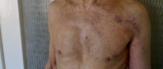
- Proximity of the electrode to the myocardium (for example, in thin patients or after resection of the lower lobe of the left lung - see clinical case);
- Incorrect direction of ventricular depolarization (for example, with blockade of the left bundle branch, ventricular extrasystole, etc.).
Depending on the device that records the ECG and its settings, when printing onto tape, the device can automatically change the recording amplitude to accommodate complexes without overlap.
For example, in the ECG below, the device printed leads V1-V3 with an amplitude of 10 mm/mV, and leads V4-V6 with an amplitude of 5 mm/mV.
This can be seen both from the control millivolt before recording and from the indicated recording amplitude on top of the tape.
In this example, the recording amplitude did not change and was 10 mm/mV. At the same time, the more “sweeping” QRS in the chest leads overlapped and became difficult to read. In this situation, the ECG is recorded either in three leads instead of six (each lead is given more space), or the recording amplitude is reduced to 5 mm/mV, as in the example above.
Devices that record an ECG on a narrow tape in one lead, in some cases (as in this patient with complete blockade of the LBP) do not fit the high-amplitude QRS on paper and the complex turns out to be “cut off” from below or from above. In this case, it is impossible to determine the depth of the S wave; it is necessary to reduce the recording amplitude and repeat the ECG recording.
An amplitude of 20 mm/mV is used when it is necessary to “stretch” the electrocardiogram vertically:
- The patient's ECG voltage is very low and it is not possible to see the morphology of the waves
- you need to accurately determine the presence/absence of wave P, and its voltage is very small
- There is concern about the presence of ST elevation in acute infarction, and the lead is of low voltage (eg, lead aVL).
In this recording, P waves only become apparent as the recording amplitude increases. Also note that not only does the wave amplitude increase, but so does the noise amplitude.
The main causes of low-voltage ECG:
- The presence of “insulating” layers around the heart: fluid (hydropericardium, hydrothorax), air (pneumothorax, emphysema), fat (obesity), etc.
- Reduced amount of viable myocardium (cardiosclerosis, cardiac amyloidosis, dilated cardiomyopathy, etc.).
Recording artifacts
Artifacts are artificial distortions in a recording. They can have different origins:
- Isoline drift (when the ECG seems to “float” outside the tape during recording) - occurs when the ECG machine is poorly grounded or when the patient breathes deeply and can simulate depression/ST elevation, for example:
- Poor contact of the electrodes with the skin (the electrode was not moistened, the patient has a lot of hair, etc.).
For example, an artifact simulating ventricular fibrillation that occurs if the electrodes on the limbs are not moistened: Artifact of electrode displacement V2: The appearance of a false isoline due to poor contact of the electrodes with the skin and blocking the recording of interference by the device (more about this clinical case of “SA blockade 2 degrees”)
- Motion artifact - can simulate various tachyarrhythmias or make the ECG completely uninterpretable due to additional muscle interference. For example, an ECG of a patient with Parkinson's disease and a rhythmic artifact of wrist movement simulating atrial flutter:
- Error in applying electrodes - can simulate a sharp change in the electrical axis of the heart, the appearance of signs of ischemia, etc.
- Recording speed artifact: associated with improper operation of the ECG machine, it is rare, but it is important to be able to recognize it. Described in a separate article: Write Speed Artifact
How is the treatment carried out?
First of all, the doctor treats the disease that provokes low voltage on the ECG.
In parallel, the cardiologist can prescribe drugs that strengthen myocardial tissue and improve their metabolic processes. Often such patients are prescribed an appointment:
- non-steroidal anti-inflammatory drugs;
- anabolic steroids;
- vitamin complexes;
- cardiac glycosides;
- calcium, magnesium and potassium preparations.
The main aspect in solving this problem remains improving the nutrition of the heart muscle. In addition to drug treatment, the patient must monitor his daily routine, nutrition and the absence of stressful situations. To consolidate the results of therapy, it is recommended to return to a healthy diet, normal sleep and moderate physical activity, if necessary, for example, in case of obesity.
The goal of therapy for this electrocardiographic manifestation is to treat the disease that caused the pathological changes on the ECG. Also the use of medications that improve nutritional processes in the myocardium and help eliminate electrolyte disturbances.
The main thing is that patients with this pathology are prescribed anabolic steroids (nerobolil, retabolil) and non-steroidal drugs (inosine, riboxin). Treatment is carried out with the help of vitamins (group B, E), ATP, cocarboxylase. Prescribe drugs containing: calcium, potassium and magnesium (for example, asparkam, panangin), oral cardiac glycosides in small doses.
For the preventive purpose of cardiac muscle dystrophy, it is recommended to promptly treat the pathological processes leading to this. It is also necessary to prevent the development of vitamin deficiencies, anemia, obesity, stressful situations, etc.
To summarize, it should be noted that such a pathological change in the electrocardiogram as a decrease in voltage is a manifestation of many cardiac as well as extracardiac diseases. This pathology is subject to urgent treatment in order to improve myocardial nutrition, as well as preventive measures to help prevent it.
There are two types: peripheral and general decrease. If the ECG shows a decrease in waves only in the limb leads, then they speak of a peripheral change; if the amplitude is also reduced in the chest leads, then this means a general low voltage.
Causes of low peripheral voltage:
- heart failure (congestive);
- emphysema;
- obesity;
- myxedema.
Overall voltage may be reduced due to pericardial and cardiac causes. Pericardial causes include:
- myocardial damage of an ischemic, toxic, infectious or inflammatory nature;
- amyloidosis;
- scleroderma;
- mucopolysaccharidosis.
The amplitude of the waves may be less than normal if the heart muscle is damaged (dilated cardiomyopathy). Another reason for deviation of ECG parameters from normal is treatment with cardiotoxic antimetabolites. As a rule, in this case, pathological changes in the electrocardiogram occur acutely and are accompanied by pronounced impairments in the functionality of the myocardium. If after heart transplantation the amplitude of the waves is reduced, then this can be regarded as its rejection.
- ECG and alcohol: doctor’s error or patient’s negligence?
- What can an electrocardiogram tell you?
- Normal and pathological ECG results in pregnant women
My report says sinus arrhythmia, although the therapist said that the rhythm is correct, and visually the teeth are located at the same distance. How can this be?
Drug treatment of the heart
Rhythm of contractions
Violation of the rhythm of contractions is called arrhythmia. The irregularity of the rhythm during arrhythmia is measured as a percentage. An irregular rhythm is indicated by a deviation in the distance between similar teeth by more than 10%. Sinus arrhythmia, that is, arrhythmia combined with sinus rhythm, may be normal for adolescents and young adults, but in most cases it indicates the onset of a pathological process.
A type of arrhythmia is extrasystole. They say it in the case when extraordinary contractions are observed. Single extrasystoles (no more than 200 per day with Holter monitoring) can also be observed in healthy people. Frequent extrasystoles that appear on the cardiogram in the amount of several pieces may indicate ischemia, myocarditis, or heart defects.
Rhythm of contractions
This option is the simplest and most understandable. It determines the number of contractions in one minute. The number of contractions may be higher than normal (tachycardia) or lower than normal (bradycardia). The normal heart rate in adults can range from 60 to 80 beats. However, the norm in this case is a relative concept, so bradycardia and tachycardia may not always be evidence of pathology.
Heart rate norms for children of different ages
| Age | Heart rate, beats/min |
| Newborns | 140-160 |
| 6 months | 130-135 |
| 1 year | 120-125 |
| 2 years | 110-115 |
| 3 years | 105-110 |
| 5 years | 100-105 |
| 8 years | 90-100 |
| 10 years | 80-85 |
| 12 years and older | 70-75 |
There are several types of heart rhythm, depending on where the nerve impulse begins to spread, causing the heart to contract:
- Sinus,
- Atrial,
- Atrioventricular,
- Ventricular.
Normally, the rhythm is always sinus. In this case, sinus rhythm can be combined with both a heart rate above normal and a heart rate below normal. All other types of rhythms are evidence of problems with the heart muscle.
Atrial rhythm
Atrial rhythm also often appears on the cardiogram. Is atrial rhythm normal or is it a type of pathology? In most cases, the atrial rhythm on the ECG is not normal. However, this is a relatively mild degree of heart rhythm disturbance. It occurs when the sinus node is suppressed or disrupted.
Rhythm emanating from the atrioventricular node. With atrioventricular rhythm, the pulse rate usually drops to less than 60 beats per minute. Causes: weakness of the sinus node, atrioventricular block, taking certain medications. Atrioventricular rhythm, combined with tachycardia, can occur during heart surgery, rheumatism, and heart attack.
Ventricular rhythm
With ventricular rhythm, contractile impulses propagate from the ventricles. The contraction frequency drops to below 40 beats per minute. The most severe form of rhythm disturbance. Occurs in acute infarction, heart defects, cardiosclerosis, cardiac circulatory failure, and in a preagonal state.
Types of voltage reduction
There are two types: peripheral and general decrease. If the ECG shows a decrease in waves only in the limb leads, then they speak of a peripheral change; if the amplitude is also reduced in the chest leads, then this means a general low voltage.
Causes of low peripheral voltage:
- heart failure (congestive);
- emphysema;
- obesity;
- myxedema.
Overall voltage may be reduced due to pericardial and cardiac causes. Pericardial causes include:
- pericardial effusion;
- pericarditis;
- pericardial adhesions.
- myocardial damage of an ischemic, toxic, infectious or inflammatory nature;
- amyloidosis;
- scleroderma;
- mucopolysaccharidosis.
The amplitude of the waves may be less than normal if the heart muscle is damaged (dilated cardiomyopathy). Another reason for deviation of ECG parameters from normal is treatment with cardiotoxic antimetabolites. As a rule, in this case, pathological changes in the electrocardiogram occur acutely and are accompanied by pronounced impairments in the functionality of the myocardium. If after heart transplantation the amplitude of the waves is reduced, then this can be regarded as its rejection.