Etiology and pathogenesis of the disease
The classification for varicose veins of the esophagus is as follows:
- Grade 1 esophageal pathology is characterized by an increase in the diameter of the veins (three mm);
- in the second degree, the size of the arteries is 3–5 mm in diameter;
- Grade 3 esophageal pathology is determined by the size of the veins (diameter more than 5 mm);
The arterial expansion of the esophagus is due to its anatomical connection with the portal vein system, as well as the spleen and other organs of the gastrointestinal tract. Phlebectasia develops against the background of diseases leading to blockage of venous networks and the formation of collaterals and aneurysms.
There are congenital and acquired varicose veins of the digestive tube. The first disease is less common than the second disease. With acquired varicose veins in the esophageal tube, the saccular arterial dilatations are significantly increased.
Causes of varicose veins
Varicose veins of the esophagus can be acquired or be a congenital pathology. The acquired form appears more often and develops against the background of portal hypertension.
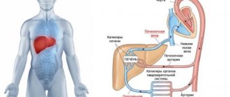
The condition is characterized by excess pressure in the portal vein, which is responsible for blood circulation from organs to the liver. Portal hypertension provokes thinning of the venous walls with an increased risk of internal bleeding.
The direct cause of the development of the disease is a blockage of blood supply.
- liver pathologies - cirrhosis, hepatitis, tuberculosis, etc.;
- vascular thrombosis;
- persistently high blood pressure;
- thrombophlebitis (Chiari syndrome);
- compression of the portal vein by tumor or cystic formations, enlarged lymph nodes, gallstones.
Upper varicose veins of the esophagus are provoked by the following reasons:
- malignant goiter;
- benign tumor (angioma);
- vascular problems in Rendu-Osler syndrome.
Less commonly, varicose veins of the esophagus develop against the background of chronically poor circulation due to cardiovascular insufficiency. More often, the pathology appears in men aged 50 years and older. The causes of the congenital form are not clear.
Gastric varicose veins can develop due to increased pressure in the portal vein. Initially, a compression process occurs in the portal vein, which provokes a blood clot or stone if a person suffers from gallstone disease.
Varicose veins of the mucous membrane of the stomach and the lower third of the esophagus are far from being as common a pathological condition as varicose veins of the lower extremities or hemorrhoidal plexus, however, a number of patients experience dilation of the veins of the mucous membrane of the gastrointestinal tract in the lower part of the esophagus and the cardiac part of the stomach of varying degrees. degree of expression.
Unlike varicose veins of the lower extremities, the causes of this condition and the factors that contribute to its progression are completely different, as are the symptoms, which are often completely absent in the first stages and appear exclusively in the terminal stages of the disease.
Varicose veins of the stomach and lower esophagus are a disease characterized by a violation of the outflow of blood from the veins of these organs and a change in their structure (vessels dilate, become unnaturally looping, long and form nodes).
Gastric varicose veins or phlebectasia is a serious pathology that requires urgent measures to be taken to solve the problem. The disease provokes protrusion of the walls of the stomach and an increase in the lumen. As a result, tortuosity is observed in the area of the vessels, and this requires proper treatment.
The main reason for the development of gastric varicose veins is portal hypertension, which provokes swelling of the veins in the esophagus. Often this condition is observed with cirrhosis of the liver, as many scars appear on it. But this is not the only reason for the manifestation of an unpleasant disease.
There are other reasons that can cause the disease:
- Liver diseases that cause impaired blood flow.
- The appearance of blood clots.
- Compression of the rotary vein by tumors.
- Vascular and heart failure.
Particular attention should be paid to diagnosing the likelihood of venous disease in patients suffering from liver cirrhosis and hepatitis. Since these organs become even more vulnerable and untimely treatment can lead to negative consequences.
Varicose veins of the esophagus and stomach: stopping bleeding and treatment
The disease in question usually affects people over 50 years of age. Varicose veins of the esophagus occur due to impaired blood circulation through the veins of the collar zone and the superior hollow vessels. The pathology has various causes and requires immediate treatment of the patient.
Types of VRVP
According to symptomatic manifestations, VVD is divided into types. Depending on the criterion, esophageal varicose veins can be:
- with bleeding;
- without one;
- congenital;
- acquired as a result of unfavorable environmental conditions and concomitant diseases.
Each form of varicose veins of varicose veins is classified according to the severity of the patient’s condition.
Severity
There are several classifications of the disease that are used in medicine simultaneously. However, most often, esophageal varices (EVV) are divided depending on the degree of dilation of the veins.
- In the first stage of esophageal varicose disease, the diameter of the veins increases to half a centimeter. At the same time, they acquire an elongated shape.
- At the second stage of varicose veins, the vessels twist, expanding up to one centimeter. Their location moves from the edges of the esophagus a little closer to the center.
- At the most serious stage of varicose veins (VVV), the patient’s venous walls become thinner and severe tension develops. The diameter already exceeds the centimeter mark. The veins come closer together, and scarlet “markers” appear on them.
Classification of varicose veins of the stomach can be carried out using other methods: for gastric varicose veins, according to A. M. Vitenas / D. I. Tamulevichute, the system of the Scientific Center of Chemistry of the Russian Academy of Medical Sciences, according to Zdenek Marzhatka.
For varicose veins of the stomach
Varicose veins of the stomach are also divided into 3 degrees.
- The first degree of varicose veins is characterized by the diameter of the varicose veins up to half a centimeter. Veins do not protrude from the mucosa.
- A sign of the second stage of VRVP is an expansion of up to 1 centimeter and a change in the shape of the patient’s veins, which become solitary-polypoid.
- At the third stage of varicose veins (VVVV), nodular polypoid growth occurs, exceeding 1 cm in diameter.
The following classification identifies another stage in the course of VRVP.
Classification according to Vitenas and Tamulevichiute
This classification of esophageal varices (EVV) is not used to characterize the gastric veins.
- In their initial form, the veins have a diameter of 0.2–0.3 cm, are characterized by a bluish tint and are directed in a straight line.
- Next, the patient develops nodular formations wider than 0.3 cm, and an uneven location of the tortuous veins is observed.
- In the third stage of VRVP, clearly visible nodules appear on the vessels, which protrude into the lumen of the esophagus, reaching the fornix of the stomach.
- Nodules of varicose veins are localized in clusters and polyps, blocking the lumen of the esophagus. With varicose veins, the mucous membrane of the node itself grows with venous processes, forming a new layer of varicose veins.
In his classification, Zdanek Mařatka is guided only by the condition of the veins, without taking into account the digital indicator.
According to Zdenek Mařatka
According to this method, at stage 1 of VRVP, the vessels run along the esophagus, protruding slightly above the mucosa. Then they grow in width and twist. At stage 3, they already cover 50% of the lumen and look like tumors.
The Academy of Sciences proposes a classification of varicose veins, which, on the contrary, takes into account only the size of the venous lumen in the vessels of the esophagus of a patient with varicose veins.
Scientific Center for Chemistry of the Russian Academy of Medical Sciences
The boundaries between the stages of pathology are the following numbers:
- 2-0.3 cm;
- 3-0.5 cm;
- more than 0.5 cm.
There are no other classifications of VRVP syndrome, or they are not accepted in official medicine.
Causes
The main cause of disease of the lower esophagus with varicose veins is stagnation of blood flow in the area of the portal vein and its thrombosis. With cirrhosis of the liver, varicose veins of the esophagus also occur quite often.
In the upper zone of the VRVP, the disease provokes a malignant goiter.
Other causes of varicose veins in the esophagus are associated with a whole range of health problems. The disease provokes:
- portal vein sclerosis;
- portal hypertension;
- liver pathologies;
- Chiari syndrome;
- various vascular diseases, especially arterial hypertension;
- tumor diseases in the esophagus.
Men, people over 50 years of age and patients with varicose veins and suffering from sudden changes in pressure are at risk of a complicated course of the disease with bleeding.
Symptoms of esophageal varicose veins
The symptoms of varicose veins may vary depending on the form of the disease and the individual characteristics of the patient with varicose veins. Common symptoms associated with varicose veins include:
- difficulty swallowing;
- heartburn;
- burping;
- slight bleeding from varicose veins of the esophagus, which will be noticeable in the stool;
- increased heart rate;
- feeling of heaviness in the sternum;
- pain syndrome;
- increased fatigue;
- general deterioration of health.
There may be other symptoms of esophageal varicose veins. In any case, the manifestations of varicose veins do not have a characteristic specificity that makes it possible to identify varicose veins in a patient.
Some categories of patients may not experience visible signs of the asymptomatic pathology of varicose veins for a long time.
Establishing diagnosis
Diagnosis of VRVP is carried out using a hardware method. The patient will also have to undergo a series of tests.
- During the first visit, a patient with varicose veins is prescribed a general blood test and tests for various indicators, including biochemistry.
- To determine varicose veins, a liver function test is performed.
- In cases of varicose veins, an analysis of ascitic fluid is performed.
- If the patient complains of symptoms characterizing esophageal varices, a fibroesophagoscopic examination may be prescribed. It involves examining internal organs with a special device. If necessary, they can also take a biopsy from the vein of a patient with varicose veins.
- At the same time, ultrasound is used as a hardware method for superficial examination of the abdominal cavity.
- For varicose veins, computed tomography is used.
- To check the preliminary diagnosis of varicose veins, the doctor may order an X-ray examination of the area with varicose veins.
Based on the diagnosis, specific treatment for varicose veins is prescribed.
Treatment options
Treatment of varicose veins of the esophagus is carried out in several ways, depending on the symptoms, degree of pathology and characteristics of the patient.
Observation by a therapist
In the early stages of varicose veins, therapy is usually limited to observation by a general practitioner.
The therapist prescribes medications for varicose veins, sets a diet and imposes restrictions regarding physical activity and bad habits.
If varicose veins of the esophagus are in a late stage, therapeutic methods act as concomitant treatment when surgery is mandatory for the patient in this case.
Operation
Surgical methods for treating varicose veins involve several techniques that are chosen at the request of the patient or based on the recommendations of a specialist. In any case, the indication for a more radical measure than conservative therapy for varicose veins of the esophagus is varicose veins of the esophagus of grade 2 and subsequent stages.
Let's look at the basic techniques.
- During sclerosis, a special solution is injected into the pathological area to promote the “resorption” of excess tissue. The procedure is carried out 4 times in one year: the main operation, repeated after 5 days, another one, a month later and on the 100th day after the start of the course.
- Another measure involves lining the veins to prevent their expansion.
- There are methods of anastomosis and portosystemic stent bypass - a special instrument is inserted into the patient's hepatic vein to improve blood circulation in this area.
- Surgical removal of the damaged area of the esophageal vein and its replacement with an implant.
Before the operation, a mandatory study is carried out to determine the possibility of applying the technique to a specific patient with varicose veins due to his individual characteristics. Therefore, it is not recommended to insist on the operation you like if doctors recommend another solution.
Stopping bleeding with endoscopy
Endoscopic sclerosis of the esophageal veins is performed in case of bleeding. During the puncture, the patient is injected with a sclerating substance, which strengthens the venous structure, preventing it from rupturing.
Treatment of varicose veins is carried out using the method of thrombosis of venous nodes. But when treating in the gastric region, no more than two nodular zones are treated in one session. Otherwise, complications are possible in the form of necrosis, perforation of the esophagus, and the formation of abscesses.
Modern method - balloon tamponade
This method is used in emergency cases of VRVP to stop bleeding. It requires a highly qualified doctor and is fraught with extremely serious complications, therefore it is not prescribed if the patient only has grade 1 varicose veins of the esophagus.
- During the procedure, a probe with a balloon is inserted.
- By inflating it, tamponade of the bleeding venous nodule is carried out.
- If the first operation does not help, the patient undergoes repeated thrombosis of the venous plexus.
In general, the effectiveness of such treatment for varicose veins is 90%. It almost completely reduces the risk of death of the patient due to the sudden onset of intense internal bleeding in the esophagus.
Treatment with drugs
The goal of drug therapy for esophageal varicose veins is to reduce the load on the patient’s vein walls due to an unfavorable environment.
This task is achieved with the help of drugs to reduce acidity in the gastrointestinal tract, astringents, and synthetic vitamins that strengthen the vascular walls in the esophagus of a patient with varicose veins.
Diet
Fractional meals for varicose veins are divided into 4-6 doses per day, the last one being taken 3 hours before bedtime.
For esophageal varicose veins (EVV), the patient’s diet should contain the following types of products:
- vitamin E-rich onions (green), chicken eggs, natural oils of vegetable origin, lettuce;
- vitamin C-rich oranges and tangerines, potatoes, fresh berries, peppers;
- nuts, black currants, grapefruit, as they contain extremely important rutin;
- flavonoids useful for varicose veins of the esophagus: cherries and sweet cherries.
A patient with esophageal varicose veins needs to drink constantly - at least a liter of clean water per day.
A diet for varicose veins of the esophagus requires exclusion from the patient’s diet:
- alcoholic products;
- black teas and coffees;
- sweet;
- flour;
- hot seasonings.
If you have varicose veins, you should eat boiled food, avoiding fried and very hot foods. Following a diet can significantly reduce the symptoms of the pathology and minimize the likelihood of complications for a patient with varicose veins.
What does traditional medicine say?
Traditional medicine recommends that patients with esophageal varicose veins use vitamins in any form.
Instead of tea and coffee, you can drink herbal tinctures and decoctions (rose hips, linden leaves, thyme, etc.), eat as many fruits and vegetables as possible that are good for the condition of the veins.
However, folk remedies for varicose veins do not exclude regular examination by a doctor and surgical treatment of the patient if necessary. These are only auxiliary methods for improving the health of a patient with esophageal varicose veins.
Complications and further prognosis
The most dangerous complication of varicose veins (VVV) is spontaneous bleeding in the patient, which can occur at any time in the late stages of the pathology.
Complications arise for a number of reasons:
- sudden jump in blood pressure;
- patient's body tension;
- sudden lifting of a heavy load;
- peptic ulcer of the esophagus;
- tumor destruction;
- prolonged fever;
- Mallory-Weiss syndrome in a patient.
A consequence such as hemorrhage often leads to the death of a patient whose condition is complicated by other liver diseases, especially cirrhosis. Even if the patient survives the first bleeding, its recurrence is possible after 1.5-2 years in the later stages of varicose veins.
In general, the prognosis for grade 1 URVP can be quite favorable. If the patient detects his disease in a timely manner and takes the necessary measures, varicose veins will progress very slowly and practically asymptomatically for the patient.
Prevention
Prevention of varicose veins includes a whole range of requirements, the most important of which are a healthy lifestyle and strict compliance with the requirements of doctors.
What should a patient pay attention to in case of esophageal varicose veins?
- The condition of the liver should be monitored.
- Watch your veins and heart.
- Undergo regular examinations and follow the order of endoscopic procedures.
- Reduce high blood pressure in a timely manner.
- Comply with diet and nutrition requirements.
- Drink multivitamin complexes.
- Do exercises without allowing high loads.
Proper prevention of varicose veins of the esophagus is the key to life with varicose veins of the esophagus. Therefore, the patient should pay special attention to this at any degree of pathology.
Treatment of varicose veins, bruised veins on the leg:
Source: //PoSosudam.ru/varikoz/vidy/varikoznoe-rashirnie-ven-pishhevoda.html
Types and degrees of varicose veins
The classification of the disease consists of the degree of damage to the veins. There are four degrees. Depending on the degree of varicose veins, treatment is prescribed. The greater the degree, the more likely surgical treatment is.
In total, there are four degrees of varicose veins of the stomach or esophagus.
- Degree 1 – no symptoms are observed, venous ectasia is isolated, which does not prevent the patient from feeling great. The first degree of the disease can only be determined using endoscopy.
- Grade 2 – vascular structures become tortuous and uneven. But at the same time, swelling does not exceed 3 mm, and the gaps narrow to insignificant distances. In this case, bleeding is rare. The problem can also be identified mainly using x-rays or endoscopy. Symptoms are not expressed.
- Degree 3 – the venous lumen noticeably narrows, the veins bulge. At this stage, the nodes are clearly visible, the tone of the walls narrows. The main symptoms are already appearing; all that remains is to notice them in time, since the risk of bleeding at this stage of the disease increases markedly. Treatment cannot be delayed at the third stage.
- Degree 4 – the nodes are clearly visible, the lumens are narrowed, the gastric mucosa is seriously thinned. A whole thread of affected vessels may diverge from a large node. All of them are so thin that bleeding can open at any moment. If this happens, the patient is in serious danger.
To diagnose venous disease and determine its degree, doctors use different research methods:
- Laboratory tests of blood, urine and feces.
- Ultrasound of the abdominal organs.
- Esophagoscopy.
- X-ray studies.
Varicose veins of the esophagus and stomach, what they are, dangers, treatment
Varicose veins can also affect internal organs. The digestive system is no exception. Varicose veins of the esophagus and stomach, or phlebectasia, is a serious disease that occurs infrequently and is practically asymptomatic.
What is phlebectasia
Phlebectasia is a pathology that is less common compared to varicose veins of the lower extremities. It is difficult to diagnose because the disease is practically asymptomatic.
In most cases, it is diagnosed only after a venous bed has ruptured and bleeding has begun.
Classification
Defects of blood vessels in the esophagus are classified by several indicators. The main thing is the severity of the disease.
Degrees of phlebectasia:
- 1st degree - symptoms are mild or completely absent. When conducting an examination at this level, the canals are expanded to 3-5 mm. In this case, single ectasia or its absence can be detected. Clear lumen, diagnosed by endoscopy;
- 2 - the first symptoms are observed. Diagnosed using radiography. The veins at this stage expand to 10 mm. The results of the study are tortuous, dilated channels in the lower esophagus. The blood supply system is greatly expanded and can occupy 1/3 of the cavity of the entire esophagus;
- 3 - capillaries dilated more than 10 mm and occupied 2/3 of the esophagus cavity. The veins swell, the nodes are visible visually. Gastroesophageal reflux begins to develop, as the mucous membrane has become very thin;
- 4 - advanced stage, at which bleeding begins. The nodes form clusters, severe damage to the mucosa is observed, and there is practically no lumen.
Of all the degrees, 4 is the most dangerous for human life.
Development mechanism
With phlebectasia, the blood outflow from the liver vessels is disrupted. However, it is significantly reduced in the portal vein during hypertension. This can further lead to thinning and deformation of the bloodstream.
Blood begins to accumulate on the walls of the vessels, which forms characteristic thickenings in this place. When blood pressure rises, the load on the capillary wall increases and it bursts.
In case of cardiac pathologies, it develops slightly. Localization occurs over the entire surface of the esophageal tube.
If the disease is caused by liver pathology, dilation of the vessels located in the lower cavity occurs.
Bleeding directly depends on the condition of the vascular tissue, the size of the node and the pressure surge.
Symptoms
In the initial stages, the disease proceeds unnoticed. In some cases, the symptoms are similar to those of gastrointestinal diseases.
Over time, progressive pathology makes the venous channels brittle and fragile, partial or complete rupture occurs, which entails bleeding. This condition becomes life-threatening.
The onset of varicose veins may be accompanied by the following symptoms:
- swallowing dry food becomes difficult;
- pain is felt in the sternum;
- the occurrence of frequent belching;
- the presence of constant heartburn.
At the stage of hemorrhage you can observe:
- dizziness;
- general malaise;
- due to loss of blood, the skin becomes pale;
- the patient is constantly bothered by diarrhea, black in color;
- constant nausea, attacks of vomiting with blood clots in the vomit.
At the first symptoms, you should immediately call an emergency ambulance team. Medical intervention in this case is necessary.
Why is it dangerous?
Diagnosing the disease in a timely manner and prescribing adequate effective treatment does not exclude the possibility of relapse. It can occur within 3 years from the onset of the disease. This is the danger of the disease.
As a result of hemorrhage, the patient loses a lot of blood. If not treated promptly, blood loss can be fatal.
The most dangerous condition is observed:
- after vomiting;
- when ulcerative formations occur;
- after straining or overeating;
- with fever and high blood pressure;
- with sudden lifting of weights.
If you monitor your condition all the time, bleeding can be predicted.
Blood loss is promoted by:
- sudden darkening of the eyes, complete loss of consciousness;
- sudden bleeding with the consistency of coffee grounds. At the same time, the blood can be either brown or scarlet;
- there is a constant tickling sensation in the larynx;
- There is a salty taste in the oral cavity.
After bleeding, treatment is suggested with surgery. Rarely, hemorrhage may occur during sleep. Its complications can be caused by decreased blood clotting and cardiac failure.
Which doctor should I contact?
If the diagnosis is known, you need to contact a phlebologist. If you have problems with the digestive system, you will need to consult a gastroenterologist.
If the clinic does not have a phlebologist, you can contact an angiologist. This specialist has a broader specialization. An angiologist deals not only with veins, but also with all capillaries, arteries, and any vessels.
If you are not sure about varicose veins, you should first contact your local physician. When the diagnosis is confirmed by the therapist, he gives a referral to a gastroenterologist, phlebologist or angiologist.
The surgery is performed by a vascular surgeon and gastroenterologist.
Diagnostics
To make an accurate diagnosis, it is necessary to carry out a number of tests:
- biochemical and general blood tests;
- Ultrasound of the abdominal cavity;
- radiography;
- esophagoscopic examination.
Therapy methods
Therapy directly depends on the degree of damage to the esophageal veins. In cases where the disease is diagnosed due to hemorrhage, treatment is aimed at preventing blood loss.
Therapeutic measures:
- fixation of affected vessels with a probe;
- electrocoagulation of affected channels;
- prescribing drugs that constrict blood vessels and restore blood circulation;
- blood transfusion is performed.
If hemorrhage occurs due to cirrhosis of the liver, treatment is directed towards the treatment of the underlying disease.
In this case, treatment is aimed at restoring liver tissue. Also, measures are being taken to prevent relapse.
Therapeutic treatment:
- antacids and astringents are prescribed;
- Vitamin therapy is prescribed.
Surgery may also be prescribed:
- devascularization - removal of the affected arteries;
- sclerotization - injection of a hemostatic solution into the affected area. The procedure is carried out 4 times a year;
- bandage - installation of rubber discs at expansion points;
- portosystemic shunting - connection of the portal and hepatic beds to normalize pressure.
For patients suffering from cirrhosis, surgery is contraindicated, so they undergo endoscopic ligation of the affected vessels.
The principle of the procedure is ligation of blood vessels with elastic rings or nylon threads.
In addition, for varicose veins of the esophagus and stomach, a diet is prescribed.
At this stage, it is very important to follow certain nutritional rules. The food consumed must include fiber, vitamins B and C.
Therapy with folk remedies
In combination with traditional therapy, traditional therapy methods can be used. In this case, consult your doctor before using them. In the treatment of folk methods, it is recommended to use medicinal compositions from rose hips and red rowan.
For the composition you need to take 1 tbsp. l. rowan berries and 1 tbsp. l. rose hips, add 500 mg of boiling water and simmer over low heat for 5 minutes. Next, the drink is filtered and cooled.
Take the composition ½ cup 4 times a day.
Signs signaling the development of varicose veins in the stomach
As described above, gastric varicose veins at the initial stage show practically no signs of themselves. And if symptoms do appear, they are more similar to gastrointestinal pathologies.
But if you take a closer look at this problem, the differences can still be identified in a timely manner:
- severe vomiting mixed with biological fluid and black vomit;
- frequent vomiting with pink mucus in it;
- pain in the abdomen;
- rapid heart rate, which is accompanied by frequent interruptions.
How is the disease diagnosed?
Varicose veins in the stomach can be diagnosed through examination using certain instruments. For such purposes, the following methods are used:
- collection of biological fluid;
- collection of the liver complex for research;
- Ultrasound examination of the abdominal area.
It is worth noting that such an examination should only be carried out by a qualified specialist, since in some cases it is necessary to insert a probe, which can damage the thin walls of the stomach and thereby cause bleeding.
Signs signaling the development of varicose veins in the stomach
As a rule, the pattern of bleeding from the upper gastrointestinal tract, often massive, develops suddenly, not accompanied by pain. Signs of shock may be detected. Blood loss usually occurs from the lower part of the esophagus, less often from the fundus of the stomach. Blood loss from gastric varicose veins can also be acute, but more often - subacute or chronic.
Bleeding into the gastrointestinal tract due to liver dysfunction contributes to the increase in portosystemic encephalopathy.
At the initial stage of the disease there are no specific symptoms. Therefore, it is difficult to determine the problem. But as the problem develops, the patient experiences certain signs of phlebectasia.
- Heaviness and discomfort in the chest.
- Enlarged abdomen due to accumulation of fluid in the abdominal cavity.
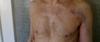
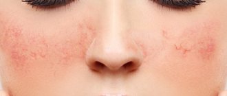
- A “jellyfish head,” as it is called, may appear in the anterior part of the abdominal wall. This is a pattern of veins dilated in the esophagus.
- Shortness of breath as a result of activity.
- Severe heartburn regardless of food intake.
- Swallowing dysfunction.
It’s great if the patient pays attention to the listed signs and consults a doctor. Diagnosis of the disease based on the listed symptoms will make it possible to take urgent measures to treat gastric varicose veins.
But situations when patients come to the doctor at this stage are rare. Most often, phlebectasia manifests itself already at the moment of vein rupture.
When bleeding begins, the signs of the problem become more pronounced.
- Vomiting with bloody discharge.
- Black stool, as well as noticeable admixtures of blood in it.
- Decreased blood pressure.
- Cardiopalmus.
- State of shock.
Bleeding from varicose veins can be minor, but most often it is massive. Treatment must be started immediately, as the problem can lead to serious deterioration or death.
Symptoms
At the first stage of the disease there is no pronounced discomfort; it is usually attributed to a violation of the diet and its errors, or to the symptoms of a gastrointestinal disease.
Heartburn appears, a mild indigestion, for which people often do not consult a doctor. When the disease progresses to the second stage, symptoms appear: belching, heartburn. Difficulty swallowing. Enlarged abdomen - ascites, accumulation of fluid in the abdominal cavity. Heaviness and burning in the chest
Complications of the disease are accompanied by: impaired swallowing reflex, bleeding, vomiting with blood, dark feces. In this case, urgent assistance is needed. Symptoms are different for everyone; diagnostics, a thorough medical history, and collection of all necessary tests are required.
Survey
Pathanatomy offers the study of micropreparations, tissue sections, and their laboratory study to identify the pathogen. The macroscopic specimen shows how the vessels are damaged and the esophageal mucosa is changed; specialists need this data to perform operations.
If a disease is suspected, the patient is prescribed tests and medical procedures. In differential diagnosis, other diseases that give similar symptoms are excluded. Qualitative research contributes to successful diagnosis. They include:
- General blood analysis. Shows the number of leukocytes and platelets in the blood
- Blood chemistry. Cholesterol content, liver enzymes, bilirubin level, protein amount, blood type and Rh.
- Endoscopy. The most detailed and reliable diagnosis is fibroesophagoscopy. Internal examination of the walls of the esophagus using an endoscope. The procedure allows you to find out the condition of the veins, the esophageal mucosa, the cause of bleeding, and reveals signs of other diseases with similar symptoms: cancer, ulcers.
- X-ray. Contrast radiography is used. To do this, the patient uses a radiopaque agent barium sulfate internally. When filling the esophagus, barium has a white light in the image and clearly highlights all the structures of the esophagus. Damaged vessels are clearly visible, in the form of oval filling defects.
- Duplex scanning of blood vessels. This is a dual ultrasound examination consisting of B-mode and Doppler. In modern medicine, the method of color Doppler mapping has appeared. The color determines the direction and intensity of blood flow. The use of these two methods allows you to examine the anatomy of blood vessels and veins, find out the degree of their expansion, see the presence of nodes and ectasia
Gastric varicose veins have a poor clinical picture. Symptoms are often similar to other gastrointestinal pathologies. However, there are distinctive features:
- Bloody vomiting with black vomit, which indicates the opening of gastric bleeding. Vomiting may be frequent and contain pink mucus.
- Increased heart rate with frequent interruptions.
- Abdominal pain.
The initial stages of gastric varicose veins are often accompanied by severe heartburn, which, although it causes discomfort, does not cause the need to go to the doctor. As the pathology develops, gastric bleeding begins. This condition is accompanied by symptoms such as:
- ascites, when free extrudate accumulates in the peritoneum, which provokes a strong increase in the size of the abdomen;
- profuse, bloody vomiting that does not cause pain;
- tachycardia with erratic and rapid pulse;
- hypotension, characterized by a sharp decrease in blood pressure;
- hypovolemic shock, accompanied by a sharp decrease in the effective volume of circulating blood.
Dilatation of the esophagus, as well as dilated veins of the esophagus, are most often an advanced disease of the gastrointestinal tract. The initial period of development of the pathology is not characterized by an increased degree of activity. Thus, the disease most often develops asymptomatically, it does not cause any specific inconvenience, allowing a person to coexist normally.
The development of pathology occurs until bleeding begins. In this case, the disease manages to expand to large-scale forms. In this case, it makes sense to talk about grade 3 varicose veins.
Symptoms of VRP can sometimes appear as follows:
- heaviness in the chest;
- belching;
- esophagia and dysphagia;
- heartburn;
- malaise;
- weakness.
In some cases, a feeling of tightness may indicate the presence of a pre-mortem hemorrhage. So, according to statistics, 3 out of 5 deaths are observed with varicose veins. Most often, it turns out that all three cases of death were not subjected to sufficient research or sought help too late.
The pathology in most cases occurs without visible symptoms. The first obvious signs appear when bleeding from the esophagus opens. When blood flow is obstructed, the veins begin to unbalancedly expand in the form of nodules, lengthen, and become tortuous. Due to the thinning of the walls, they rupture, which leads to esophageal bleeding.
Sometimes signs of venous dilatation of the esophagus develop rapidly, like the pathology itself, but more often the disease proceeds very slowly, not appearing for many years.
Diagnostics
Detection of varicose veins is possible only through instrumental examination. For this purpose the following methods are used:
- General and clinical blood tests, which are necessary to assess the general condition of the patient.
- Functional and hepatic examinations to determine coagulopathy.
- X-ray with contrast (barium sulfate), carried out to assess the functionality of the digestive tract.
- Esophagogastroscopy, used to visualize the condition of the internal walls of the stomach. The method is highly accurate, but requires increased attention and accuracy, since the affected tissues are fragile and the probe can cause bleeding.
- Ultrasound of the abdominal organs, which is necessary to confirm the diagnosis.
- Endoscopy.
- Studies to detect coagulopathy.
Varicose veins of the esophagus can be detected using x-ray examination. More accurate data is obtained by esophagoscopy, but it must be done very carefully, since injury to the vein can lead to bleeding.
Diagnostic tests are carried out to identify the cause of the disease and determine its extent. So, most often, with varicose veins of the esophagus, the following measures are suggested:
- fibroesophagoscopy - helps to identify the causes of bleeding, determines the degree of dilatation of the veins, the condition of the walls, and the presence of ruptures.
- X-ray of the esophagus - determines the nature of the dilation of the veins;
- laboratory tests - biochemistry, APTT, liver test;
- esophagoscopy.
Stage 1 and 2 illnesses are sometimes somewhat difficult to diagnose. This may well require additional research and analysis.
Important: The first trip to the doctor should take place immediately after the first suspicious symptoms are detected. If the doctor did not hospitalize you and prescribed a second appointment, then you cannot ignore it.
Dilated veins of the esophagus can only be determined using hardware, since visible symptoms are often absent. Usually prescribed:
- all types of plasma studies - to determine the patient’s condition;
- functional and liver tests - to study coagulopathy;
- Ultrasound and radiography - to clarify the pre-diagnosis;
- fibroesophagoscopy - to determine the causes of blood loss.
If there are underlying pathologies, an additional examination is performed to determine the cause of their development.
There are three treatment approaches:
- therapeutic;
- medicinal;
- surgical.
The first two regimens are used in the early stages or after effective control of bleeding. Late stages are treated only surgically, as the risk of death associated with hypovolemic shock is high. The main approaches to treating gastric varicose veins are presented below.
Treatment
Pathology is most often detected at a late stage, which often leads to hypovolemic shock or death. The patient is required to undergo immediate hospitalization and resuscitation. The first step is to stop the bleeding, stabilize the body’s condition, replenishing the blood volume.
Drug treatment
Treatment of varicose veins involves squeezing the problem area. This is how you get rid of bleeding. The effectiveness of treatment is 90%. When operating on damaged vessels of the esophagus, relapses are possible. Often another surgery is performed. To increase the chances of recovery, the following methods are used:
- Injection of a special solution into the vein that has ruptured to reduce the outflow of blood.
- Monthly repetition of actions aimed at stopping possible internal bleeding.
- The use of drugs that affect gastric juice to reduce pressure on the walls of blood vessels.
- Astringents that stop possible bleeding.
- Multivitamin complexes to increase vascular elasticity.
Surgical intervention
Measures are carried out in several ways: damaged veins are ligated using a rubber bandage, intrahepatic material is shunted to reduce pressure. A special device is inserted into the liver to prevent blood from accumulating in the cavities, preventing bleeding from occurring. Actions are carried out using an X-ray machine to avoid possible errors.
The third method is splenorenal shunting. Used to prevent fluid accumulation. A shunt connects certain veins, preventing hemorrhage.
Folk remedies
A person who has been diagnosed begins to think about how to cure varicose veins. Treatment is complex and long. Unfortunately, it does not always bring results. If esophageal varicose veins are diagnosed as a result of a concomitant disease, the chance of complete recovery is low.
Specialists can only maintain the vital function of the body. Prevention is better than cure. Physical activity, combating obesity, proper nutrition, timely treatment of concomitant diseases will give a chance to avoid varicose veins. Treatment of varicose veins of the esophagus includes various measures:
- Therapeutic diet. Most often, varicose veins are a consequence of liver disease. It is very important to follow a proper diet so as not to aggravate the condition of the body. The doctor will select a therapeutic diet, taking into account individual characteristics. Dieting should become a way of life to maintain quality of life.
- Taking medications. Antacids, vitamins, hemostatic and vasoconstrictor drugs. Diuretics, drugs to reduce portal pressure.
- Blood transfusion. In case of bleeding, blood or plasma transfusion occurs in cases that threaten the patient's life. This procedure is carried out after surgery.
- Sclerotherapy Injection of a special solution into a vein. The substance is a sclerosant, causing the destruction of the vein. Using an esophagoscope, a probe is inserted and an injection is made into the vein on both sides.
- Intrahepatic shunt. A catheter is inserted into the neck through a vein, with which a stent is installed. A metal cylindrical device that connects the hepatic and portal veins. The operation reduces the pressure in the liver.
- Splenorenal shunt. A surgical operation during which the splenic vein is connected to the kidney vein, reducing blood pressure in the liver.
- Surgery (cessation of blood supply). In case of acute disease, bleeding, varicose veins are surgically removed.
- Liver transplantation. Prescribed against the background of advanced liver diseases.
The first two regimens are used in the early stages or after effective control of bleeding. Late stages are treated only surgically, as the risk of death associated with hypovolemic shock is high. The main approaches to treating gastric varicose veins are presented below.
- Introduction of plasma substitutes.
- Endoscopic ligation of varicose veins (backup method - sclerotherapy).
- Intravenous administration of octreotide.
Measures to combat hypovolemia and hemorrhagic shock. In case of coagulation disorders (for example, increased MHO), it is necessary to transfuse 1-2 doses of fresh frozen plasma and administer 2.5-10 mg of vitamin K intramuscularly. In the presence of cirrhosis of the liver with gastrointestinal bleeding, the risk of bacterial infection increases; Prophylactic antibiotics are indicated: norfloxacin or ceftriaxone.
T. to
During endoscopy, it is always possible to detect varicose veins; the main methods of treatment are endoscopic interventions. Endoscopic ligation is preferred over injection sclerotherapy.
At the same time, octreotide is administered intravenously. Octreotide increases splanchnic vascular resistance by suppressing the release of vasodilatory hormones of internal organs (in particular, glucagon, a vasoactive intestinal polypeptide).
The standard dose is 50 mcg intravenous bolus, followed by infusion at a rate of 50 mcg/hour. Administration of octreotide is preferable to the previously used vasopressin and terlipressin due to the lower incidence of adverse events.
The patient should understand that treatment can only weaken some symptoms of the disease, but, alas, it is not yet possible to get rid of varicose veins completely; complete recovery of patients from varicose esophagus has not been observed in world medical practice.
The main emphasis in therapy is on preventive measures that can save the patient from the possible onset of bleeding. The main 3 principles of treatment are:
- Therapy aimed at eliminating the disease that led to varicose veins. This could be hepatitis, angina pectoris, cirrhosis of the liver, etc.);
- Adjusting the patient's lifestyle. Physical activity and fatigue (including emotional fatigue) are completely excluded from the patient’s daily routine; emphasis is placed on observing strict rules of personal hygiene;
- The patient must follow a diet. When prescribing therapeutic nutrition, the doctor focuses his attention on the disease that led to varicose veins. Based on this diagnosis, a specific table is assigned.
Medical therapy includes a number of medications that can be prescribed to the patient. Among them are 3 main types of drugs:
- Antacids are substances that help reduce the acidity of gastric juice;
- Vitamin substances;
- Astringents.
If the patient has suffered from blood loss, the doctor prescribes the following methods of maintaining the body:
- Transfusion of blood, red blood cells, plasma;
- Vasoconstrictor drugs;
- Drugs aimed at stopping bleeding;
- Inserting a probe into the esophageal tube to stop bleeding;
Application of thrombin or adhesive film to the affected areas.
The most optimal treatment method is surgery. It usually involves ligating the splenic artery, lining the veins, removing some vessels from the esophageal tube, and other procedures.
Doctors also often use ligation of esophageal varices. Surgical treatment is more acceptable, since the mortality rate with this method of therapy is three times lower than in the case of conservative treatment.
Unfortunately, the disease is most often discovered as a result of internal bleeding, which can cause hypovolemic shock and even lead to death. In such cases, the patient requires emergency hospitalization and resuscitation measures. As soon as possible, you need to stop the bleeding and replenish the blood volume.
After the patient’s condition has been stabilized, the underlying disease is treated and procedures are aimed at reducing portal vein pressure and preventing bleeding.
Now the following basic methods are used to solve the problem:
- Sclerotherapy, which consists of endoscopic injection of an adhesive solution into the vessels of the stomach or esophagus. This procedure is repeated, depending on the severity of the disease, once a week or month, until a scar forms.
- Ligation of varicose veins using a rubber bandage. The method is more effective than sclerotherapy.
- Intrahepatic shunting. Reducing pressure by introducing a stent under X-ray control into the middle zone of the liver to connect the hepatic and portal veins.
- Splenorenal shunt. It is produced to prevent bleeding by combining the splenic vein and the vein of the left kidney using a shunt.
- Drug treatment - the use of vasoconstrictor drugs (Vasopressin), nitrates to reduce pressure in the portal vein (Nitroglycerin), as well as Somatostatin (or its analogue Octreotide) to reduce blood pressure in internal organs and narrow dilated blood vessels (taken for a long time).
infovarikoz.ru
Regardless of the severity of varicose veins, the disease is considered incurable. But it cannot be ignored, since late diagnosis and identification of causes, untimely emergency care or improper therapy can lead to death.
Treatment of varicose veins of the esophagus is complex, comprehensive, combining conservative, medicinal and surgical (if detected late) therapy.
Today, modern medicine offers three types of therapeutic therapy with which a person can be relieved of this defect, namely:
- therapeutic method;
- treatment with drugs;
- surgical intervention.
The first two treatment methods are relevant only when the disease is at the initial stage of development or after bleeding has been blocked.
As for advanced stages of development, in this case only surgical intervention is used, since there is a high risk of death for the patient. As a rule, the following treatment methods are used for such purposes:
- sclerotherapy;
- surgical intervention.
Sclerotecrapia
With this technique, a special substance is used, which is injected into the veins affected by the defect to glue them together; this procedure is performed using an endoscope.
- Comprehensive examination
- Eliminate the underlying disease
- Probe
- Antacids
For varicose veins of the esophagus or stomach, a competent approach is needed. After determining the degree of the disease, it is important to find the causes of the development of the pathology. The doctor prescribes the necessary medications to strengthen the liver and blood vessels. The patient is also advised to adhere to a diet and not give up physical activity.
Medicines for gastric varicose veins are mainly prescribed as follows:
- Drugs whose action is aimed at narrowing blood vessels and stopping bleeding.
- Colloidal solutions.
- Astringents.
- Antacids.
- Vitamins.
Conservative therapy with the prescription of medications is a long-term process. Surgery is considered more effective. Electrocoagulation of damaged vessels and squeezing them during bleeding, which is carried out using a special probe, helps to quickly improve the patient’s condition.
Unfortunately, no matter what treatment methods are attempted, it is impossible to cure the disease. Therefore, all measures are aimed exclusively at preventing the further development of pathology. In the first stages, maintaining the condition of the esophagus and stomach is much easier than in the third and fourth stages of the disease. And the appearance of bleeding further worsens the prognosis.
Varicose veins of the stomach - symptoms, causes, treatment methods
Gastric varicose veins develop when the blood supply to the organ is disrupted. The disease provokes changes in the structure of the vascular network, loops or nodes appear, expansion or contraction occurs. Pathology can appear with liver diseases, mainly with cirrhosis. A constant symptom is heavy bleeding.
Symptoms of varicose veins
The first stage of the disease does not manifest itself in any way in the body, so it is difficult to determine the changes that are occurring. As the pathology develops, the following symptoms may appear:
- heaviness, dull chest pain;
- enlarged abdomen due to a large volume of accumulated fluid;
- the appearance of noticeable dilated veins on the abdominal wall;
- shortness of breath with minimal physical exertion;
- heartburn, independent of the foods consumed;
- difficulty swallowing food.
It is important to notice the presence of signs and consult a doctor in time. During treatment, urgent measures are necessary in order to have time to eliminate possible damage to the body.
Attention
: It is rarely possible to determine the presence of varicose veins in time. The disease manifests itself mainly when veins rupture. Bleeding makes all signs of the disease more noticeable.
With bleeding, accompanying symptoms are possible:
- vomiting with drops of blood;
- blood in the stool and turning it black;
- decreased blood pressure and increased heart rate;
- shock.
Bleeding is often profuse, although there are exceptions. If any of the signs are detected, treatment should begin on time. Untimely assistance leads to severe deterioration of the condition, and death is possible.
Causes of gastric varicose veins
The cause of gastric varicose veins is portal hypertension. The veins dilate due to increased pressure, and the outflow of blood is impaired. The condition may occur due to a blood clot compressing a vein. The following diseases of the pancreas or liver may affect:
- Cirrhosis or chronic hepatitis.
- Sarcoidosis.
- Malignant or benign tumor.
- Aneurysms of the arteries of the spleen or liver.
- Fibrosis.
The disease can be inherited and can appear at any time. With cirrhosis of the liver, the structure of the organ changes, the tissues grow and contribute to the cessation of its work. Varicose veins occur, being a serious complication of the first disease.
Treatment
Pathology is most often detected at a late stage, which often leads to hypovolemic shock or death. The patient is required to undergo immediate hospitalization and resuscitation. The first step is to stop the bleeding, stabilize the body’s condition, replenishing the blood volume.
Drug treatment
Treatment of varicose veins involves squeezing the problem area. This is how you get rid of bleeding. The effectiveness of treatment is 90%. When operating on damaged vessels of the esophagus, relapses are possible. Often another surgery is performed. To increase the chances of recovery, the following methods are used:
- Injection of a special solution into the vein that has ruptured to reduce the outflow of blood.
- Monthly repetition of actions aimed at stopping possible internal bleeding.
- The use of drugs that affect gastric juice to reduce pressure on the walls of blood vessels.
- Astringents that stop possible bleeding.
- Multivitamin complexes to increase vascular elasticity.
Diet
If you have varicose veins, you must follow basic nutritional rules. You need to eat at least 4 times a day.
The load on the digestive system is significantly reduced, as the amount of food taken in at one time is reduced. It is forbidden to remain for a long time without food, or to be even a little hungry.
It is worth eating at the same time so that the gastric mucosa gets used to a constant routine. Skipping meals on a set schedule is not recommended.
Food must be chewed thoroughly; there is no need to rush when consuming it. To develop such a habit, it is worth removing all distractions: TV, books, the Internet. Avoid eating before bed. The last time to eat is about 2 hours before meals, no later.
The diet does not have clear rules; experts usually prescribe general nutritional recommendations. If you have stomach varicose veins, you should not eat the following foods:
- fatty foods, fried foods, fast food and the like;
- meat, poultry, fish are not prohibited for consumption, but are not recommended in large quantities;
- smoked products, pickles that can retain water in the body;
- high fiber foods;
- fresh vegetables and fruits - they must be heat treated;
- alcoholic or carbonated drinks, as well as caffeinated drinks;
- substances that affect the cardiovascular system to any degree.
Important
: The disease is dangerous due to the possibility of heavy bleeding into the stomach cavity. It is necessary to exclude any products that can put a strong burden on the body. Do not consume substances that will irritate the digestive system.
Risks and forecasts
Often, deaths from varicose veins of the stomach occur due to late access to doctors. Complex pathologies that arise during the development of the disease quickly lead to deterioration of the condition. Problems with blood vessels, complicated by heavy bleeding and cirrhosis of the liver, are eliminated only at the initial stage of development.
Mortality in the presence of the disease is 50%. In approximately 79% of cases, internal bleeding can be stopped in time. Return of the disease with successful treatment is possible in 55% of cases. Given the statistics, do not forget about proper nutrition. The measures taken will delay or prevent the onset of the disease.
Varicose veins of the stomach are considered an extremely dangerous disease that can arise even from simple overeating. Everyone should carefully monitor their diet, the amount of food consumed and the quality of purchased products so that the consequences of the disease do not turn out to be an unpleasant surprise.
Source: https://ritmserdca.ru/varikoz/varikoznoe-rashirnie-ven-zheludka.html
Operation
The main goals of treating the disease:
- compensation of hypovolemia;
- suppression of hemorrhagic shock.
For patients with blood clotting dysfunction, it is recommended to administer up to 2 doses of plasma and vitamin K intramuscularly. If grade 1 or 2 dilation of the esophagus is detected, treatment includes endoscopic hemostasis.
Endoscopic artery suturing is more effective than injection therapy. At the same time, Octreotide helps improve visceral vascular resistance (by inhibiting the release of corresponding hormones).
The drug is the most suitable of the group for the treatment of food varicose veins. For recurrent bleeding, a solution of calcium chloride and Vikasol intramuscularly are used intravenously.
If the above treatment fails and the disease progresses, urgent transjugular intrahepatic portosystemic shunt (TIPS) is performed to lower portal pressure, stopping hemorrhage.
The essence of the method is to introduce special medical glue into the affected vessels endoscopically. A certain frequency of the procedure is required, which is determined by the doctor individually. More often, the technique is applied once every 7 or 30 days, and is completed when a permanent scar is formed.
The essence of the method is to regularly take such means as:
- "Vasopressin" - to restore the normal state of narrowed blood vessels;
- nitrate containing “Nitroglycerin” - to lower pressure in the portal vein;
- "Somatostatin" or "Octreotide" - to lower blood pressure in the internal organs and restore the normal state of dilated blood vessels.
To treat gastric varicose veins, it is important to maintain proper nutrition. The basic principles are as follows:
- Fractional meals in small portions - up to 6 times a day.
- The last snack is 3 hours before going to bed.
- Increasing in the diet the amount of foods rich in vitamins such as:
- vitamin E (greens, yolk, corn or sunflower oil);
- vitamin C (berries, potatoes, fresh peppers, all types of citrus fruits);
- rutin (nuts, tea, grapefruit, currants);
- bioflavonoids (cherries);
- plant fibers (legumes, fresh vegetables and fruits).
- Organize plenty of drinking - up to 2.5 liters of water per day.
- Complete rejection of harmful products:
- alcohol;
- concentrated black tea, coffee;
- sweets and sugar;
- hot seasonings and spices;
- flour products.
- Preferred culinary processing is boiling, baking in the oven, stewing, steaming.
- Dishes must be warm.
- injection of a special solution into the affected vein and lumen;
- repetition of the manipulation after 5, 30, 90 days.
A sustainable effect is achieved by performing the procedure 4 times a year.
Other techniques used:
- bypass surgery, when an intrahepatic stent is inserted connecting the portal and hepatic veins;
- anastomosis, when a connecting bridge is created to bypass the problem area;
- lining of veins (alloying with tying of vessels with 1-3 elastic rings or nylon loops);
- devascularization, when the affected vessels are excised and replaced with a prosthesis.
Treatment of pathology
Since gastric varicose veins are not considered a self-occurring disease, there is no therapy for it. The use of certain medications makes it possible to reduce portal hypertension, so they are used comprehensively, exclusively as prescribed by the doctor. If it is possible to eliminate the cause of hypertension, proper treatment or surgery is carried out; in most cases, the only cure is liver transplantation.
Drug treatment is carried out using the following means:
- Vasopressin - normalizes the condition of narrowed veins.
- Nitrates included in Nitroglycerin due to a decrease in pressure in the portal vein.
- Somatostatin or Octreotide - reduce blood pressure in internal organs.
Help is provided at the onset of hemorrhage, for which a Blackmore probe is inserted into the digestive tract, through which the veins are compressed and the bleeding stops. At the same time, treatment is given to increase blood clotting.
Phlebectasia becomes a severe exacerbation of portal hypertension due to the fact that at one stage or another it provokes the formation of gastrointestinal hemorrhage, accompanied by the loss of a large volume of blood and threatening life.
In parallel, assistance with bleeding still remains symptomatic, and complete therapy involves eliminating portal hypertension directly. This is not acceptable in every case; for this reason, the prevention of both portal hypertension and phlebectasis is of particular importance, because it turns out to be very difficult to overcome existing disorders.
Prevention
The essence of preventive measures is to maintain healthy veins. To do this you should:
- monitor the condition of the liver;
- follow the doctor's recommendations exactly;
- regulate high blood pressure in a timely manner;
- follow the rules of a healthy lifestyle (giving up bad habits, proper nutrition).
Preventive measures for varicose veins include treating liver disease in the early stages. To do this, you need to regularly visit your doctor and follow his recommendations. For example, high portal pressure is reduced by certain medications that are intended for the heart and blood vessels. These could be beta blockers.
You should also be careful about your quality of life, which includes following a diet if you have liver disease. The diet includes eating four to six times a day, and the last meal should be no later than three or four hours before bedtime.
It is best to consume boiled foods that are steamed. You should not eat very hot or very cold food. It is necessary to ensure that the acidic environment of the stomach does not enter the esophagus. To do this, the head of the bed needs to be raised ten centimeters. While eating, you do not need to watch TV or read; food should be well absorbed by the body.
The goal of preventive measures is to maintain the normal condition of blood vessels. For this:
- it is important to monitor the condition of the liver;
- follow the doctor's recommendations;
- treat pathologies in a timely manner;
- regulate high blood pressure;
- lead a healthy lifestyle without bad habits;
- eat right and follow a routine;
- harden up, do massage and perform light exercises to strengthen the body;
- take multivitamins.
The essence of preventative measures is to keep the veins healthy. To do this you will need to follow a few simple rules, namely:
- do not overload your liver and monitor its condition;
- follow all the advice of your doctor;
- quickly regulate high blood pressure.
And don’t forget one simple rule: a healthy lifestyle is the key to a long, disease-free life. Therefore, every person should monitor their health.
Complications and prognosis
Varicose veins of the stomach are very dangerous due to their complications. If massive bleeding develops, death cannot be ruled out. If the disease is detected in the early stages, the prognosis is quite favorable.
Unfortunately, the pathology is prone to relapse, and even after successful treatment it can return. Relapses are observed in 55% of cases. For this reason, patients should observe preventive measures and carefully follow all specialist recommendations.