Dilatation of the veins of the esophagus. ICD I85.0 (I85.9)
The walls of the esophageal veins are thin and easily stretchable, so an increase in pressure in them quickly leads to dilatation, deformation and bag-like bulging of part of the vessel. This is also facilitated by the fact that the veins of the esophagus are surrounded by loose, pliable connective tissue.
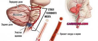
The anatomy of the venous system of the esophagus is quite complex. Blood from this organ flows to the three most important veins of the body. Impairment of blood flow in any of them can cause the appearance of phlebectasis in the esophagus. Most often, phlebectasis occurs in the lower parts of the tubular organ, as a result of disturbances in the v. system. portae (portal vein).
Various liver diseases (hepatitis, thrombosis, sclerosis, cirrhosis) lead to blood flow slowing down and pressure in the vein increasing (portal hypertension). The outflow of venous blood from internal organs, including the esophagus, slows down sharply, resulting in stagnation in the venous system.
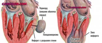
The venous vessels of the esophagus, not adapted to such a volume of blood and pressure, stretch, their walls become thinner, the lumen of the veins becomes uneven, the veins lose their elasticity and bulge in a bag-like manner. Varicose nodes appear. The mucous membrane of the esophagus above the affected vessels becomes thinner and inflamed. All this creates conditions for damage to the integrity of the venous wall and the occurrence of bleeding - the most dangerous complication of varicose veins, threatening the life of the patient.
Treatment with traditional methods
You should always contact a gastroenterologist first. If varicose veins are detected in the first 3 stages, therapy is aimed at preventing the development of bleeding and reducing the level of pressure in the circulatory system. If bleeding is detected, then surgery is used.
Drug therapy includes the following:
- Beta blockers and nitroglycerin drugs are used to reduce blood pressure. The choice of medications is made only by the attending physician based on test results.
- Mandatory vitamin therapy.
- Colloidal preparations are also used.
- The doctor will prescribe hemostatic medications and agents that narrow the veins.
- If necessary, blood transfusion is performed.
Surgical endoscopic intervention includes the following methods:
- The electrocoagulation method allows you to remove damaged areas of blood vessels using electric current.
- A special bandage may be installed to stop minor bleeding. During the procedure, the doctor takes small rubber discs and places them over the damaged vein.
- Endoscopic doping, that is, ligation of varicose vessels. For this purpose, special ring attachments (ligature) or nylon loops are used. They are placed on the damaged vein in quantities from 1 to 3.
- With the help of sclerotherapy, bleeding can be stopped. The operation is based on the introduction of a special sclerosing solution into the vessel, which glues the damaged elements together, as a result of which the blood stops.
- Shunting into the liver. The surgeon inserts a stent into the middle of this internal organ, connecting the veins of the liver and the portal vessel. Additionally, the progress of the operation is monitored using an X-ray machine.
- Splenorenal bypass is based on the connection of the splenic vein with the renal vessel. Thanks to this, the pressure decreases and the blood stops.
- Thanks to balloon tamponade, problematic vessels are compressed using a probe, which is equipped with a special balloon. The balloon is inserted into the stomach and begins to inflate, as a result of which the esophagus is stretched, but it does not inflate. This technique is not in great demand, as it is considered very dangerous. Although effective.
If you want to visually see what the equipment and instruments used for endoscopic alloying look like, watch this video.
If esophageal varicose veins, caused by cirrhosis of the liver, are detected in the initial stages, treatment involves the injection of stem cells into the liver. They are removed from human bone marrow and grown artificially.
VRVP classification
All phlebectasias of the esophagus are divided into congenital and acquired.
- Congenital phlebectasis of the esophagus is a rare pathology, accompanied by many other severe abnormalities. The disease is associated with genetic predisposition and disorders that occur during pregnancy at the time of organ formation.
- Acquired are the result of impaired blood flow in large veins and, as a consequence, phlebectasis in the veins of the esophagus. Esophageal varices are common in liver cirrhosis.
Depending on the size of varicose veins (varicose nodes) and the severity of the disease, four degrees of varicose veins are distinguished.
| Degree of disease development | Condition of veins and location of phlebectasias | X-ray characteristics | Condition of the esophagus | Clinical manifestations of pathology |
| 1st degree | Single dilated veins | Not defined | Satisfactory | Doesn't appear |
| 2nd degree | The veins of the esophagus dilate, become tortuous, and moderately enlarged varicose nodes appear. | An unclear venous contour is revealed. You can often see areas of stretched or tortuous veins. | The esophageal mucosa is not changed, its integrity is not compromised. | The clinic is not specific. Some patients experience discomfort when swallowing food. |
| 3rd degree | The lumen of the vessel narrows, the veins take on a serpentine shape, and the first angioectasia (dilation) appears. | In the middle of the esophagus and above, bulging vessels are detected. Distinct protrusion of venous nodes into the esophageal cavity. | The mucous membrane is thinned, erosions and foci of inflammation are detected on it. The mucous membrane is easily injured. | Belching and pain in the epigastrium and retrosternal space appear. Signs of micro- and sometimes macro-hemorrhage appear. |
| 4th degree | Veins are twisted, collected in nodes with numerous angioectasia | The esophagus is narrowed. Polypoid and cluster-shaped phlebectasias are identified. | The esophageal mucosa is thinned, with many erosions and inflammatory foci. | The patient has a clinical picture of severe esophagitis. A characteristic salty taste of blood appears in the mouth. The development of massive bleeding is very likely. |
Possible complications
The disease can either manifest itself or occur in a closed, latent form. Tumor formations appear, which increase in size over time and become tense. The skin underneath becomes thinner, and ulcerative processes begin.
This leads to varicose veins, and when squeezing or lifting the limbs upward, the unpleasant symptoms decrease. Phleboliths - round formations - are present in the later stages. The disease progresses slowly, pain, motor dysfunction and trophic problems are possible.
With intestinal pathology, pain occurs in the stomach and liver. As the disease progresses, the symptoms intensify, and the walls of the esophagus and stomach become thinner. This is fraught with disruption of the gastrointestinal tract.
Causes of VRVP
The cause of VRVP is congestion in the esophageal veins and increased pressure in them. The main factor for this is disruption of blood flow and increased pressure in one of the three main veins.
Pathologies most often occur in the v. system. portae. This is due to the fact that almost any disturbance of blood flow in the system of this vein leads to portal hypertension. Moreover, an obstacle to blood flow can occur in any area v. portae:
- the lower ones are thrombosis of the splenic vein, congenital narrowing of the portal vein;
- hepatic – liver cirrhosis, active hepatitis, hepatocellular carcinoma, schistosomiasis;
- upper – constrictive pericarditis, right ventricular failure.
With portal hypertension at the initial stages of development of varicose veins, phlebectasias are localized in the distal part of the organ, and only as the disease progresses, the veins of the stomach and the veins of the middle part of the esophagus are involved in the process.
VRVP can form when compressed by the superior vena cava (hypertrophied organ, enlarged lymph nodes, tumor). In this case, varicose veins of the esophagus will develop in the upper part of the organ.
In rare cases, VRVP occurs as a result of disturbances in the entire circulatory system (severe degrees of heart failure). In this case, blood circulation in all organs is disrupted. As a result of a violation of the outflow of blood from the esophagus, deformation of the veins is observed along the entire length of the organ, although the size of the nodes will be significantly smaller than with portal hypertension.
Causes
Having analyzed what phlebectasia is, you can draw your own conclusions about what leads to the development of pathology. Doctors call the main reasons for the development of the disease:
- disruption of normal valve operation;
- problems with blood flow;
- presence of tumor or scars.
Phlebectasia of the esophagus occurs as a result of problematic blood flow in the hepatic veins. The pathology is provoked by cirrhosis of the liver or blood clots in the organs.
Dilated veins of the esophagus: symptoms
For a long time, the pathology does not manifest itself in any way. The first symptom of varicose veins is often bleeding. Sometimes hemorrhagic manifestations are preceded by unpleasant sensations in the retrosternal region (behind the sternum). More often, symptoms of esophagitis appear within a few days (sour belching, heartburn, difficulty swallowing solid food).
More often, hemorrhage is immediately preceded by overeating and sudden physical exertion. Although sometimes bleeding occurs during sleep.
The severity of hemorrhagic syndrome can be very different, from minor bleeding to massive blood loss that threatens the patient’s life. But even minor, constant bleeding can lead to serious consequences. Patients are constantly worried about:
- weakness;
- nausea;
- vomiting streaked with blood;
- melena.
The result of this is exhaustion, sudden weight loss, and iron deficiency anemia.
With massive blood loss, when blood flows out in a stream from damaged vessels, the condition immediately becomes critical, with rapidly increasing symptoms:
- suddenly the patient feels severe weakness, accompanied by a faint or faint state;
- severe sweating;
- nausea followed by vomiting large amounts of blood with clots;
- Blood pressure drops sharply, tachycardia appears, often accompanied by various kinds of arrhythmias, and the patient loses consciousness.
Bleeding from esophageal varices
Bleeding from esophageal varices can begin at any stage of the disease.
Provoking factors are:
- sudden rise in weight;
- increased blood pressure;
- rising temperature to a feverish state;
- excess food intake;
- concomitant diseases of the digestive tract.
Before bleeding develops, the patient may complain of a tickling sensation in the throat, soreness, and a taste of salt in the mouth. Further vomiting of blood or blood mixed with blood is possible. If bleeding or bloody vomiting develops, you must immediately call emergency medical help. Self-medication is unacceptable. As a medical emergency, you can drink cold water.
Diagnostics
Diagnostic measures are aimed at identifying the cause that caused varicose veins, as well as determining the stage of varicose veins, which determines the treatment strategy for the patient.
Mandatory diagnostic measures.
Laboratory research:
- study of peripheral blood (hemoglobin, red blood cells, hematocrit);
- study of venous blood (blood sugar, bilirubin and its fractions, AST and ALT, amylase, creatinine, total protein);
- coagulogram.
- Ultrasound examination of the liver
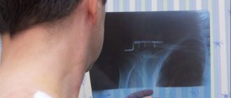
- X-ray examination with contrast of the esophagus (allows you to determine the nature of the dilation of veins and varicose nodes).
- fibroesophagoscopy (the main diagnostic method that allows you to determine how much the veins are changed, the presence of damage and hemorrhages, the condition of the venous nodes).
Classification and diagnosis
The classification of portal vein dilatation is based on several criteria. The first is localization. Based on this feature, intra-, post-hepatic and mixed forms are distinguished. In the intrahepatic form, the branches of the portal vein are affected; in the posthepatic form, the vessel itself is affected. In the case of a mixed type, the pathology affects both the vein and its branches.
There is a classification based on the pressure in the vessel itself. There are three stages:
- from 250 to 400 mm.
- from 400 to 600 mm.
- more than 600 mm.
Diagnosis begins with a history and general examination. The doctor palpates the liver and spleen and determines whether they are enlarged. Other research methods are also prescribed:
- General blood and urine analysis.
- Blood chemistry.
- Ultrasound.
- Contrast X-ray.
- Immunological tests for suspected viral infections.
Did you know the symptoms of liver vein thrombosis?
Not really
To clarify the diagnosis, scintigraphy, computed tomography or magnetic resonance imaging, or diagnostic laparoscopy may be needed.
Treatment
If a diagnosis of varicose veins is established, then, depending on the degree of phlebectasis in the organ, appropriate treatment is immediately prescribed.
Basic principles of management of patients with esophageal varicose veins:
- treatment of the disease that caused varicose veins;
- delay the progression of varicose veins as long as possible;
- slow down the occurrence of hemorrhagic complications as much as possible;
- quickly stop the bleeding that has begun and rehabilitate the victim as fully as possible;
- use all means to prevent the recurrence of hemorrhages.
Varicose veins of the esophagus I degree
There are no clinical manifestations suggesting esophageal varicose veins at this stage of the disease. The veins are slightly dilated, their lumen is free. Nodes are just beginning to form, there may be 1-2 of them. The only method to detect the disease at this stage is esophagoscopy.
At this stage, the main method is conservative treatment, the patient is supervised by a gastroenterologist, therapy is mainly aimed at treating the disease that caused varicose veins. In addition, the patient is prescribed:
- diet therapy with strict adherence to the diet;
- develop a work and rest regime with the exception of heavy physical activity;
- warn about the need to eliminate bad habits;
- prevention of esophagitis: antacids (Gaviscon, phosphalugel), PPIs (omeprazole, rabeprazole), IGRs (famotidine, nizatidine), prokinetics (domeperidol, itopride).
Esophageal varicose veins, grade 2
In the second degree of pathology, the veins are dilated and tortuous, but their lumen is free. The mucous membrane is slightly changed or completely intact (not affected). At this stage, the disease can be detected not only by endoscopy, but also by contrast radiography of the esophagus. Clinical manifestations are determined mainly by the clinical picture of esophagitis:
- belching with a bitter or sour aftertaste, heartburn;
- discomfort in the retrosternal area;
- symptoms of dysphagia to varying degrees.
There are no bleedings with stage 2 VRVP.
A patient at this stage of the disease is usually prescribed:
- drugs that increase the pH of gastric juice;
- hemostatic drugs;
- vitamin complexes containing rutin and tocopherol to strengthen the vascular wall;
- iron-containing and vasoconstrictor drugs.
Grade 3 varicose veins of the esophagus
At this stage of phlebectasis, the veins are changed, serpentine in shape, their lumen is narrowed, the tone of the walls of the venous vessels is reduced, multiple nodes are clearly visible, and angioectasias are detected. The mucous membrane is changed, its integrity is damaged, multiple foci of inflammation and erosion appear on the surface. Gastroesophageal reflux is pronounced. With grade 3 VRVP, the risk of bleeding is very high. Hemorrhage can be caused by a variety of reasons:
- lifting weights;
- straining;
- feverish conditions;
- sudden rise in blood pressure.
The main goal of drug treatment is to prevent bleeding by:
- drug therapy for reflux esophagitis (diet, antacids, PPIs, IGRs, vitamins);
- transfusions of plasma, red blood cells, blood;
- administration of vasoconstrictor drugs.
If bleeding begins, it is necessary to stop it as soon as possible and carry out restorative therapy:
- tamponade of bleeding esophageal veins using a double-balloon Blackmore probe is a temporary measure, more often used in case of massive bleeding; inflated balloons press against the bleeding vessel, as a result the bleeding stops;
- ligation of varicose veins of the esophagus is ligation of bleeding veins (sometimes electrocoagulation of bleeding vessels is performed);
- restoration of blood volume, patients are immediately prescribed a transfusion of blood substitutes, and, if necessary, blood;
- measures are taken to prevent infection of the damaged mucosa and blood vessels.
After the bleeding has stopped and hemodynamic parameters have become stable, surgical treatment is prescribed to prevent relapses.
There are many methods of surgical treatment of varicose veins of the esophagus:
- anastomosis to reduce pressure in the affected vessel;
- prosthetics of venous vessels;
- suturing and removal of affected veins of the esophagus.
The method of surgical treatment and the extent of the operation are individually selected for each patient.
Stopping massive bleeding, and even more so preventing recurrent bleeding, is a very difficult task. Everything becomes more complicated if the cause of varicose veins is cirrhosis of the liver, a severe, constantly progressive disease. It is impossible to stop its progression with the current level of medical knowledge, which means it is impossible to reduce the level of constantly progressing portal hypertension. Even such a radical method as a liver transplant does not always lead to the desired results. And with the progression of portal hypertension, varicose veins progress, which is why patients with cirrhosis of the liver often die from bleeding from the esophagus.
Symptoms
Harbingers of the development of esophageal varicose veins are the following signs:
- constant heartburn;
- frequent belching;
- pain in the sternum;
- it becomes difficult for the patient to swallow dry food.
When bleeding from the esophagus, the following symptoms exist:
- nausea and vomiting, and the vomit contains blood clots;
- constant diarrhea with a black tint;
- the skin turns pale as blood is lost;
- weakening of the body;
- dizziness.
In this case, it is necessary to urgently call an ambulance because surgical intervention is required.
The main complication of esophageal varicose veins is bleeding and blood loss, which leads to death.
Prognosis of VRVP in liver cirrhosis
The prognosis for VRVP is very serious, however, if the disease is detected in the early stages (stage I, and in some cases stage II), then it becomes possible not only to prolong the patient’s life, but, provided the patient follows all medical recommendations and proper treatment, for a long time maintain ability to work. In cases where the pathology is detected at stages III-IV, the prognosis is disappointing, especially in cases where hemorrhagic complications have begun. The main reason for this is that the diseases that cause varicose veins of the esophagus, especially liver pathology, are currently poorly treated, which means that varicose veins of the esophagus will progress all the time.
Effective treatment
Therapy of the disease can be carried out in surgical or gastroenterological departments. It is very important to treat not only the pathological condition itself, but also to influence the causes of its occurrence.
Treatment regimen
Patients are actively treated conservatively, consisting of taking antacids (Almagel, Rennie, etc.), hemostatic medications, hepatoprotectors and vitamins. In addition, each patient should follow a diet, physical therapy and proper rest.
The development of bleeding from venous vessels is an indication for the transfusion of fresh frozen blood plasma, the prescription of calcium and vitamin K. It is also necessary to carry out surgical treatment, which consists of esophagoscopy, searching for the source of hemorrhage and clipping the ruptured vessel or its electrocoagulation.
Placement of the Blackmore probe
An effective method of therapy is the placement of a Blackmore probe, which allows you to compress the bleeding veins and stop internal bleeding. Despite the availability of these treatment methods in most medical institutions, the mortality rate of patients with hemorrhages remains at 50%.
Successful stoppage of bleeding is an indication for continued surgical treatment. Conservative methods are ineffective. The operation consists of applying additional shunts between the portal system and the vena cava system. This allows you to relieve blood flow in the blood vessels and significantly reduces the risk of recurrent hemorrhage.
The choice of a specific method of surgical intervention always remains with the surgeon, who, after examining the patient, determines his indications and contraindications.
Self-medication for this condition is unacceptable due to the severity of the disease and high mortality due to the development of internal bleeding.
Drug therapy
One of the first drugs used to stop variceal bleeding was vasopressin.
It causes a pronounced contraction of the arterioles of the internal organs and a decrease in portal blood flow. The use of vasopressin leads to stopping bleeding in 55% of cases, but side effects (myocardial ischemia, decreased cardiac output, cardiac arrhythmias, hypertension, hyponatremia, etc.) are observed in 20-30% of patients [1]. Due to the above side effects, the drug is currently practically not used. It is administered intravenously at a dosage of 0.2-0.4 U/min until bleeding stops and 12 hours after, then the drug is discontinued by gradually reducing the dose over 24-48 hours. In an attempt to reduce the side effects of vasopressin, nitroglycerin was given concomitantly. Thus, Tsai UT et al., comparing the results of using intravenous infusion of vasopressin (19 patients) and vasopressin in combination with sublingual administration of nitroglycerin (20 people), observed complications in 17 patients from the first group (6 severe) and 7 patients from the second group (2 severe) [21]. There were no significant differences between the number of patients in whom hemostasis was achieved.
Terlipressin is a synthetic analogue of vasopressin. Dosage 2 mg every 4-6 hours (intravenously), for 24-48 hours. In one study, the use of this drug in 80 people with variceal bleeding made it possible to achieve hemostasis in 80% of cases; complications occurred in 38.8% of patients (6.2% severe) [4].
Somatostatin increases resistance in the splanchnic arteries and reduces portal blood flow and portal pressure. It is administered as a bolus of 250 mcg followed by intravenous infusion at a rate of 250-500 mcg per hour. In the work we mentioned earlier, its introduction made it possible to achieve hemostasis in 84% of cases, and complications were observed in only 4 out of 81 people [4].
A synthetic analogue of somatostatin - octreotide, known in our country as sandostatin, is administered intravenously at a rate of 25-50 mcg per hour (sometimes first prescribed as a bolus at a dose of 50 mcg) for up to 5 days. Jenkins et al. reported that octreotide and sclerotherapy were equally effective for variceal bleeding, with hemostasis achieved in 85% and 82% of cases, respectively [11]. It should be emphasized that in both groups, the additional use of balloon tamponade for 12 hours to achieve hemostasis was not considered as a sign of ineffectiveness of the intervention. Without such additional treatment, bleeding was controlled in only 26% and 18% of patients randomized to sclerotherapy or octreotide, respectively. In another study, administration of octreotide, combination of octreotide with endoscopic therapy, and endoscopic therapy alone resulted in bleeding control in 69%, 97%, and 93% of cases, respectively [16]. The results of octreotide therapy vary widely, with Silvain, et al., reporting the achievement of hemostasis in 55% of patients receiving it [17], and according to Sung et al., the use of this drug effectively controlled bleeding in 84% of cases [19]. This is probably due to the use of different doses of the drug, as well as the different severity of the underlying pathology of esophageal varices. Based on a meta-analysis of studies on the use of octreotide in acute episodes of variceal bleeding, it was concluded that it is more effective than vasopressin or terlipressin and comparable to sclerotherapy, but it was emphasized that further research is needed to determine the dose, route of administration and duration of use of the drug [2 ].