Structural features
The celiac trunk is the main vessel (artery) that supplies the organs of the upper peritoneum. It departs from the aorta in the area of its diaphragmatic opening, which is formed by the tendons of the left and right legs of the diaphragm. They are connected by the median arcuate ligament of the diaphragm (MADL) and the vertebral bodies. Normally, the SDSD should be above the mouth of the trunk, and if it is below the mouth, this leads to stenosis and circulatory disorders. The celiac trunk has 3 main branches:
- Left gastric. It is located in the left curvature of the stomach, giving off branches to the abdominal part of the esophagus. Supplies blood to part of the stomach and esophagus.
- Common hepatic artery. It nourishes the duodenum (with the help of a branch to it), then goes to the liver (the proper hepatic artery). This organ is divided into right and left. The right one gives a branch to the gallbladder.
- Splenic. It supplies blood to the spleen and part of the pancreas through small branches.
The trunk is very short (up to 2 cm) and wide (diameter 1 cm), in some people its length can be even 10 mm.
Treatment
3.1 Conservative treatment
Symptomatic drug treatment is only auxiliary therapy.
There is no evidence that non-interventional or drug treatments are effective.
Drug therapy for SCS partially stabilizes the symptoms of mesenteric ischemia in 29%.
The development of any symptoms of gastrointestinal dysfunction indicates progression of mesenteric ischemia and the need to discuss revascularization.
Conservative therapy is possible after decompression and revascularization.
3.2 Surgical treatment
Revascularization of the celiac trunk in case of symptomatic SCS and single-vessel lesion:
- relief of symptoms of CMI,
- improving quality of life,
- normalization of weight and nutrition,
- prevention of acute mesenteric ischemia and intestinal infarction, rupture of aneurysms.
Key principles of treatment for SCS:
- restoration of normal blood flow in emergency situations,
- eliminating the source of constant irritation of the solar plexus,
- elimination of vasoconstriction.
The main options for SKChS:
- dissection (decompression) of the median arcuate ligament or medial crura of the diaphragm,
- vascular operations with reconstruction or bypass of the celiac trunk,
- endovascular operations,
- operation of destruction of the semilunar ganglion.
Open surgical decompression
For symptomatic SCS , decompression of the celiac trunk with splanchnic ganglionectomy :
- optimal dissection of the median arcuate ligament in the absence of deformation of the joint,
- replacement of a deformed joint or its shunting (strangulation) of the joint,
- the best results of a combination of decompression with ganglion resection ,
- improvement of long-term results when supplemented with paravasal desympatization .
Laparoscopic decompression:
- rapid relief of pain syndrome,
- reduction of postoperative stay,
- reduction in the incidence of early surgery-associated complications and blood loss,
- complete rehabilitation and pain relief may require at least 1 month,
- high risk (0-26%) of conversion to an open correction option.
Primary treatment for symptomatic KSCS:
- laparoscopic decompression of the emergency;
- open decompression emergency.
Reconstructive surgeries
For degenerative strangulation changes in the emergency after decompression:
- replacement of the changed section of the artery,
- bypass of the altered section of the artery,
- in comparison with isolated decompression, a moderate improvement in treatment results - a decrease in the frequency of relapses and reinterventions.
Endovascular and hybrid surgeries:
- endovascular stenting is not used without prior decompression of the emergency joint,
- risk of deformation, fracture, migration of stents,
- risk of thrombosis of the stented artery in the early or late period.
In case of restenosis of the joint or return of symptoms of the joint after isolated decompression - endovascular stenting :
- a hybrid approach is preferable - stenting immediately after decompression or short postoperative observation,
- use of bare metal stents.
Open surgery if laparoscopic decompressive and/or endovascular treatment fails.
3.3 Other treatment
Pain therapy in adults
Premedication for sedation and emotional stability:
- on the evening before surgery, tranquilizers and antipsychotics (lorazepam, thioridazine, sulpiride);
- before introducing the patient into the operating room, opiates and/or benzodiazepines (trimeperedine and/or diazepam/midazolam).
Operating anesthesia:
- induction of anesthesia (midazolam/diazepam/propofol and fentanyl);
- maintenance of anesthesia (midazolam/diazepam/propofol and fentanyl).
Combined anesthesia with halogen-containing gas anesthetics is preferable.
In the early postoperative period:
- 1-day opiates (trimeperedine or morphine IM every 4-8 hours),
- from day 2 NSAIDs;
- while the severe pain syndrome of trimepedine/morphine/fentanyl persists.
Causes of celiac trunk compression syndrome
The main cause of KSChS is a congenital anomaly associated with the presence of a bridge of fibrous tissue. It can be compressed by the legs of the diaphragm, as a result of which the lumen of the vessel decreases. The development of acquired stenosis can be provoked by:
- diseases of the pancreas associated with its enlargement;
- proliferation of neurofibrous tissue;
- atherosclerotic plaques;
- blood clots;
- increased volume of lymph nodes;
- tumors;
- sclerotic changes in blood vessels.
KSChS can also be associated with compression of the trunk, inflamed organs located nearby.
Important! Congenital CSF is associated with changes in chromosomes or exposure to adverse factors during pregnancy.
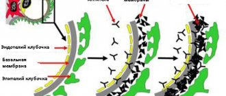
Pathogenesis of Dunbar syndrome
The development mechanisms are associated with impaired hemodynamics in the celiac trunk and its arteries. They are responsible for blood supply:
- duodenum;
- pancreas;
- liver;
- stomach;
- spleen;
- gallbladder;
- parts of the intestine.
Arteries have a dense network of vessels and anastomoses, so a decrease in blood flow in one unpaired vessel leads to disruption of nutrition of all gastrointestinal organs.
A short excursion into anatomy
The celiac trunk is an artery that supplies all organs of the upper abdominal cavity. It branches off from the aorta where the aortic opening of the diaphragm is located. The celiac trunk itself is a rather thick and very short vessel - only 2 cm. But three important branches go from it:
- The gastric artery supplies the stomach at its lesser curvature and the esophagus.
- Hepatic, which supplies blood to the liver and duodenum. Further, it has branches going to the gallbladder, the greater curvature of the stomach and the pancreas.
- The splenic artery, also through small vessels, gives part of the blood to the pancreas and provides the main nutrition to the spleen.
The celiac trunk forms a branched vascular network with a large number of connections - anastomoses. Stenosis (narrowing) of this artery reduces the flow of blood to the organs, causing ischemia - oxygen starvation.
Symptoms of celiac stenosis
Signs are pain, dyspeptic, neurovegetative disorders, but the main thing is pain. According to statistics, 98% of patients with KSChS complain about it. In most cases, it is felt in the epigastric region, in the navel, ilium, hypochondrium, and lower abdomen. More often the pain is constant, the person feels fullness, heaviness, and in more rare cases an attack occurs.
Pain appears or intensifies 15 minutes after eating. The intensity may depend on the volume of food. Because of the fear of pain, people often stop eating and lose weight. In addition to food, pain can be triggered by physical activity - fast running, lifting weights, staying in a bent position for a long time. Stress and clothes with a tight waistband can also cause pain.
Dyspeptic disorders are the second most common complaint in KSChS. These include:
- burping;
- bitterness in the mouth;
- nausea and vomiting;
- heartburn;
- bloating.
Symptoms of intestinal dysfunction often appear in the form of diarrhea and constipation. Neurovegetative pathologies accompany an attack of pain or occur independently. Manifest:
- heartbeat;
- difficulty breathing;
- headaches;
- shortness of breath;
- increased sweating;
- throbbing in the abdomen;
- weakness;
- sensitivity to temperature changes;
- decreased performance.
Clinical picture
The main complaint of patients with KSChS is abdominal pain. This complaint is presented by 97[4] to 100%[1] of examined patients. In most cases, pain is localized in the epigastric region (epigastric region), less often - in the umbilical region, hypochondrium, iliac regions, lower abdomen or throughout the abdomen. In some patients the pain is constant, in others it is paroxysmal.
Most often, the occurrence or intensification of pain is associated with food intake, its quantity and nature. Pain occurs 15-20 minutes after eating, subsides after 1-2 hours and depends on the amount of food taken. Fear of renewed pain often forces patients to limit the amount of pain. Sometimes patients associate the onset of an attack with eating cold, spicy, or sweet foods.
The second complaint of patients with KSChS is dyspeptic disorders. These include, first of all, a feeling of heaviness and fullness in the epigastric region, which worries almost all patients. Some authors consider it as the equivalent of pain. Dyspeptic symptoms also include belching, heartburn, nausea, vomiting, a feeling of bitterness in the mouth, etc.
The third complaint of patients with KSChS is neurovegetative disorders. They can appear independently or accompany a painful attack. These include headaches, palpitations, increased sweating, shortness of breath, difficulty breathing, a feeling of pulsation in the abdomen, poor tolerance of heat and cold, etc. Almost all patients note general weakness, fatigue and decreased performance [1].
Diagnosis of the disease
The diagnosis of KSChS is difficult to make, since the disease does not have specific symptoms, but is “masked” as other gastrointestinal pathologies. Sometimes gastritis, pancreatitis, enterocolitis, duodenitis or hypochondritis are suspected and treatment for them is begun. Positive dynamics are not achieved, symptoms do not disappear. During the initial examination, the doctor detects pain during palpation of the abdominal area, and during auscultation, records a systolic murmur in the area of the abdominal aorta. Patients with KSChS, as a rule, have an asthenic physique and pale skin.
In what situations is ultrasound examination required?
Ultrasound of the abdominal aorta is recommended in the following cases:
- Patients over 40 years of age with persistent episodes of high blood pressure;
- With a syndrome of abdominal pain of unknown origin, which can be pulsating (an alarming sign);
- In the presence of intermittent lameness and pain in both legs;
- For erectile dysfunction in men.
An ultrasound scan of the abdominal aorta will help identify the following pathological changes:
- Atherosclerotic lesions of the vascular wall;
- Aneurysm of the wall of the abdominal aorta (abnormal expansion of the vessel cavity with thinning of the wall);
- Stenotic deformities of the celiac trunk;
- Occlusion of the lumen of the vessel (as a result of the proliferation of atherosclerotic plaques, leading to narrowing or complete closure of the lumen of the artery);
- The presence of pathological bends of the aorta (pathological tortuosity);
- An aortic aneurysm that dissects (the process affects almost all layers of the vessel wall).
The physician can visualize the celiac axis by obtaining transverse sections from the epigastrium.
The procedure begins with applying a special gel to the area under study, which ensures the best contact of the ultrasound device sensor with the surface of the patient’s body. The doctor moves the sensor from the xiphoid process to the branch of the celiac trunk to the artery of the liver and spleen.
The celiac trunk has the appearance of a vessel reaching 4 cm in length, which extends from the anterior wall of the aorta at an angle. Transverse scanning involves examining the entire length of the celiac trunk, as well as the proximal part and the orifices of the common arteries of the liver and spleen.
The celiac trunk with its visceral branches have high peripheral resistance. All changes in the length and frequency of ultrasonic waves are displayed on a computer monitor in the form of a graph, which is carefully analyzed by a doctor.
To achieve the best visualization, Doppler ultrasound is performed in a darkened room. The whole procedure takes no more than half an hour.
Since clinical manifestations do not always allow us to unambiguously assume the existence of this pathology, diagnosis turns out to be complex and not always organized in a timely manner. Initially, the patient turns to a gastroenterologist, who carries out a full range of diagnostic measures to assess the condition of the gastrointestinal tract.
Since the disease is disguised as various disorders in the body, the patient is often examined for a long time by other specialists, but the cause of the pain and other symptoms is never found (unless complications already develop). Patients with Dunbar syndrome, as a rule, have many diagnoses, but not a single course of therapy has brought relief.
Among the examination data and physical tests, the doctor’s attention should be drawn to the following signs:
- pallor, sudden weight loss;
- pain on palpation of the abdomen;
- systolic murmur during auscultation of the abdomen in the projection of the celiac trunk.
Among the instrumental methods to confirm the diagnosis are:
- angiography of peritoneal vessels;
- CT angiography;
- Ultrasound of the abdominal aorta with Doppler measurements and its branches.
Angiography is the most informative method and immediately notes stenosis of the celiac trunk, as well as dilation of the vessel immediately after the site of stenosis. Since this method is invasive, in recent years it has often been replaced by CT angiography, which is more easily tolerated by patients. To differentiate the disease from other pathologies, an examination of the stomach and intestines is required, including X-ray, ultrasound of the abdominal organs and pelvis. Women will need to consult a gynecologist to rule out gynecological diseases.
Treatment for stenosis of the celiac trunk of the abdominal aorta
The main method of KSChS is surgical. For minor symptoms and hemodynamic disturbances, observational tactics are allowed. The patient should be examined 2 times a year for timely detection of disease progression. During this period, conservative treatment of concomitant diseases associated with complaints is prescribed.
Indications for surgery are: KSChS, confirmed by angiographic images, and the presence of clinical symptoms. The essence of the intervention is the removal of ligaments, neurofibrous tissue, and the legs of the diaphragm, which compress the trunk. The operation is mainly performed by laparoscopy. Its advantages:
- low degree of trauma;
- minor pain syndrome in the postoperative period;
- low risk of complications (adhesive disease, hernias);
- absence of postoperative wounds.
The laparoscopic method also has disadvantages. The surgeon is limited in movement, it is difficult for him to assess scar changes in the walls of the arteries. The area of the celiac trunk is dangerous and anatomically complex; it contains large arteries and the aorta. If these vessels are accidentally damaged, severe bleeding will occur, which is difficult to stop during laparoscopy.
Another type of operation is upper midline laparotomy (open access). During it, you can visually and palpably assess the condition of the vessels and internal organs of the abdominal cavity. Disadvantages: traumatic, long postoperative period, risk of developing postoperative hernias, adhesions. In addition, the operation is accompanied by blood loss.
Operation
Surgery for compression stenosis of the celiac trunk is necessary in almost every case, because it is simply impossible to restore normal blood supply in other ways. Surgical treatment tactics are selected based on the degree of advanced disease and its course. The general condition of the patient and the presence of other diseases of the internal organs must be taken into account. The further prognosis also depends on well-chosen treatment.
Indications for surgical treatment are:
- constant pain after eating,
- relief when refusing food,
- weight loss,
- angiographic signs of stenosis,
- absence of severe concomitant diseases and mental disorders.
Surgical intervention is aimed at releasing the celiac trunk from compression. For this, an endoscopic or abdominal version of the operation to excise the median arcuate ligament of the diaphragm can be used.
If during the examination indications are revealed for simultaneous removal of the gallbladder or removal of stones from it, then this can also be performed together with decompression. In case of severe or prolonged stenosis, dilation of the artery and installation of a vascular prosthesis or stent may be required.
Rehabilitation in the postoperative period
After laparoscopy, patients do not require a long recovery period. They are discharged after 3-4 days home for outpatient treatment. After abdominal surgery, patients remain in the hospital for up to 10 days. Doctors recommend limiting physical activity for a month and sticking to the recommended diet. To restore the functioning of organs and relieve pain, enzyme preparations (Mezim forte, Panzinorm) and antispasmodics (No-shpa) are prescribed. After 1-2 months, a repeat blood flow study is necessary to determine the effectiveness of treatment. In the future, it is repeated every six months.
Nutrition for Dunbar syndrome
Compression stenosis causes disruption of digestive function, so it is necessary to follow a diet before and after surgery. In order not to overload the gastrointestinal tract, meals should be fractional, up to 6 times a day. The last dose is 2 hours before bedtime. In case of severe pain, switch to pureed foods. They must be boiled or steamed.
During exacerbations, vegetables and fruits should not be consumed raw, but only boiled or baked. Meat and fish are allowed only low-fat varieties. Dairy products are low in fat, sour ones should be completely excluded. Alcoholic and energy drinks are strictly prohibited. It is better to drink tea with chamomile, rose hips, and mint. The duration of the diet is determined by the doctor.
Important! Before and after surgery, you need to avoid fatty, spicy, salty, fried, pickled foods, and limit the consumption of coffee and strong tea.
Consequences, preventive measures
The development of Dunbar syndrome results in hypoxia of the gastrointestinal tract. Nutrition and oxygen deficiency can manifest as gastritis, peptic ulcers, duodenitis, enteritis and colitis. If blood flow in the liver and pancreas is impaired, hepatitis and pancreatitis can develop.
Preventive measures include annual examination and proper treatment of diseases that may be the impetus for the development of acquired KSChS. There is no prevention for congenital Dunbar syndrome. Atherosclerotic changes can lead to it, therefore, it is necessary to adhere to dietary standards and monitor cholesterol levels annually.