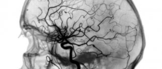
Selective cerebral angiography (SCA) is an x-ray method for studying individual areas of the cerebral circulatory network. It is based on contrasting blood vessels followed by radiography of the vascular pattern. It is a clarifying diagnosis in cases where the results of CT or MRI suggest damage to the cerebral vessels.
On a note! Selective cerebral angiography allows you to correctly diagnose, determine a treatment regimen for cerebral vessels and prescribe effective medications, determine the technique and tactics of surgical treatment for vascular pathologies.
How can you see the blood vessels of the brain?
Cerebral angiography is an X-ray method for visualizing cerebral vessels, which consists of staining the vascular bed with previously injected contrast. This is a highly effective and modern diagnostic method that allows you to make an accurate diagnosis.
The method of visualizing blood vessels using a contrast agent has been known to medicine for about a century. Back in 1927, a neurologist from Portugal began using this method, and it came to Russia in 1954. Despite such a long use, cerebral vascular angiography has changed significantly over this time, becoming more sophisticated.
The essence of the method
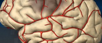
In order for the radiologist to see the vessels of the brain, an injection of an iodine-based X-ray contrast agent (Triiodtrust, Ultravist) is performed into one of the cerebral arteries. Injection is possible either into a vessel in the brain or through a catheter through an artery on the periphery, for example, the femoral one. Without this procedure, cerebral angiography of the cerebral vessels will be ineffective, since the arteries will be poorly visible on the image.
Next, two x-rays are taken, in frontal and lateral projection. After which the radiologist writes his report.
Types of cerebral angiography
There are several classifications of this type of examination. It is divided depending on the method of administration of the drug, as well as on the number of vessels that are included in the examination.
The following types of this examination are distinguished depending on the method of injection of the X-ray-containing substance:
- puncture or direct - contrast is injected directly into the brain vessel using a puncture;
- catheterization or indirect - contrast is administered using a catheter through the femoral artery.
Depending on the extent of the vessels that can be visualized, the following types of angiography are distinguished:
- general angiography - the entire vascular network of the brain is visible;
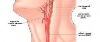
- selective cerebral angiography of the brain - one of the pools can be examined (in total, there are two blood supply pools in the brain: vertebrobasilar and carotid);
- superselective angiography - individual small-caliber vessels are visualized in one of the pools. It is used not only as a diagnostic method, but also for treatment, in which immediately after visualizing the location of the thrombus or embolus in the vessel, it is removed.
Indications
A doctor's referral is required to examine the brain using cerebral angiography. This diagnostic method is not carried out only at the request of the patient.
The main indications are:
- suspicion of the presence of a cerebral aneurysm (sac-like bulging of the artery wall);
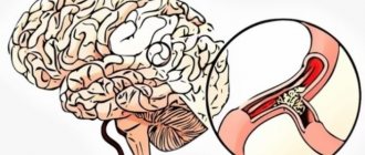
- determining the degree of narrowing of the lumen of the vessel by atherosclerotic plaques (narrowing of more than 75% significantly aggravates blood circulation in the brain and is an indication for surgical intervention);
- control of the location of clips pre-installed on the vessels;
- diagnosis of arteriovenous malformation (pathological connections between arteries and veins; usually congenital);
- suspicion of the presence of tumors, while the angiogram visualizes a change in the normal vascular pattern at the site of the tumor;
- visualization of the arteries of the brain during volumetric processes in it (tumors, cysts) in order to establish the location of the vessels relative to each other;
- suspicion of the presence of a brain angioma (a benign tumor formed by the vascular wall);
- lack of information when using other neuroimaging methods (CT, MRI), but in the presence of patient complaints and symptoms of the disease.
List of detected deviations
Since angiography of neck vessels in the form of both MRI and CT has a high degree of detection of the diagnosed picture, the likelihood of obtaining a clear name for the pathology during the study increases several times.
Diagnostics can reveal:
- tumor growth;
- consequences of serious head and neck injuries; hematomas, aneurysms;
- narrowing and dilation of blood vessels, atherosclerotic growths;
- complications caused by surgery;
- atherosclerosis, thrombosis, vascular obstruction;
- preconditions or consequences of stroke.
Atherosclerotic lesion is one of the frequently identified pathologies
Contraindications
Carrying out both indirect and direct cerebral angiography has a number of contraindications:
- Allergy to iodine and iodine-containing substances. In this condition, the contrast can be replaced with gadolinium. If there is an allergy to other contrast components, it is necessary to completely abandon this examination method.
- Kidney and liver failure in the stage of decompensation. These conditions lead to impaired removal of contrast from the body.
- Severe chronic diseases.
- Acute inflammatory diseases, as the symptoms of infection may worsen.
- Age up to two years, as radiation impairs the growth and development of the child.
- The period of pregnancy and breastfeeding, since X-ray radiation has a detrimental effect on the fetus.
- Mental illnesses during exacerbation.
- Bleeding disorders (hemophilia, thrombocytopenic purpura), which increases the possibility of bleeding after contrast administration.
Possible consequences
Despite the risk of complications, angiography is considered an informative and safe diagnostic method.
- Extravasation of contrast agent. Extravasation is the accidental entry of an iodine-containing substance into the subcutaneous tissue surrounding the vessel. This can happen when the vein wall is damaged or when the vein cannot withstand the increased pressure when contrast is administered. Extravasation of up to 10 ml of the substance is usually not accompanied by any complications, but if its amount exceeds 10 ml, an inflammatory process in the fatty tissue may develop.
- Allergy to iodine. In most cases, an allergic reaction to iodine occurs spontaneously (anaphylactic type reaction). Itching, redness of the skin and swelling of the tissues appear at the site of drug administration, then suffocation, weakness, and a drop in blood pressure occur, which can result in anaphylactic shock. Therefore, if such symptoms appear, the procedure is immediately stopped, after which the person is provided with emergency assistance. Currently, a new generation of contrast agents without iodine has appeared, due to which the list of contraindications for angiography has decreased.
- Acute renal failure. Before angiography, a kidney examination is required, since the contrast agent is eliminated from the body through the kidneys. In the presence of renal pathology, the introduction of a large volume of the substance can lead to rapid progression of kidney dysfunction, up to acute failure and the need for dialysis (hardware purification of the blood from metabolic products).
Despite some disadvantages, angiography is a very informative diagnostic tool and is considered a relatively safe technique. According to statistics, any complications are observed only in 5% of cases. When performing non-invasive angiography, all of the above risks are absent.
Preparing for the examination
Since the examination method is X-ray with the introduction of a contrast agent, you need to carefully prepare for cerebral angiography. Preparation includes the following steps:
- A maximum of 5 days before the examination, take a general blood and urine test (to determine the condition of the kidneys and exclude the presence of infectious diseases), a coagulogram (to determine the coagulation function of the blood).
- Do electrocardiography and phonocardiography (to exclude heart diseases).
- Do not drink alcohol for at least two weeks before the examination.
- Do not take medications that affect blood clotting for at least a week before angiography.
- 1-2 days before the examination, perform an allergy test with contrast, which is carried out by administering 0.1 ml of the drug to the patient and further observing the reaction on the skin. If redness, rash, and itching do not appear on the skin, then the test is negative and angiography is possible.
- Do not eat anything 8 hours before the examination and do not drink anything during the last 4 hours.
- It is possible to take tranquilizers or herbal sedatives if there is significant anxiety. However, it should be remembered that these medications can only be taken as prescribed by a doctor!
- If necessary, shave the contrast injection site.
- Remove all jewelry and other metal objects before angiography.
- Immediately before undergoing the examination, medical personnel must explain to the patient the methodology, goals and possible risks of this examination method.
What should the patient do?
Before talking about how the study is carried out and what it is, cerebral angiography of the cerebral vessels, it is necessary to consider the issue of properly preparing the patient for the examination. The attending physician must provide the following preparatory steps:
- Conducting a diagnostic examination, including a general and biochemical blood test, fluorographic examination of the lungs, electrocardiography, analysis of the blood coagulation system.
- Consultations with a general practitioner and anesthesiologist.
- Carrying out a test for sensitivity to iodine-containing medications. This test is carried out by intravenous administration of 1 ml of contrast agent, followed by assessment of the patient’s condition and identification of clinical symptoms of an allergic reaction.
- Conversation with the patient about the upcoming study.
Before performing an angiography, the attending physician should discuss the nuances of this study with the patient, as well as explain to him the necessary actions after the procedure.
In addition to the doctor’s actions, the patient is recommended to follow the following tips:
- the last meal should be taken no later than 10-12 hours before the study;
- During the procedure, it is necessary to remove various earrings, rings, dentures, etc.
Following these recommendations allows you to ensure high safety of the procedure and prevent the development of negative consequences.
Methodology
Before conducting the examination, the doctor must obtain written consent from the patient. After placing a catheter in a peripheral vein, necessary for immediate administration of drugs, the patient is premedicated. He is administered painkillers and a tranquilizer to achieve maximum patient comfort and relieve pain. The patient is connected to special devices to monitor his vital functions (oxygen concentration in the blood, pressure, heart rate).
Next, the skin is treated with an antiseptic to prevent infection, and contrast is injected into the carotid or vertebral arteries during direct angiography, and into the femoral artery during indirect angiography. If indirect angiography is performed, a catheter is also inserted into the femoral artery, which is pushed through the vessels into the desired artery in the brain. This procedure is completely painless, since the inner vascular wall has no receptors. The movement of the catheter is monitored using fluoroscopy. Most often, indirect angiography is performed.
When the catheter has reached the required location, a contrast volume of 9-10 ml is injected into it, having previously been warmed to body temperature. Sometimes, a few minutes after the administration of contrast, the patient is bothered by a feeling of heat and the appearance of an unpleasant metallic taste in the mouth. But these feelings quickly pass.
After the contrast is administered, two x-rays of the brain are taken - in the lateral and direct projections. The images are assessed by a radiologist. If there are still uncertainties, it is possible to reintroduce contrast and take two more photographs.
At the end, the catheter is removed, a sterile bandage is applied to the insertion site, and the patient is monitored for 24 hours.
Varieties
Depending on the site of administration of the contrast agent, angiography can be:
- general - a contrast agent is administered by catheterization into the abdominal or thoracic aorta;
- selective - the drug is injected directly into the arteries of the brain;
- superselective - the branches of the main arteries of the brain are contrasted.
In addition, there are different visualization methods:
- Classical angiography is the oldest method using conventional radiography, which today is used less and less. In classical angiography, under local anesthesia, a puncture of the carotid artery is performed, into which a contrast agent in a volume of 10-12 ml is injected, heated to body temperature. Then x-rays are taken in two projections with an interval of 1-2 seconds, which allows you to evaluate the different phases of cerebral blood flow.
- CT angiography is one of the modern methods for studying cerebral blood supply. In this case, a contrast agent in a volume of about 100 ml is injected through a catheter into a vein in the elbow. After this, images of the brain are taken in several sections, then a computer program reconstructs a three-dimensional image with visualization of the vascular bed.
- MT angiography - with this technique, instead of X-rays, the properties of a magnetic field are used. The state of blood vessels and circulatory phases are studied by tracking energy changes in tissues. Magnetic resonance angiography can be performed with or without a contrast agent, with the second option being used more often.
Each technique has its own advantages and disadvantages. The required examination option is selected by the doctor taking into account individual indications.
Characteristics of types of angiography (table)
| Advantages | Flaws | Contraindications | |
| Classical |
|
|
|
| Computer |
|
|
|
| Magnetic resonance |
|
|
|
Possible complications
Adverse reactions and complications during cerebral angiography of cerebral vessels do not occur often, up to 3% of cases. However, such reactions may occur, and the patient must be informed about them. Among the main possible complications are the following conditions:
- allergic reactions: from mild - redness of the skin, itching, rashes, to severe - Quincke's edema and anaphylactic shock;
- development of cerebral stroke due to arterial spasm;
- seizure attack;
- bleeding at the puncture site;
- penetration of contrast into the soft tissue surrounding the vessel, which can lead to inflammation;
- nausea and vomiting.
Features of CT angiography
Since the angiography method has been used for more than a century, it is constantly being improved. A more modern and high-quality method for visualizing cerebral vessels is cerebral CT angiography. Although in general the survey method is similar to the traditional one, there are some peculiarities:
- It is carried out not using an X-ray machine, but using a tomograph. Also based on the passage of X-rays through the human body, it takes a large number of images at once, layer by layer, which makes it possible to more accurately visualize the vessels and surrounding tissues.
- The image turns out to be three-dimensional, which allows you to view the vessel from all sides.
- The contrast injection is given into a vein, not an artery.
- There is no need to keep the patient under observation after the procedure.
CT angiography is a more effective and safe method of vascular imaging.
Principles for interpreting results
If the angiographic examination procedure itself is performed by an angiographist, then the angiogram is transferred to a neurosurgeon, angiosurgeon or phlebologist for interpretation.
Images of the vascular system obtained using angiography make it possible to detect pathologies in the form of tumors, cysts, microstrokes and others.
The specialist evaluates the picture that depicts arteries and veins. The presence of smooth outlines and uniform narrowing of the lumens, the absence of neoplasms, signs of bleeding and fluid accumulation is considered normal.
The interpretation is based on the fact that the volume of radiation transmitted by tissues is determined by their density. The amount of radiation transmitted gives different colors in the pictures. The densest bone material will appear white.
The cerebrospinal fluid and blood vessels are shown in black. The brain matter is depicted in shades of gray of varying degrees of intensity. Based on an analysis of the size, structure, and location of the vessels, their condition is assessed and possible pathologies are identified.
If the vessels are displaced on an angiogram, the cause may be a tumor, brain swelling, or poor flow of cerebrospinal fluid. Studying the network of vessels feeding the tumor allows us to clarify its location and assess the admissibility of surgical intervention.
With an aneurysm, the wall of the vessel bulges or widens. The image makes it possible to measure its parameters. The presence of atherosclerotic plaques, malformations, and vessel spasm can be determined by changes in the diameter of the vessel or its lumen.
Features of MR angiography
MR angiography is even more informative than CT. It allows you to see soft tissues that are difficult to visualize on CT. It is carried out using a magnetic resonance imaging scanner and is not an x-ray method, unlike other angiography methods. This avoids exposure to radiation.
Another advantage is good visualization even without the use of contrast, which is why MR angiography without contrast can be used for allergy sufferers.
The main contraindication for use is the presence of any metal objects in the body (artificial pacemakers, prostheses, implants, metal clips on blood vessels).
Perhaps selective cerebral angiography of the brain has already become commonplace and routine for doctors. It may be inferior in effectiveness to CT and MRI angiography. However, being more affordable and not requiring special high-tech equipment, it is still actively used 100 years later for diagnosing brain diseases.