The study of cerebral structures is a fundamental task within the framework of a general assessment of health status. The event is especially important if there are suspicions of pathological processes in the nervous tissues. Or when symptoms of a disorder are present.
There are five main methods to check the blood vessels of the brain, a short list looks like this:
- Ultrasound of cerebral structures. Used to investigate functional and organic disorders. A kind of gold standard in diagnosing vascular disorders.
- Electroencephalography. It allows us to identify functional disorders of the entire brain. Arteries, veins and small structures are not visualized. But information arrives indirectly. The technique is usually not used as the only one. Much more often - in the system.
- Rheoencephalography. It resembles the previous diagnostic method, but there is one feature. During this procedure, doctors pass a low-intensity electrical impulse through the structures of the brain. The vessels react to the pathogen.
- MRI of the central nervous system. Used for fine tissue imaging. Including blood vessels. As a rule, it is assigned in a system with other activities.
- Finally, angiography is used. It's essentially an X-ray, only targeted. Directed for special diagnostics of the condition of cerebral vessels. There are several modifications to this procedure.
When and how are such studies carried out? The main thing is what can be revealed during diagnostics?
General indications
There are many reasons for special events. It is worth highlighting a small list of situations when you cannot do without the described techniques.
Headache
Generalized indication. In more detail, intense or extremely strong discomfort. Or another option is long-term, persistent pain. Moreover, if the severity of the manifestation increases over time.
In these cases, a variety of diseases and conditions are possible. From a banal spasm due to overexertion to complete destruction of blood vessels or progression of an aneurysm. The latter are deadly problems. Therefore, there is no point in delaying diagnosis.
Flickering before the eyes
Photopsias. The simplest hallucinations in which a person sees flashes, flying flies and other “artifacts” of the image. As a rule, in this case the blood supply to the occipital lobe is disrupted.
The classic process is called vertebrobasilar insufficiency. It is caused by damage to the vertebral arteries. That is, in addition to the main study, it would be nice to evaluate the condition of the neck vessels. This is a slightly more labor-intensive task, but it does not present any great difficulties.
Dizziness
An examination of the cerebral vessels is carried out in order to identify possible problems with trophism of the hemispheres (nutrition). Impaired gait, constant sensation of the image rotating before the eyes. All these are frequent signs of processes such as encephalopathy and hydrocephalus.
Using the methods described above, you can quickly identify the nature of the disorder and prescribe treatment.
Disorders of higher nervous activity
There are a huge number of options here.
- Problems with behavior and mood. Usually indicates damage to the frontal lobes.
- Epileptic seizures, hallucinations - objective and mental, voices in the head, other verbal hallucinosis. Occurs with damage to the temporal lobes.
- Vision problems - the occipital lobe suffers.
- Coordination disorders - cerebellum, extrapyramidal system.
Diagnostics will allow us to put an end to the issue of the origin of the disease.
Jumps in blood and intracranial pressure
It is also necessary to check the vessels of the head in case of persistent hyper- or hypotension; it may well be that the primary link of such a pathological process concerns the brain, and not the hormonal background (endocrine status). Doctors can come to this conclusion only after a complete diagnosis.
Convulsions
Occurs when different lobes of the brain are affected. Especially often - temporal, frontal. Tonic-clonic seizures are possible, as in epilepsy.
In this case, you need to carefully examine the patient. To exclude other neurological diseases. Not associated with circulatory disorders.
Fainting
A business card of insufficient blood supply to nerve structures. There are both full-fledged syncope episodes and conditions preceding this. With classic symptoms: a burning sensation in the head, the unreality of what is happening, insufficient perception of sound, etc.
Fainting is almost guaranteed to indicate brain pathology. At a minimum, this is a reaction to weather conditions.
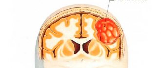
Suspicions of neoplastic processes in cerebral structures
In other words, tumors. Exactly what type can be found out only if you perform an MRI, and then a biopsy and histological examination of the tissue. However, initial measures allow one to suspect something is wrong. Findings like these are invaluable because diagnostics make it possible to detect neoplasia in its early stages.
Attention:
Unfortunately, not all tumors of nerve tissue depend on the timing of detection. Some are not treated from the very beginning.
Paresis and paralysis
Arms, legs, on one or both sides at once (which happens extremely rarely). Such manifestations occur with damage to the frontal lobes and less often than other structures of the central nervous system. It makes sense to check the cerebral tissue inside and out.
Declining intelligence
The study of cerebral vessels is necessary in cases of impaired cognitive and monistic functions.
Especially if the person before the onset of the pathological process had high or more than average intelligence. The reason is most likely a violation of cerebral blood flow.
The list is incomplete. The specialist can prescribe activities at his own discretion and discretion. It all depends on the case.
Angiography of cerebral vessels
Angiography is the most accurate and most informative examination of cerebral vessels, accompanied by the administration of a contrast agent. It will allow you to learn about the functional state, characteristics of blood flow, changes in vascular patency, and localization of the disorder.
This study checks for the following pathologies:
- aneurysm;
- arteriovenous malformation;
- vascular stenosis;
- blockage of blood vessels;
- neoplasms.
Angiography is also performed in the postoperative period to control the location of clips installed on the vessels.
There are 3 methods of angiography: classical, CT angiography, magnetic resonance imaging.
Classical and CT angiography is not performed in cases of increased sensitivity to contrast agents and anesthetics, impaired hemostasis, pathology of internal organs, acute infectious diseases, exacerbation of mental disorders, changes in thyroid function, pregnancy, lactation.
Classical (X-ray angiography)
The procedure examines the aorta, large vessels (carotid arteries), and small arteries.
Preparation includes giving up alcohol 2 weeks in advance. It is forbidden to buy or drink 4 hours before the procedure. In some cases, hydration is given to facilitate the removal of the contrast agent from the body. They do fluorography and electrocardiogram. It is possible to prescribe antihistamines.
During the procedure, the patient is placed on a special table, fixed, and connected to a cardiac monitor. The femoral (possibly carotid or vertebral) artery is punctured, a catheter is installed into it, through which a contrast agent (mainly iodine) is injected, and an examination is carried out. X-ray television records everything that happens in the vessel. Upon completion of the study, a bandage is applied to the puncture site for a day. In the absence of contraindications, it is recommended to drink as much water as possible.
Duration: 60-180 minutes, performed only in a hospital. The advantage is the accuracy of the results. Disadvantages: invasiveness and radiation exposure.
CT angiography
This study is a type of computed tomography of the brain, but to obtain a clearer picture, a contrast agent is injected through a catheter into the cubital vein. It is performed on an outpatient basis using a computed tomograph. A week before the procedure, a creatinine test is taken.
The tomograph takes images layer by layer with a distance between layers of about 1 mm in different projections. As a result, the doctor receives many images, which are later combined into three-dimensional images of the structure of the blood vessels. The study allows you to combine images of arterial and venous blood flow.
Advantages of CT angiography: absence of shadows, high information content. Disadvantages: invasiveness and x-ray exposure.
An additional contraindication is third degree obesity.
Magnetic resonance
This type of angiography is a type of MRI. Magnetic fields are used instead of X-rays. With 4D angiography, not only a three-dimensional image is obtained, but also the dynamics of blood flow are visualized. In rare cases, a contrast agent may be administered.
After the patient has taken off his jewelry and glasses, he is placed on the table and moved into a closed capsule. It is important to ensure immobility, so if the examination is performed on young children, they are given light anesthesia.
Duration: 30 minutes. The procedure is performed on an outpatient basis.
Advantages: no radiation, accuracy of information, possibility of carrying out in case of sensitivity to iodine-containing substances.
Contraindications include installed pacemakers and metal-containing implants, heart failure, pregnancy, mental disorders, third-degree obesity, claustrophobia.
Ultrasound
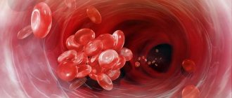
A standard, routine study, it is prescribed in most of the described cases. The method is based on the ability of the tissues of arteries, veins, and capillaries to reflect a high-frequency signal. The device converts these answers into a picture.
In this way, the doctor can assess the condition of the cerebral structures, the speed of blood flow and its quality.
There are several other indications for which an ultrasound scan is needed:
- Osteochondrosis. Degenerative-dystrophic disorder. Accompanied by gradual compression of the vertebral arteries. Changes in the vertebrae themselves. The condition progresses gradually, but inevitably. Therefore, it is necessary to systematically check the vessels. If necessary, medications are prescribed to speed up blood flow.
- Suffered a stroke. Dangerous emergency condition. Accompanied by severe disorders of higher nervous activity. It makes sense to conduct Doppler ultrasound and standard ultrasound to assess the condition of the blood vessels and the degree of brain damage. And then - the speed and quality of recovery. Already during the rehabilitation period.
- Diabetes. Sugar. It primarily affects blood vessels. Coronary, which nourish the heart, retina, kidneys, and also the brain. These are the main target organs. Diagnostics are carried out in the system. The study of the vessels of cerebral structures is just part of the whole.
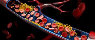
- Suspicion of atherosclerosis. Blockage of arteries with cholesterol plaques. The condition carries enormous danger. If you overlook the disorder in the early stages, a stroke will occur. At least before this, encephalopathy will occur.
The condition should not be neglected also because at an early stage the plaque can be removed with drugs. At a later stage, only surgery will be the solution.
Read more about cerebral atherosclerosis in this article.
- Hypertension. In this condition, blood pressure systematically increases. The task is to check how suitable the vessels are for work. Their functional state, if necessary, is restored with medication. Drugs are prescribed.
Modifications of simple ultrasound are also used. For example, Doppler ultrasound (USDG). This method will allow you to clearly visualize tissues and vessels. Shows blood flow speed.
Functional tests are carried out. The patient is asked to turn his head, bend his neck, etc. The nature of the response of the vessels indicates a particular condition. For example, about vertebral artery syndrome, etc.
It makes sense to conduct a study of the vessels of the brain and neck.
Features of EEG in children
It is not easy to conduct an EEG of the brain in a small child. He is frightened by a large number of wires, a strange cap, unfamiliar surroundings, people, and devices. It is quite difficult to convince your baby that he will have to lie still for some time. Young children are examined during sleep. Before the study, they need to limit the rest time so that he is tired and wants to sleep before the study. On the day of the examination, babies are woken up 4-6 hours before the usual wake-up time. Children of primary school age - 6-8 hours in advance, and children over 12 years old are not allowed to fall asleep at all at night.
You can wear the cap while inventing stories about knights rushing into a campaign with monsters. You can practice beforehand by wearing pool caps or real helmets.
Be sure to wash your hair before the procedure. For girls to wear their hair down, let them imagine that today she is a mysterious princess, waiting for a brave knight to save her from the clutches of a monstrous dragon.
A couple of hours before the examination, feed the child, and when leaving the house, do not forget to take a toy, book or treats with you so that you have something to do while waiting.
EEG can be performed on children at home, in a hospital or clinic. The best way is to spend it at home in the evening before going to bed - at 8-9 pm. A familiar atmosphere will calm the baby and allow you to get more accurate results.
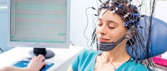
Encephalography
Also called EEG. Another routine activity for diagnosing cerebral vessels.
A special apparatus is used. It is capable of recording even the weakest electrical impulses of a few hertz. Maximum - 100 Hz.
The research is simple. A special corset equipped with sensors is put on the head. Then turn on and configure the device.
The device takes readings and displays them in the research protocol. Everything takes about 10-15 minutes. The patient does not experience any problems, discomfort or unpleasant sensations.
Attention:
The technique practically does not give direct answers. But you can suspect something is wrong by the intensity of the rhythm.
As a rule, standard encephalography is carried out in a system with other measures. EEG copes well with the functional picture of the disorder. That is, in order to determine how actively the brain itself works.
This is important, for example, after a stroke or long-term encephalopathy.
In addition to general indications, there are also specific ones for EEG:
- Epilepsy. Classic neurological diagnosis. As a rule, it has nothing to do with vessels. The brain produces too powerful signals. So the attack begins. Electroencephalography clearly sees such foci. Areas of increased activity.
- TBI. For example, concussions and others. EEG is used to exclude functional disorders.
- Stroke. Acute circulatory disorder in the central nervous system.
- Inflammatory processes. For example, meningitis or encephalitis. Characterized by high aggressiveness. If they are not treated promptly, the diagnoses lead to brain damage. Impairment of intelligence and other functions, features of higher nervous activity.
The technique is simple, cheap and accessible to most patients.
How to prepare
You need to prepare for the examination. Tell your doctor if you are taking certain medications. Some of them affect brain activity and should be discontinued 3-4 days before the examination. These medications include anticonvulsants and tranquilizers.
On the eve of the EEG and on the day of the test, you should not consume caffeine-containing products and energy drinks: coffee, tea, chocolate, energy drinks. You can't drink alcohol. These foods have a stimulating effect on the brain, and the brain's encephalogram will be distorted.
It is advisable to eat a few hours before the examination.
It is recommended to wash your hair, but do not apply hairspray, styling foam, or other cosmetics. The fats and other components they contain can worsen the contact of the electrodes with the scalp. Braids and dreadlocks will have to be unbraided, and earrings and jewelry removed.
During the procedure, you need to remain calm and not be nervous. Nothing bad happens, and the procedure is completely harmless.
Rheoencephalography
Similar research in nature. But there is a serious difference. If during an EEG, sensors take readings of the electrical activity of the brain itself, during rheoencephalography, doctors stimulate the work of the central nervous system. Using small current pulses.
It is safe and does not pose a health risk. The patient does not experience any discomfort. Based on the results of the examination, we can talk about something specific.
Rheoencephalography is completed within 10-15 minutes, like a standard EEG. It is impossible to say which method is more accurate and effective. Because they are used to register different values.
As in previous cases, there are additional indications for the procedure:
- Vascular diseases. Any. Be it aneurysms or malformations. Of course, doctors do not receive visual data, but based on the results of a functional study and the rhythms of the brain, a lot can be said. This is what doctors do.
- Evaluation of the effectiveness of the treatment. Usually conservative. Medication. In general, the same indication can be applied to other methods. But this one is the most effective in terms of results and accuracy.
- Atherosclerosis. As already mentioned, blockage of the arteries of cerebral structures with plaques. The greater the layering, the worse the situation is on rheoencephalography. The signal is poor. Therefore, the stimulation of cerebral structures is insufficient.
EEG and this technique are used in combination. This way doctors can describe the full picture of what is happening.
Magnetic resonance imaging
Magnetic resonance imaging, or MRI, allows you to fully diagnose the location and nature of lesions, as well as accurately determine the direction of blood in different parts of the brain.
In addition, this examination of the brain using magnetic resonance imaging brings quite good results in the case of vascular diseases.
For example, if a person gets sick with cerebral atherosclerosis. In addition, magnetic resonance imaging is of great help in the case of a critical period of stroke, since this particular procedure is capable of detecting thromboembolic infarction, as well as ischemic patterns of blood circulation.
MRI
Tomography is the most advanced imaging method. The device takes layer-by-layer images and then combines them. The result is a clear picture.
Attention:
Unlike CT, it does not create radiation exposure and is ideal for visualizing soft tissues, which include blood vessels and the brain itself.
If necessary, contrast-enhanced MRI is prescribed. A drug based on gadolinium is used. But for studying the vessels themselves, this is not necessary, since blood as such is a powerful natural contrast.
MRI is prescribed if other methods have failed. Or they are not informative enough.
There are special indications for this procedure:
- Suspicion of a tumor. Vascular disorders may be the result of compression by the formation. Gold standard for diagnosing neoplastic processes. Basically, the technique is prescribed for probable cancerous structures.
- A contrast study shows the size and shape of the tumor. Its exact location and degree of blood circulation. Conclusions can be given regarding the rate of cell division. Proliferative activity of the tumor. A clear picture is emerging.
Unfortunately, a biopsy must be performed to confirm the result. This is usually done directly during the operation. A tissue sample is collected and sent for histological evaluation. The laboratory gives the answer.
- Possible vascular formations. Two types: Malformations and aneurysms. Arterial wall protrusions. Both conditions are extremely dangerous and without treatment are fraught with complications. Up to massive bleeding and brain death. Accordingly, the death of the patient himself.
- Developmental defects. Congenital or acquired. In addition to malformations. For example, an open circle of Willis. And other anomalies. They are not always dangerous, but to establish this, one cannot do without research.
MRI is an indispensable method.
Contraindications
Electroencephalography has no contraindications, but in young children it is best performed during sleep. In case of exacerbation of mental illness, you need to wait for the condition to normalize. The study should be carried out with caution in people with injuries and wounds to the head.
Electroencephalography is a safe, informative research method that allows you to reliably diagnose some brain diseases. An EEG of the brain shows what underlies loss of consciousness, fatigue, insomnia, and headaches. It is used in children and adults in any condition.