Features of phlebectasis of the jugular vein
This is a congenital malformation that develops in approximately 1 in 10 thousand children. It begins to appear at the age of 2 - 5 years. When straining, coughing, or crying, there is a noticeable bulge in his neck. It is caused by the accumulation of blood and stretching of the weakened wall of the jugular vein. This weakening is associated with impaired development of the vein in the embryonic period.
1- internal; 2- external jugular veins; 3- common carotid artery
There are pathologies of the internal and external jugular (jugular) veins. Internal - a wide vessel that collects blood from the internal parts of the skull. The outer one is thinner; venous vessels flow into it from the outer surface of the head. There is also an anterior vein, which is a collector for venous blood from the neck and sublingual region. All these vessels are paired; they flow into the subclavian veins.
All veins are equipped with developed valves that prevent blood from flowing in the opposite direction. This is possible when the pressure in the chest cavity increases, when venous blood normally flows back to the head in small quantities. When a child screams or cries, the neck veins or vessels on the surface of his head may swell. This happens symmetrically.
With congenital weakness of one of the valves, blood flows into the affected vein more intensely, and then with tension it is clear that its increase is much greater on one side. This symptom is the main sign of phlebectasia.
We recommend reading the article about subclavian thrombosis. From it you will learn about the reasons for the development of subclavian thrombosis, symptoms of a problem in the artery in the acute form of venous blockage, as well as methods of diagnosis and treatment. And here is more information about thrombophlebitis of the veins of the face and neck.
Ectasia on the right side, what is it?
The external jugular artery is smaller and its purpose is to drain blood from the outside of the neck and head. Catheters are inserted into this vessel to administer medications. The trunk of the transverse veins of the neck flows into the external one, connecting with the suprascapular vein. The anterior jugular vein is one of the smallest among them. Its origin is located in the chin area.
The internal vein drains most of the blood from the head. It has a diameter from 11 to 21 mm. The diagram of its location and tributaries is as follows.
Having its origin at the cranial jugular foramen, it goes down, forming the sigmoid sinus, and further to the clavicle. Near the place where the subclavian vein joins it, which is formed by the fusion of the external vessel with the axillary one.
The internal vein has a thickening called the inferior dilatation, above which the valves are located.
With connections in the suprasternal interaponeurotic space, the anterior veins form the jugular venous arch. Intracranial tributaries are sinuses of the dura mater into which veins leading to the brain flow.
They are venous collectors. The sinus connects with the trunks and with the venous plexuses.
An important transverse sinus is located in the groove of the occipital bone, in the area of the plexus of the occipital vascular trunk with other vessels.
In the space between the pterygoid muscles and the branch of the lower jaw there is the pterygoid venous plexus. From here, blood flows through a network of large vessels, to which the anastomoses of the facial vein connect.
The superior thyroid vein passes near the artery of the same name and reaches the facial and internal jugular venous trunks. The lingual veins are the dorsal and deep veins of the tongue. At the large horn of the hyoid bone they merge into one trunk of the lingual vein.
Jugular is characterized by the presence of a developed anastomosis.
Vascular trunks are critically necessary for the functioning of the human body. The functions are:
- The removal of blood saturated with carbon dioxide and other waste products from the brain towards the heart.
- Formation of blood circulation in the brain area.
When screaming, stressing, or crying, all people, from babies to adults, can swell blood vessels, often on the right. This is the norm, although it often worries new parents. Problems with blood vessels often occur in old age, but in the presence of congenital defects they can appear at a young age. Changes include:
- Thrombosis.
- Vessel dilation.
- Consequences of inflammation (phlebitis).
- Congenital defects, dilatation.
Phlebectasia
Due to the deep location of the vessel inside, it is difficult to distinguish ectasia. Violations of the vascular trunk are visible to the naked eye from the outside. Phlebectasia of the right internal jugular vein is common. It can be almost invisible. There may be unpleasant sensations in the neck, especially strong when screaming. Severe ectasia can change the voice and make breathing difficult.
Among the main causes of the disease:
- Trauma, bruise.
- Passive lifestyle.
- Problems with valves.
- Heart diseases.
- Leukemia.
- Neoplasms.
- Abnormal functioning of the endocrine system.
The cause of the disease is often an inflammatory process in the middle ear and tissues of the mastoid process. If a blood clot becomes infected, its particles can spread throughout the body along with the infection.
With thrombophlebitis, the patient feels pain, swelling, swelling occurs, accompanied by symptoms of intoxication. The spread of infection may be accompanied by tachycardia, rash, fever, and shortness of breath.
The cause of phlebitis may be:
- injury or bruise;
- infection;
- distribution of the drug in the tissues around the vessel.
- Problems with blood clotting.
- Consequences of operations, installation of catheters.
- Neoplasms.
- Long period of immobility.
- Use of hormones.
- Pathologies of internal organs, inflammation and infections.
Methods of treating pathologies
It is an antipyretic, analgesic and anti-inflammatory drug. Used after surgery or injury to relieve pain and swelling. There are contraindications: individual sensitivity to the components of the drug.
Lowers temperature, relieves inflammation, has an analgesic effect. Ibuprofen cannot become addictive; it does not have a depressant effect on the central nervous system.
Jugular vein catheterization
- there are no other methods of administering medications into peripheral vessels;
- infusion therapy is coming;
- examinations are required;
- detoxification.
Photo of the jugular vein in the neck
The information presented in the article is for informational purposes only. The materials in the article do not encourage self-treatment. Only a qualified doctor can make a diagnosis and make recommendations for treatment based on the individual characteristics of a particular patient.
https://sovets.net/13872-yaremnaya-vena.html
Jugular vein and its diseases
The jugular veins belong to the superior vena cava system and are responsible for the outflow of blood from the head and neck. Another name for them is jugular. These are three paired vessels: internal, external, anterior.
A little anatomy
The internal jugular vein has intracranial and extracranial tributaries. Intracranial - these are the sinuses of the dura mater with the veins of the brain, orbits, hearing organs, and skull bones flowing into them.
Extracranial veins are vessels of the face and the outer surface of the skull that flow into the internal jugular along its course.
The extracranial and intracranial veins are connected to each other by ligaments that pass through special cranial foramina.
The internal jugular vein is the main line that drains blood saturated with carbon dioxide from the head. This vein, due to its convenient location, is used in medical practice to place catheters to administer medications.
The second most important is external. It runs under the subcutaneous tissue along the front of the neck and collects blood from the outer parts of the neck and head. It is located close to the surface and is easily palpable, especially noticeable when singing, coughing, screaming.
The smallest of the jugular veins is the anterior jugular, formed by the superficial vessels of the chin. It runs down the neck, merging with the external vein under the muscle connecting the mastoid process, sternum and clavicle.
Functions of the jugular veins
These vessels perform very important functions in the human body:
- They provide reverse blood flow after it is saturated with carbon dioxide, metabolic products and toxins from the tissues of the neck and head.
- Responsible for normal blood circulation in the cerebral sections.
Catheterization
According to doctors, puncture and catheterization of the internal jugular vein is preferable to that of the subclavian veins, due to fewer complications, such as bleeding, thrombosis, and pneumothorax.
Scheme of jugular vein catheterization
Main indications of the procedure:
Catheterization of the internal jugular vein is contraindicated if:
- history of surgery in the neck area;
- blood clotting is impaired;
- there are ulcers, wounds, infected burns.
There are several access points to the internal jugular vein: central, posterior and anterior. The most common and convenient of them is the central one.
The technique for vein puncture using central access is as follows:
- The patient is placed on his back, his head is turned to the left, his arms are along his body, the table on the side of his head is lowered by 15°.
- The position of the right carotid artery is determined. The internal jugular vein is located closer to the surface parallel to the carotid vein.
- The puncture site is treated with an antiseptic and limited with sterile napkins, lidocaine (1%) is injected into the skin and subcutaneous tissue and the search for the location of the vein begins with an intramuscular search needle.
- The course of the carotid artery is determined with the left hand and the needle is inserted 1 cm lateral to the carotid artery at an angle of 45°. Slowly advance the needle until blood appears. Inject no deeper than 3-4 cm.
- If it is possible to find a vein, the search needle is removed and a needle from the set is inserted, remembering the path, or the needle from the set is first inserted in the direction found by the search needle, then the last one is removed.
Catheter placement is usually done using the Seldinger technique. The injection technique is as follows:
Pathologies of the jugular veins
The main diseases of these veins include pathologies characteristic of all large vessels:
- phlebitis (inflammation);
- thrombosis (formation of blood clots inside vessels that obstruct blood flow;
- ectasia (expansion).
This is an inflammatory disease of the vein walls. In the case of the jugular veins, three types of phlebitis are distinguished:
There may be several causes of phlebitis of the jugular vein:
For uncomplicated phlebitis (without suppuration), local treatment is prescribed in the form of compresses and ointments (heparin, camphor, ichthyol).
Heparin ointment is used for phlebitis to prevent the formation of blood clots
Purulent phlebitis requires a different approach. In this case the following are shown:
- anti-inflammatory drugs (Diclofenac, Ibuprofen);
- medications that strengthen the walls of blood vessels (Phlebodia, Detralex);
- drugs that prevent thrombosis (Curantil, Trental).
If therapeutic methods do not bring results, the affected area of the vein is surgically excised.
Jugular vein thrombosis
With thrombosis, a blood clot forms inside the vessel, which obstructs blood flow. Jugular vein thrombosis can be congenital, acquired or mixed.
Hereditary risk factors include:
- special structure of veins;
- antithrombin-3 deficiency;
- blood clotting disorder;
- lack of proteins C, S.
Mixed ones include an increase in the blood of some coagulation factors, fibrinogen and homocysteine.
For jugular vein thrombosis, medications and anticoagulant treatment are prescribed, and in rare cases, surgery is performed.
This is what swelling of the jugular veins looks like in patients
Among the drugs shown:
- anti-inflammatory;
- painkillers;
- phlebotonics;
- anticoagulants (Cardiomagnyl, Thrombo ACC, Warfarin, injection of heparin under the skin in acute forms).
In addition, a low-cholesterol diet is prescribed.
In some cases, thrombectomy (removal of a blood clot with excision of tissue) and thrombolysis, which involves resolving blood clots, may be required.
Congenital defects of the jugular vein
Congenital diseases include hypoplasia (underdevelopment) and jugular vein aneurysm.
Conclusion
https://serdec.ru/spravochnaya-informaciya/yaremnaya-vena-zabolevaniya
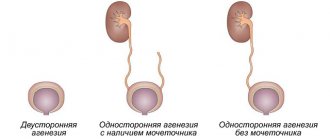
Reasons for changes in the right, left, and both veins
The cause of phlebectasia is the weakness of the connective tissue of its valves. The pathology can manifest itself in a child, but quite often it occurs in women during menopause and in the elderly. This is due to increased processes of structural change under the influence of age-related or hormonal changes. In these cases, jugular phlebectasia may occur with equal probability on either side or even bilaterally.
Dilatation of both neck veins is a sign of severe heart disease with insufficient functioning of the left ventricle. This can occur in chronic lung diseases or severe heart defects, such as mitral stenosis.
In addition to the anatomical weakness of the venous valves, the cause of the disease can be a tumor that compresses the overlying part of the vessel. In this case, it matters which side the lesion occurred on :
- right-sided jugular phlebectasia can be observed with a significant increase in the cervical lymph nodes on the right or soft tissue tumors in this area;
- Accordingly, damage to the left jugular vein should alert doctors to any pathology of the lymphatic vessels on the left.
There is no list of diseases that cause phlebectasia. In each case, the doctor examines the patient individually, identifying all the features of his body.
Causes
It should be noted that phlebectasia does not depend on the age of the patient; it can equally occur in both an adult and a child.
Causes of dilatation of the jugular vein:
- neck injuries, traumatic brain injuries, head and cervical contusions, concussions;
- spinal and back injuries, rib fractures leading to general venous stagnation;
- long forced, uncomfortable posture, sedentary work without a break;
- vascular diseases, heart failure, heart defects, coronary and hypertension;
- benign and malignant tumors of internal organs, blood cancer;
- diseases of the spine and back muscles, in which the patient takes a forced position to alleviate the condition, for example, osteochondrosis;
- endocrine diseases.
Often, with the development of dilatation of the jugular vein, there are several factors that cause the disease.
Symptoms of the disease
In boys, the pathology occurs 3 times more often than in girls . Often, simultaneously with the expansion of the vein, there is also its tortuosity.
Outwardly, the pathology proceeds almost unnoticeably. Typically, patients consult a doctor between the ages of 8 and 15 years with complaints of a bulge on one side of the neck, which is caused by dilation of the external jugular vein. Initially, it manifests itself only as swelling on the side of the sternocleidomastoid muscle of the neck when it is tense.
Then, as it progresses, this formation increases with crying, straining and other conditions that increase pressure in the chest cavity and impede the normal venous flow of blood through the subclavian and superior vena cava to the heart.
Disruption of the normal outflow of blood from the tissues of the head is accompanied by clinical symptoms that first appear in childhood:
- headache;
- episodes of dizziness;
- sleep disturbance;
- fast fatiguability;
- poor performance at school;
- nosebleeds of unknown origin;
- feeling of suffocation, pressure on the neck;
- nausea and vomiting;
- fainting.
The incidence of such symptoms ranges from 10 to 40% and forces the patient to consult a doctor . In other cases, if the disease is asymptomatic, a person may live his entire life and not know that he has such a vascular anomaly.
The larger the lumen of the expansion, the more often the patient is bothered by something. This is due to the volume of blood return and the development of venous stagnation in the tissues of the head.
Phlebectasia - causes, development, classification
Phlebectasia is a dangerous disease that disrupts the functioning of the entire body. It is important to identify the disease in its early stages to reduce the risk of needing subsequent surgery.
What it is
The disorder is an expansion of the vein. In this case, the condition can be either congenital or acquired. If the first is diagnosed after birth, then the second can be caused by pathologies of the vein valves, reflux of blood from deep veins to the superficial, as well as disruption of the blood circulation process caused by tumors or scars.
The disease is accompanied by disruption of the valves and dilation of the veins. Literally from Latin the name is translated as “extension”.
Causes
Having analyzed what phlebectasia is, you can draw your own conclusions about what leads to the development of pathology. Doctors call the main reasons for the development of the disease:
- disruption of normal valve operation;
- problems with blood flow;
- presence of tumor or scars.
Phlebectasia of the esophagus occurs as a result of problematic blood flow in the hepatic veins. The pathology is provoked by cirrhosis of the liver or blood clots in the organs.
Classification
Classified according to the location of the disease:
- phlebectasia of the jugular vein;
- esophagus;
- lower extremities.
By origin they are divided into:
- congenital phlebectasia;
- acquired.
Doctors distinguish stages of development based on the clinical picture.
- The first degree is detected during endoscopy by chance - the expansion does not exceed 3 millimeters, the lumens are not darkened, ectasia is not present or is small.
- The second degree is characterized by venous adjustment - strong expansion, for example, if the problem concerns the esophagus, then their volume occupies about a third of the organ cavity.
- The third stage is determined by enlargement and swelling of the veins, the presence of nodule formations, occupies more than half the volume of the esophagus, and there is a deterioration in the condition of the mucosa.
- The fourth stage threatens with thinning of the walls of the mucous membrane, and in other organs there are cluster-shaped formations and bleeding.
The prognosis is favorable in the first 3 stages. The neglected fourth can be fatal, especially phlebectasia of the IJV.
Manifestations
Pathology of the esophagus occurs at stages 1-2 asymptomatically. The patient is not bothered by pain. Sometimes there is a burning sensation in the chest and shoulder blades, and belching with a sour taste. As the disease progresses, pain symptoms are observed in the chest, esophagus, stomach, and liver.
At stage 4, vomiting of blood occurs, and purple blood clots are present in the stool. Swallowing will be impaired. Constant loss of blood leads to anemia, a person feels weak, cannot work or move normally, loses weight, feels short of breath and has a strong heartbeat.
Phlebectasia in a child is usually diagnosed as congenital. Enlarged and tortuous venous ducts are noticeable and can be localized anywhere.
It appears either from birth or in the first months of life. The color of the skin changes, the integument becomes cyanotic. Next, tumor formations appear.
Diagnostic methods
If you suspect jugular phlebectasia, you should contact a vascular surgeon who will conduct an appropriate angiological examination. To assess the severity of the process caused by impaired venous outflow, a consultation with a neurologist and ophthalmologist (fundus examination) is scheduled.
The screening method, that is, quick preliminary diagnosis, is ultrasound duplex scanning. It allows you to identify the following signs :
- location and structure of the formation, its size;
- direction of blood flow, its nature (laminar, that is, linear, or turbulent, that is, swirling);
- patency of veins, condition of their walls and valves.
Then the patient is prescribed the following research methods:
- blood tests, urine tests, ECG;
- X-ray examination of the chest and cervicothoracic spine;
- Ultrasound triplex scanning in B-mode;
- Dopplerographic determination of linear and volumetric blood flow velocity through the veins;
- X-ray contrast venography (filling the lumen of the vein with a substance that does not transmit x-rays);
- computer and magnetic resonance imaging to accurately determine all characteristics of the lesion.
According to phlebography, 4 types of disease are distinguished:
- limited circular expansion in combination with tortuosity of the vein;
- limited circular expansion;
- diffuse circular expansion;
- lateral extension, or aneurysm.
Depending on the data obtained, the surgeon plans the type of operation.
Treatment of jugular vein phlebectasis
Phlebectasia is not only a cosmetic defect. It leads to disruption of the blood supply to the brain and disrupts its functions. In the future, this condition may progress. Therefore, it is best to have surgery at 7–10 years of age.
Types of surgical interventions:
- circular resection (removal) of the extension;
- longitudinal resection;
- casing (strengthening the walls of the vessel) with a polymer mesh;
- resection of the extension with vessel plasty.
All these types of interventions are equally effective and allow you to finally restore normal blood flow. The operation is performed under general anesthesia and lasts about 2 hours. The recovery period is short. These tissues are well supplied with blood and heal quickly.
Features of the pathology
Dilatation of the jugular vein (phlebectasia) usually occurs due to disruption of the valves located along the entire length of the vein. For one reason or another, the valves are no longer able to control venous blood flow. As a result, blood begins to accumulate in large volumes in the vessel, expanding its walls and disrupting the functioning of an increasing number of valves.
The next important factor is the discharge of blood from deep veins to superficial ones. Such an unnatural redistribution of blood provokes dysfunction in the activity of the entire venous system, which also leads to the development of vasodilation.
The jugular vein includes several branches - two internal, anterior and external. These vessels play an important role in the functioning of the body - carrying blood away from the brain and neck. The location too close to the brain forces us to take any pathologies of the jugular vein with particular seriousness.
Possible complications
After surgery on the jugular veins, in the near future, 8 - 9% of patients experience stenosis or thrombosis of the vessel. Doctors are good at managing these complications. The use of modern medications can reduce the incidence of complications to a minimum.
There were no complications noted in the long-term postoperative period.
If surgery is necessary, refusing it will lead to adverse consequences.:
- prolonged headaches;
- inability to engage in intense physical activity;
- poor performance at school;
- increased severity of other symptoms;
- increase in cosmetic defect in the neck area.
A rare, but most dangerous complication is injury or rupture of a dilated venous vessel. In this case, intense bleeding occurs, requiring emergency medical attention. This condition occurs with large (up to 10 cm or more) dilations.
Even the smallest phlebectasias serve as a source of improper blood flow, so over time they can thrombose. This is dangerous because a blood clot enters the heart, and through its right ventricle into the pulmonary circulatory system. The result is such a serious and often fatal condition as pulmonary embolism.
How is the treatment carried out?
If varicose veins appear or the IJV is swollen, then therapeutic procedures must be started as soon as possible. In case of deviation, an integrated approach is required, thanks to which it will be possible to normalize blood circulation and eliminate the pathological signs of ectasia. If there is no exacerbation, then no special treatment is carried out, and the patient is constantly monitored.
The damaged vein fragment is removed through surgery if conservative therapy does not produce results.
Enlargement of the jugular vein often requires symptomatic treatment, during which drugs are prescribed that improve blood circulation and reduce pain. It is important to reduce physical activity during and after therapy, which aggravates the course of the pathology. If ectasia progresses rapidly and conservative measures are powerless, then surgical intervention is performed. During manipulation, the surgeon eliminates the problem area of the jugular vein, and connects healthy areas into a single vessel.
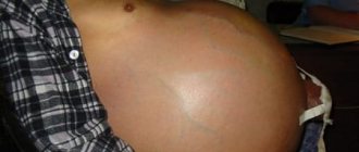
Is it possible to give birth with moderate phlebectasia?
During childbirth, the pressure in the chest cavity increases, which creates additional stress on the dilated vein. Therefore, the question of managing the birth process depends on the severity of phlebectasia.
A pregnant woman should consult a vascular surgeon.
You can give birth with this disease in any case. Depending on the severity of the pathology, natural childbirth, exclusion of the period of pushing, and anesthesia can be performed.
In case of particularly severe phlebectasis and other concomitant diseases, a cesarean section is indicated.
The issue of childbirth tactics is decided for each woman individually. If she underwent surgery for this disease in childhood, there are no restrictions for normal childbirth.
Prevention of development
Primary prevention for this disease has not been developed since it is congenital and its cause has not been established. Only general advice on bearing a child is given - healthy eating, proper rest, taking multivitamins for pregnant women.
If a child has had surgery for this disease, he then undergoes an annual ultrasound of the neck veins to ensure normal recovery.
If no surgical intervention was performed, if the size of the defect is small, it may subsequently shrink or disappear on its own. To do this, it is necessary to strengthen the neck muscles: massage and physical therapy are indicated. Situations that increase intra-abdominal and intrathoracic pressure should be avoided :
- severe prolonged cough;
- constant constipation;
- lifting weights;
- intense physical activity.
We recommend reading the article about vein catheterization. From it you will learn about the advantages and disadvantages of the method, indications and contraindications for vein catheterization, as well as the Seldinger technique, rules for caring for the catheter and how to remove the catheter from the vein. And here is more information about thrombophlebitis of the superficial veins.
Phlebectasia of the jugular veins is a congenital dilatation of the veins of the neck caused by weakness of their valves. It causes a cosmetic defect and also leads to disturbances in venous blood flow in the brain, head, and neck. The main method of treatment is surgery at the age of 7–10 years.
Preventive measures
Prevention of the disease involves the following measures:
- reducing stress on the body and the cervical spine if there is a predisposition or primary symptoms of the disease;
- timely treatment of pathologies that provoke dilatation of the jugular vein;
- systematic planned examination by a specialist;
- proper lifestyle, physical activity, balanced diet.
People who have a hereditary predisposition to this pathology need to be especially careful.
It should be understood that vein abnormalities are very difficult to prevent, but the disease can be stopped and eliminated in the early stages. For this reason, a regular visit to the hospital will avoid serious health problems in the future.
Vessels
Superior Jugular Vein Bulb In Latin: How To Treat High Location
Feb 03, 2020 Kokh V. A.
11899
Vessels
Posterior (Anterior) Trifurcation of the Right Internal Carotid Artery: What is It?
Feb 03, 2020 Kokh V. A.
40185
Vessels