Fetal circulatory system
When studying fetal circulation, several anatomical and physiological factors should be noted.
The normal adult circulation consists of a series of circuits of blood flow through the right heart, lungs, left heart, systemic circulation, and back to the right heart. The fetal circulation is a parallel system with cardiac output from the right and left ventricles directed to different vessels. For example, the right ventricle, which accounts for about 65% of combined output, pumps blood through the pulmonary artery, ductus arteriosus, and descending aorta. Only a small part of its output passes through the pulmonary circulation. The left ventricle supplies blood primarily to tissues supplied by the aortic arch (for example, the brain). The fetal circulation is a parallel circuit characterized by channels (ductus venosus, foramen ovale, ductus arteriosus) that provides the flow of more highly oxygenated blood to the upper half of the body and brain, less highly oxygenated blood to the lower half of the body, and low oxygenated blood to the non-functioning lungs.
The umbilical vein, carrying oxygenated blood (oxygen saturation reaches 80%) from the placenta to the fetal body, penetrates the portal system. Part of the umbilical-portal blood passes through the microcirculation of the liver, where oxygen is released. From there, the blood flows through the hepatic veins into the inferior vena cava. In the fetal circulation, most of the blood bypasses the liver through the ductus venosus, which directly enters the inferior vena cava, which also receives unsaturated (25%) venous blood from the lower half of the body. The blood that reaches the heart through the inferior vena cava is approximately 70% oxygenated (maximally highly oxygenated blood). About one-third of the blood returning to the heart from the inferior vena cava flows primarily through the right atrium, mixing with blood from the superior vena cava, then through the foramen ovale into the left atrium, where it mixes with the relatively small volume of venous blood from the lungs. Blood flows from the left atrium into the left ventricle, then into the ascending aorta.
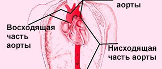
From the proximal aorta, which carries the most oxygenated blood (65%) from the heart, branches arise to supply blood to the brain and upper half of the body. Most of the blood returning through the inferior vena cava enters the right atrium, where it mixes with unsaturated blood returning through the superior vena cava (25% oxygen saturation). Blood from the right ventricle (oxygen saturation - 55%) enters the aorta through the ductus arteriosus. The descending aorta supplies the lower half of the body with blood that is less oxygenated (about 60%) than the blood coming to the brain and upper half of the body.
The role of the ductus arteriosus should be especially noted. Blood in the fetal circulation from the right ventricle enters the pulmonary trunk, most of which, due to high vascular resistance, bypasses the lungs through the ductus arteriosus and enters the descending aorta. Although the descending aorta gives branches to the lower half of the fetal body, the bulk of the blood from it flows to the umbilical arteries, which carry oxygen-free blood to the placenta.
Features of fetal blood circulation
Structure of the placenta
What distinguishes the blood circulation of a fetus from that of an adult? — A lot, and we will try to talk about these distinctive features.
- During the prenatal period, the “mother-placenta-fetus” system functions. The placenta is also called the baby's place. Through the vessels of the umbilical cord, not only nutrients and oxygen enter the fetal bloodstream, but also toxic substances, medications, hormones, etc.
- Arterial blood from the mother to the fetus is delivered through the umbilical vein, and the venous blood of the fetus, saturated with carbon dioxide and metabolic products, returns to the placenta through two umbilical arteries.
- There are three canals in the fetal circulatory system: the ductus arteriosus, the ductus venosus (Arantius) and the patent foramen ovale. This anatomy of the fetal vascular bed creates conditions for parallel blood flow, unlike in adults. Blood from the right and left ventricles enters the aorta (hereinafter referred to as the systemic circulation).
Fetal circulation - The pulmonary circulation is open, but the lungs do not perform the function of gas exchange and accept a very small amount of blood due to high resistance. This resistance is created by non-functioning respiratory structures - the alveoli. The respiratory function during this period is performed by the placenta. The pressure levels in the aorta and pulmonary artery are approximately the same.
- About half of all blood entering the fetus's body ends up in the liver. Only this organ receives the most oxygenated blood (up to 80%). All other organs receive mixed blood. More oxygenated blood supplies the liver, upper limbs and brain. Blood with less oxygen flows to the rest of the fetus's body.
- A feature of the blood circulation of the fetus is the presence of fetal hemoglobin in its blood. It differs from adult hemoglobin in that it is able to bind to oxygen much better. This circumstance is very important, since the fetus is sensitive to a lack of oxygen in the blood. Even despite the low partial pressure of oxygen in the bloodstream, fetal hemoglobin delivers this essential element of their blood to the mother as much as possible.
Symptoms of impaired blood flow
If the impaired blood flow is compensated, then the woman does not feel any abnormalities, but learns about them only after the examination.
Pronounced manifestations occur in acute form and chronic decompensation:
- the motor activity of the fetus increases sharply or completely disappears (at 28 weeks, normal development is accompanied by ten movements per day), this symptom requires immediate contact with an obstetrician-gynecologist;
- a slow increase in abdominal circumference, detected during monthly examination and measurement in the antenatal clinic (associated with excessive formation or lack of amniotic fluid);
- late toxicosis;
- high blood pressure;
- large weight gain;
- swelling in the legs;
- the appearance of protein in the urine.
Fetal circulation: diagram and description
At the end of the fifth week, the primary, or yolk, system begins to work. It consists of arteries and veins, which are called umbilical-mesenteric. This type of blood circulation refers to rudimentary forms. As the embryo develops and its systems improve, it loses its significance.
Diagram of blood circulation in the fetal body:
- From the placenta, blood enters the umbilical vein, from where it goes to the liver. Most of the blood is drained into the inferior vena cava through the ductus venosus. Before the portal of the liver, the umbilical vein merges with the portal vein, which is still underdeveloped.
- After passing through the hepatic venous system, the blood enters the inferior vena cava and mixes with the flow, which is discharged through the ductus venosus. The next point is the right atrium, where blood from the superior vena cava, collected from the entire body, enters.
- The structural features of the fetal heart do not allow the blood to mix completely. From the superior vena cava, blood flows into the right ventricle and then into the pulmonary artery. From the inferior vena cava it enters the left atrium through the foramen ovale.
- Part of the blood from the LA penetrates into the bloodstream of the lungs, which are not yet working, and is then discharged into the LA. The remaining flow through the PDA enters the descending aorta, from where it is carried throughout the lower half of the body.
- In the LA there is a mixing of two blood streams. Oxygenated blood enters the ascending aorta, from where it is delivered to the brain and the entire upper half of the body.
- Blood, which lacks oxygen, is delivered to the chorionic villi through the umbilical arteries.
Attention!
This closes the circle of fetal blood circulation. The peculiarities of the cardiac structures and fetal blood circulation provide the developing fetus with adequate nutrition and saturate its tissues with oxygen.
The structure of the circulatory system of the unborn child ensures the necessary gas exchange. This is necessary due to the lack of pulmonary respiration. The blood circulation of a fetus differs from that of an adult:
- carbon dioxide, decay products are removed from the body through the umbilical arteries (this is the shortest route);
- some of the blood passes through the small circle, but its parameters do not change;
- the main blood circulates in a large circle, which provides the oval window, arterial, venous ducts;
- a mixture of venous and arterial blood enters the fetus’s body;
- pressure in the aorta and pulmonary artery is low.
Fetal circulation: mother and child – a single system
In a growing fetus, there are two physiological shunts, i.e. places of communication between circles. Without them, fetal development would be impossible. Blood from the maternal placenta flows through the umbilical vein of the fetus into its inferior vena cava, where, mixing with its venous blood from the lower half of the body, it fills the right atrium. From here the main flow goes through the open oval window, i.e. a hole (defect) in the interatrial septum into the left atrium and left ventricle and further into the large circle. This is the first physiological, natural shunt. From the left ventricle, part of the blood goes to the aorta and vessels of the head and upper half of the body. And that part of the blood that in the right sections passed into the right ventricle through the tricuspid valve, and then into the pulmonary artery, goes into the descending aorta through the second physiological shunt - the patent ductus arteriosus: the normal connection of the pulmonary artery and the aorta.
These are natural shunts, “shunts to save” the growing fetus. Without them, the fetus is not viable, and when they close prematurely, severe congenital defects occur. In the surgery of congenital heart defects, artificial (temporary or permanent) creation of such shunts is one of the widely used treatment methods. But more on that later.
Both physiological shunts close
normally soon after birth, and then both circulation circles begin to function in the mode in which they will work for the rest of their lives. But let’s assume that, in addition to the natural communication between the systemic and pulmonary circulation at the capillary level, something else remains, for example, through a hole in the interventricular or interatrial septum, or in the form of an unclosed arterial duct.
If such a message remains, then the blood flow from any chamber appears in two ways: one is normal, i.e. provided by nature, the second is through a defect or through an open shunt. The blood will partially flow along the second path, since it is easier there, there, in a small circle, there is much less resistance. A shunt is formed “from left to right”: from a large circle to a small one.
Reset from left to right
When a certain volume of blood with each contraction deviates from the normal path and goes from the left to the right, then naturally two problems arise: a lack of blood in the large circle and an overflow of the small circle. The large circle does not suffer: complex compensation mechanisms are quickly activated. But the small circle has a harder time.
a certain amount to live, much less grow.
oxygen, which must be
constantly
, and this delivery is carried out by the heart.
At first, it copes with this, although the conditions in which it must work are far from normal. Its right chambers (depending on the level at which there is communication - atrial, ventricular or great vessels) are filled with blood, increasing in size. The lungs also become filled with blood due to the expansion of their large and small arteries. The left departments also do not remain unaffected, because they perform the work partially in vain. A “vicious circle” arises - an expression that most closely matches our situation. At the same time, the blood entering the systemic circulation is completely saturated with oxygen
, and the color of the child’s skin and mucous membranes is normal.
Right to left shunt and cyanosis
Now let's imagine the opposite situation. Venous, dark blood, which has given oxygen to the tissues, somehow, bypassing the lungs, enters the left side of the heart, the aorta and the arterial system. In other words, in a born child, blood circulates like a fetus, i.e. without a small circle and breathing lungs. But the maternal placenta is no longer there, and with it, there is no source of oxygen. If there are no open paths of communication between the circles, and venous blood is not oxidized anywhere and mixed with arterial blood, then life is impossible, and the child will not be viable. Fortunately, this happens very rarely. But, if there is a message, then through it part of the blood still gets into the small circle and into the lungs, the other part will remain undersaturated. This will be expressed in cyanosis of the skin and mucous membranes. The degree of cyanosis can be very different, as well as the time of its visible manifestation. It can be slightly noticeable or pronounced. Sometimes only others and doctors notice it. The degree of cyanosis depends on the amount of blood that passes through the lungs, and on the degree of its mixing with undersaturated blood in the cavities of the heart, i.e. on the size and level of defects in its septa, as well as on the resistance to blood flow on the way from the heart to the pulmonary arteries and alveoli. The greater this resistance, the less venous blood will enter the small circle and end up in the arteries, and the larger the defect in size, the better the blood will mix in the cavities and the less “cyanosis” will be.
After the birth of a child, the heart, as with defects with a left-to-right reset, works under overload, especially its right sections, and we will talk about this when we describe individual defects. But here we want to emphasize that the very existence of cyanosis can be dangerous, since insufficient oxygen content in arterial blood causes its thickening, an increase in the number of red blood cells and can lead to blockage of small vessels of the body, including the brain, with all the ensuing consequences.
Concept of cross dumping
In some situations, when the septal defects are large enough and the resistance to blood flow is almost the same at the exit of both ventricles, blood may partially flow through the defect in both directions during different phases of the cardiac cycle. That is, at some period of time during one contraction there is a reset from left to right, and in another period during the same cycle, but after a few fractions of a second there is a reset from right to left.
In such cases, they speak of “cross-discharge,” and the degree of oxygen desaturation of the arterial blood will depend on the predominant direction of blood flow. Accordingly, the degree of cyanosis will be visible and pronounced.
Let us say here that defects with such “cross-discharge” most often include very complex, combined defects, including combinations of different disorders of heart development.
Obstructions to blood flow
Congenital obstructions to normal blood flow usually occur due to abnormal development at the junctions of the heart chambers with each other or with the great vessels. Most often this applies to valves. The narrowing is called “stenosis” if it is caused by changes in the valves, and when it concerns the aorta, it is called “coarctation”.
We will discuss this in detail below, but here I would like to note a few points regarding blood flow. Since by the eighth week of intrauterine life of the fetus the heart is basically formed and blood circulation is already occurring, the effect of narrowing and obstruction of normal blood flow affects the early stages of embryo development. If there are no more defects, then the ventricles have to work with increased load, which will result in thickening of the walls, reduction in the size of the cavity, and underdevelopment of the heart chambers. After birth, these phenomena only progress and can become life-threatening already in the first days of a child’s life.
If such obstacles are combined with defects in the septum, then it is easier for the heart to work, because there are other paths for blood in which there is less resistance and the flow chooses such paths of less resistance.
But we have already come close to the classification of vices, i.e. to what defects there are and what happens to the child, whether the heart copes with them and how.
Quoted from the book by G. E. Falkovsky, S. M. Krupyanko. The heart of a child. A book for parents about congenital heart defects
How to get treatment at the Scientific Center named after. A.N. Bakuleva?
Online consultations
Methods for diagnosing disorders
Ultrasound examination, as well as Dopplerometry, helps to determine how serious the problems are with the blood flow and what damage the fetus has. Modern technologies make it possible to check various vessels not only of the mother, but also of the fetus.
There are certain features that indicate circulatory disorders
The doctor pays attention to them during examinations:
- the placenta becomes thinner;
- infectious diseases are present;
- condition of amniotic fluid, deviations from the norm (if any).
Using Doppler, the doctor can determine 3 stages of blood flow disorders:
- At first, minor deviations occur. Blood flow to the uterus, fetus and placenta is maintained.
- At the second stage of disorders, all blood circulation circles in the fetus are affected.
- The third stage is considered critical.
The procedure can be performed on all pregnant women, regardless of the period. This is especially true for women at risk, for whom there is a high probability of serious problems. Additionally, along with Doppler measurements, laboratory blood tests are also performed.
Features of blood circulation after birth
After the birth process, physiological changes occur in the child's body. They are necessary for his organs to begin to function independently. Tying the umbilical cord breaks the mother-placenta-fetus connection. With the first breath, the pulmonary system comes into operation. This reduces the pressure in the small circle by five times. The need for the ductus arteriosus disappears.
After the pulmonary circle starts, the synthesis of vasodilators begins - substances that promote vasodilation. There is an increase in blood pressure compared to the pressure in the lung vessels. In the first days of a child’s life, mechanisms for closing the common aortic duct and reducing bypass vessels are activated.
Fetal circulatory disorders
Often, fetal circulatory disorders begin with a pathology in the mother’s body that affects the condition of the placenta. Doctors note that placental insufficiency is observed in a quarter of pregnant women today. If the expectant mother is not attentive to herself, she may not even notice the threatening symptoms. It is dangerous that in this case the fetus may suffer from a deficiency of oxygen and other useful and vital elements. This threatens developmental delays, premature birth, and other dangerous complications.
What leads to pathology of the placenta:
- Thyroid diseases, arterial hypertension, diabetes mellitus, heart defects.
- Anemia – moderate, severe.
- Polyhydramnios, multiple pregnancy.
- Late toxicosis (preeclampsia).
- Obstetric, gynecological pathology: previous voluntary and medical abortions, malformations, uterine fibroids).
- Complications of the current pregnancy.
- Blood clotting disorder.
- Urogenital infection.
- Exhaustion of the maternal body as a consequence of lack of nutrition, weakened immunity, increased stress, smoking, alcoholism.
A woman should pay attention to
- frequency of fetal movements – changes in activity;
- belly size – does it correspond to the term;
- Pathological discharge of a bloody nature.
Placental insufficiency is diagnosed by ultrasound with Doppler measurements. In the normal course of pregnancy, it is done at 20 weeks, in case of pathology - from 16-18 weeks.
As the period of pregnancy increases during the normal course of pregnancy, the capabilities of the placenta decrease, and the fetus develops its own mechanisms for maintaining adequate vital functions. Therefore, by the time of birth, he is already ready to experience significant changes in the respiratory and circulatory system, allowing him to breathe through his lungs.
The fetal circulation occurs through the placenta, which receives 60% of the combined ventricular output, and after birth most of it is sent to the lungs.
Newborn's heart
After birth, the baby takes its first breath. This ensures the expansion of the lungs and the beginning of breathing with their help. Against this background, blood from the right ventricle rushes into the pulmonary trunk and enters the vessels of the organ. The ductus botallus begins to close and gradually becomes completely overgrown with connective tissue.
An increase in pressure in the right atrium causes blood flow through the oval window to stop. It is gradually overgrown with a muscular septum in which the conduction system of the heart is located. This reflects the end of changes in the baby's blood circulation.
Features of blood circulation during pregnancy appeared as a result of evolution. They allow the internal organs and brain of the fetus to receive sufficient oxygen and nutrients.
Any disturbances in the structure of the heart and blood vessels lead to congenital anomalies of varying severity. Moreover, if anatomical features remain after birth, this also leads to the appearance of pathologies that require treatment.
Diagram of blood flow in the heart in the fetus and after birth
This article is the first part of a series about the heart and blood circulation. Today’s material is useful not only for general development, but also for understanding what heart defects there are. For a better presentation, there are many drawings, half of them with animation.
Diagram of blood flow in the heart AFTER birth
Deoxygenated blood
from the whole body is collected in the right atrium through the superior and inferior vena cava (superior - from the upper half of the body, lower - from the lower). From the right atrium, venous blood enters the right ventricle through the tricuspid valve, from where it enters the lungs through the pulmonary trunk (= pulmonary artery).
Scheme
: vena cava? right atrium ? [3-leaf valve] ? right ventricle? [pulmonary valve] ? pulmonary artery.
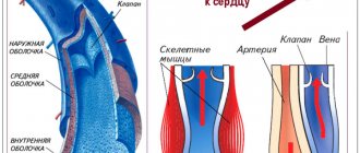
Structure of the adult heart
(picture from www.ebio.ru).
Arterial blood
from the lungs through 4 pulmonary veins (2 from each lung) it is collected in the left atrium, from where it enters the left ventricle through the bicuspid (
mitral
) valve, and then is released into the aorta through the aortic valve.
Scheme
: pulmonary veins ? left atrium? [mitral valve] ? left ventricle? [aortic valve] ? aorta.
Pattern of blood flow in the heart after birth
(animation). Superior vena cava - superior vena cava. Right atrium - right atrium. Inferior vena cava - inferior vena cava. Right ventricle - right ventricle. Left ventricle - left ventricle. Left atrium - left atrium. Pulmonary artery - pulmonary artery. Ductus arteriosus - ductus arteriosus. Pulmonary vein - pulmonary vein.
Diagram of blood flow in the heart BEFORE birth
For adults, everything is simple - after birth, the blood flows are separated from each other and do not mix. In the fetus, blood circulation is much more complicated, which is due to the presence of the placenta, non-functioning lungs and gastrointestinal tract. The fruit has 3 features:
- open oval foramen
(foramen ovale, “forAmen ovale”), - patent ductus arteriosus
(ductus arteriosus, ductus arteriosus) - and open ductus venosus
(ductus venosus, “ductus venosus”).
The foramen ovale connects the right and left atria, the ductus arteriosus connects the pulmonary artery and aorta, and the ductus venosus connects the umbilical vein and the inferior vena cava.
Consider the blood flow in the fetus.
Fetal circulation pattern
(explanations in the text).
Oxygen-enriched arterial blood from the placenta flows through the umbilical vein, which runs in the umbilical cord, to the liver. Before entering the liver, the blood flow is divided, and a significant part of it bypasses the liver along the ductus venosus
, present only in the fetus, and goes into the inferior vena cava directly to the heart. Blood from the liver itself through the hepatic veins also enters the inferior vena cava. Thus, before flowing into the right atrium, the inferior vena cava receives mixed (venous-arterial) blood from the lower half of the body and the placenta.
Through the inferior vena cava, mixed blood enters the right atrium, from where 2/3 of the blood passes through the open foramen ovale
enter the left atrium, left ventricle, aorta and systemic circulation.
Oval hole
and
ductus arteriosus
in the fetus.
Movement of blood through the foramen ovale
(animation).
Movement of blood through the ductus arteriosus
(animation).
1/3 of the mixed blood entering the inferior vena cava is mixed with all purely venous blood from the superior vena cava, which collects blood from the upper half of the fetal body. Next, from the right atrium, this flow is directed to the right ventricle and then to the pulmonary artery. But the lungs of the fetus do not work, so only 10% of this blood enters the lungs, and the remaining 90% through the arterial (botal) duct
are discharged (shunted) into the aorta, worsening its oxygen saturation. 2 umbilical arteries depart from the abdominal aorta, which in the umbilical cord go to the placenta for gas exchange, and a new circle of blood circulation begins.
Interesting to know
Liver
The fetus is the only organ of all that receives pure arterial blood from the umbilical vein.
Thanks to the “preferential” blood supply and nutrition, by the time of birth the liver manages to grow to such an extent that it occupies 2/3 of the abdominal cavity
and, in relative terms, weighs 1.5-2 times more than that of an adult.
Arteries to the head and upper body
extend from the aorta above the level of the confluence of the ductus arteriosus, so the blood flowing to the head is better oxygenated than, for example, the blood flowing to the legs.
Like the liver, the head of a newborn is also unusually large and occupies 1/4 of the entire body length
(in an adult - 1/7).
The brain
of a newborn makes up
12 - 13% of body weight
(in adults 2.5%). Probably, young children should be unusually smart, but we cannot guess this due to a 5-fold decrease in brain mass.