Determination methods and standards
Duration is measured through special tests. Most popular by:
- Sukharev;
- Duque;
- Lee White;
- Morawice.
In a hospital setting, it is possible to conduct any study in a short period of time. The clotting rate is different for each test.
Method according to Sukharev
Conducting a study related to the duration of bleeding according to Sukharev, you will need to take capillary blood. The required material is placed on the glass.
Special glass is used
Blood is taken from the glass for analysis until it thickens. The time is measured until the device can no longer draw blood. The normal value is 30 – 120 seconds.
Lee White method
For this study of the rate of bleeding control, venous blood is required. Enough material is taken to fill 3 test tubes. Preheated to body temperature (approximately 37 degrees). Place at an angle and evaluate the clotting time of all tubes. This method is used as an express method for assessing coagulability. Clotting of 1 ml of blood normally occurs in 5-7 minutes.
Morawitz method
The simplest method of determination is the Morawitz method. You will need laboratory glass, blood from your finger. The technique is simple: the blood is placed on a glass and the time is noted before it becomes coagulated. To determine the state of the coagulation system, the norm of bleeding duration is used. Each age has its own norm. It is necessary to take into account the individual characteristics of a person when conducting an examination.
It is important to know! Blood type plays a role in the study. The coagulation of capillary and venous blood is different.
Duque method
The method involves piercing the earlobe. Bleeding is then assessed using filter paper. The time according to the Duque method ranges from 1 to 4 minutes - the normal value. If blood is present for more than 4 minutes, there are clotting problems.
No special tubes needed
When conducting research, a special puncture needle is required. Artificial bleeding must be spontaneous. Bleeding is considered to have stopped when no traces of blood are visible on the paper. Paper is applied every minute. The Duques bleeding assessment is a good way to get an accurate result.
Other methods
There are several ways to determine the duration of bleeding. We can distinguish: thrombin generation test, thrombodynamics, thromboelastography. Several methods can be used in research. This allows you to get a more accurate result.
When taking a coagulogram, you must adhere to all doctor’s instructions in order to get an accurate result.
Meaning and purpose of analysis
The test allows you to identify the main disturbances in the hemostasis system. It is prescribed if there is a suspicion of a blood clotting disorder - prolongation of bleeding time or its reduction.
The main indicators for conducting the Duke test are:
- Phlebeurysm.
- Assessment of the state of hemostasis is most often used in conjunction with other methods for checking the functioning of the circulatory system before surgery.
- Diagnosis of thrombocytopathies of various types and origins.
- Disorders of the hematopoietic organs (bone marrow, spleen, liver).
- Consequences of taking a number of medications, for example, cytostatics and anticoagulants, antibiotics.
The analysis is not completely exhaustive. It only indicates the presence of disturbances in the hemostatic system and allows you to begin to conduct more in-depth research to find the root cause of the problem.
Preparation and procedure
Blood for analysis is taken from a finger or earlobe
The analysis itself is extremely simple. Compared to other tests, a deeper puncture of the skin is performed - up to 3 mm. Most often, the earlobe or fingertips are used, that is, places with a large number of small blood vessels.
After the puncture is made, the resulting drops of blood are removed with special paper at intervals of half a minute until the bleeding stops completely. At the same time, the exact time is recorded.
The test is performed with a Frank needle - a hollow tube with a trigger, a sharp tip and a spring. The peculiarity of the needle is that it allows you to control the depth of the puncture. No more than 1 ml of blood is taken for analysis, so it is completely safe for human health.
Blood clotting indicators
In a detailed blood test, there are a number of indicators that play an important role and directly affect the duration of bleeding. Capillary and venous blood is suitable for laboratory research. The result must be interpreted by a doctor.
Clotting time
According to Sukharev, coagulation began in 30 seconds, and ended in 3-5 minutes. The norm for children is approximately the same as for adults. If the White method is used, venous blood should clot within 7 minutes. Exceeding the indicators is a deviation. If an abnormality is detected, diagnostics are required to determine the cause.
Duration
Length
The duration of bleeding according to Duque is no more than 4 minutes. The indicator should not exceed this value, regardless of the location of the puncture (ear, fingertip). The duration of bleeding is influenced by several factors. In healthy people, bleeding often stops within the first 2 minutes.
Prothrombin time
This indicator is detected by performing a coagulogram. Blood plasma is used. Prothrombin time should be approximately 11-16 seconds. It is important that the laboratory technician performs all steps correctly.
Partial active thromboplastin time
APTT is an indicator of the effectiveness of internal coagulation. The normal value is 25-39 seconds. This indicator is the norm for women and men.
Thrombin time
TV values determine the presence of deviations in the final stage of coagulation. Thrombin time refers to the conversion of fibrinogen to fibrin under the influence of thrombin. The norm is considered to be 12-20 seconds.
Other indicators
Important indicators of the coagulogram are the number of platelets and fibrinogen. In adults, the platelet level is 150-400 g/l, in children - 150-350 g/l. The normal level of fibrinogen is 2-4 g/l.
Causes of prolonged bleeding
The cause of abnormal bleeding time may be hereditary or acquired.
Among the hereditary causes of bleeding disorders are the following:
You can also read: What is the blood clotting rate?
- von Willebrand disease;
- Glanzmann's thrombasthenia;
- Bernard-Soulier syndrome;
- connective tissue diseases (Ehlers-Danlos syndrome, Wiskott-Aldrich syndrome, Chediak-Higashi syndrome, hereditary hemorrhagic telangiectasia).
Among the acquired causes of abnormally long bleeding time, the most common are the following:
- long-term use of medications, especially aspirin, non-steroidal anti-inflammatory drugs, antibiotics, anticoagulants, tricyclic antidepressants, antipsychotics;
- vitamin C deficiency;
- frequent alcohol intoxication;
- uremia;
- liver failure;
- leukemia;
- myelodysplastic syndrome;
- amyloidosis.
This is why the patient should not take any blood thinning medications or alcohol for 7 days before the test as this may lead to false results.
The essence of the main analyzes
Each method used reflects a different moment of coagulation. The essence of the indicators:
- platelet level - reflects the number of cells involved in starting the coagulation mechanism;
- TV - indicates how the last stage of coagulation proceeds;
- APTT - shows the ability of plasma coagulation factors to create a clot;
- coagulability according to White - reflects the ability of blood to form a clot.
Decoding is a complex process.
Deciphering indicators on your own is problematic; you need to consult a specialist.
What is this and what does it affect?
It is very important to know how thin or thick a person’s blood is. The mechanism of coagulation depends on the physicochemical actions in the molecular system of the plasma:
The mechanism of coagulation depends on the physicochemical actions in the molecular system of the plasma:
- The main place in this is occupied by a protein called fibrinogen.
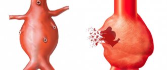
- During the reactions that take place, it is converted into fibrin (which is not able to dissolve), which falls out in the form of the thinnest threads.
- These threads can form into a dense network with small cells that trap the formed elements.
- These changes lead to the formation of a blood clot. As it exists, its base becomes compacted, the edges of the wound are tightened and healing occurs.
- As the clot tightens, it forms a yellowish liquid called serum.
Platelets, which are part of the plasma, influence the compaction of the blood clot.
To use a primitive comparison, the process of blood clotting is reminiscent of the process of turning milk into cottage cheese, when casein (milk protein) coagulates and whey is released. Over time, the wound resolves and the fibrin clot dissolves.
The plasma coagulation test is used when the presence of the following diseases is suspected:
- pathology of the liver organ,
- disorders of the immune system,
- varicose veins.
Specialists also send patients for hemostasis testing before surgery, during pregnancy, and in case of possible large blood losses.
The video explains how the blood clotting system works:
Deviations from the norm
Time may exceed normal values. Blood may clot too quickly. In this case, it is necessary to carry out diagnostics to determine the root cause. Deviations may be in favor of fast or slow clotting. Accelerated clotting may indicate intoxication, dehydration, autoimmune damage, or disruption of the endocrine system. Slow blood clotting occurs due to:
- cirrhosis;
- leukemia;
- lack of certain vitamins;
- hemophilia.
The diagnosis is made only after diagnostics.
Normally, the bleeding time is approximately the same for all people, so the deviation is extremely easy to identify.
Why is the norm broken?
Why do blood clotting abnormalities occur? What is the solid basis for this? It is worth noting some innate factors here.
In some cases, longer reaction times may be passed from parents to children. But such diseases are quite rare.
In some cases, the cause of bleeding that lasts too long may also be due to the use of a number of medications. For example, this group includes Aspirin, a number of antibiotics and other drugs. If this is the reason, then the problem is easily solved. It is enough to stop taking these medications. If the reason is different, then treatment cannot be avoided.
What processes occur during bleeding?
Since the time it takes for bleeding to stop is determined in laboratory conditions in most cases, it is necessary to know and understand the processes that accompany it. Indeed, the sheet of analysis results can indicate not only the time norm, but also hemostasis indicators.
The most accurate results can only be obtained in laboratory conditions
Hemostasis is a sequence of events that leads to the cessation of bleeding due to the formation of a special hemostatic plug of platelets and fibrin. A plug forms on the injured vascular wall from platelets and clotted blood. After platelets combine with endothelial cells and collagen in the damaged vascular wall, von Willebrand factor is released.
This is what it looks like
platelet aggregation process
under a microscope
After all factors are activated, platelet aggregation occurs. Soon after, additional platelets are taken from the bloodstream and aggregated, thanks to adenosine and thromboxane A2. Thanks to such complex chemical processes, the skin is restored after a cut or puncture. All of the above processes can be seen by the laboratory technician under a microscope, and if a failure occurs at any of these stages, the bleeding time increases.
Tests for resistance (fragility) of the vascular wall.
Roughly, increased capillary fragility is judged by a positive pinch test - the formation of a bruise when a fold of skin is compressed in the subclavian region and hemorrhages at the injection sites. More precisely, the resistance of capillaries is determined using the Konchalovsky-Rumpel-Leede cuff test (test with a tourniquet).
As modified by Borchgrevink (1971), the test is performed as follows: a circle with a diameter of 5 cm is drawn on the upper part of the palmar surface of the forearm, after which the shoulder is compressed with a cuff from a blood pressure measuring device for 5 minutes at a pressure of 90-100 mm Hg. Art. 5 minutes after removing the cuff, count the number of petechiae in the outlined circle.
The norm is up to 10 petechiae, a weakly positive test is 11-20 petechiae, a positive test is 20-30, a strongly positive test is more than 30.
Jar test (according to Nesterov): cups from Nesterov’s apparatus are placed on the palmar surfaces of both forearms, in which a vacuum of 150 mm Hg is created by suction. After 1 minute, check whether petechiae have appeared. Depending on the results obtained, the pressure is gradually reduced by 25 mmHg. Art.
Clinical and diagnostic significance. Vascular tests are positive in all thrombocytopenias with the content of blood platelets in the blood below 40-60 * 109/l, as well as in many primary and symptomatic qualitative defects and dysfunctions of platelets, including hemoblastosis, uremia, C-hypovitaminosis . Some women test positive without platelet abnormalities, especially during menstruation.
In what cases is a test prescribed?
Since platelets are the main factor in primary hemostasis, the results of the bleeding control test will directly indicate the functionality of the blood cells. Therefore, this test is prescribed to patients with suspected bleeding disorders or thrombocytopenia. A skin incision may also be done as a screening test in an outpatient setting. This is usually done in small clinics and outpatient clinics that do not have their own laboratory. This test is performed before invasive procedures in patients with suspected bleeding disorders to determine the likelihood that prolonged bleeding will occur.
The test is quick and virtually painless
Laboratory assessment of clotting time may be ordered in patients suffering from prolonged or severe postoperative bleeding. Using a microscope, the doctor will be able to recognize dysfunctional platelets and choose the right treatment.
Calculation of bleeding duration using the Duque method
The bleeding time according to the Duque method is determined a little differently: the specialist does not use a device with which to measure pressure. The Duque technique is considered less invasive. This test can be performed at home independently. The skin can be pierced in any place that does not contain large vessels. The puncture depth according to Duke does not exceed 3 mm. After the puncture, the paper filter is also manipulated, just as when using the Ivey technique.