Gallop rhythm and quail rhythm: signs of pathological tones, causes, diagnosis, treatment
From time immemorial, a doctor, when examining a sick person, relies only on his hands and ears, because imaging diagnostic methods appeared relatively recently.
And to this day, one of the most important medical procedures is auscultation, or listening with a special tube to the sounds created by the work of the heart. The heart, like any other organ, creates certain sounds when working. This is due to the fact that blood is constantly moving in the heart, passing through the valves, and the valve flaps open and close, emitting sound vibrations. In addition, at the moment of stretching of the heart muscle of the atria and ventricles, vibration is created, which is layered with the sounds of slamming valve flaps.
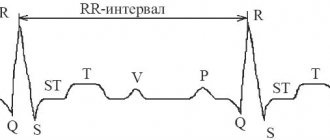
Normally, at the points of projection of the heart valves onto the anterior chest (fifth intercostal space on the left, second intercostal space on the right and left, fourth intercostal space on the left), two heart sounds are heard - the first and second. In asthenic thin people, in children and adolescents, two more tones can be heard - the third and fourth, but their absence is not considered pathology.
However, since 2004 (E. Braunwald), ideas about the nature of the origin of the first tone have changed somewhat - now it is generally accepted that such a sound is created not by the slamming of a valve, but by the impact of blood on the walls of the ventricle, when the blood first quickly fills the ventricles and then abruptly stops movement. The first sound is systolic, as it indicates systole (contraction) of the ventricles. The second sound is diastolic, as it is caused by diastole (relaxation) of the ventricles.
The second sound is formed a few hundredths of a second after the first and is formed by sounds that are created by the closure of the valves of the aorta and pulmonary trunk, as well as the oscillatory movements of the walls of these arteries.
distribution of heart sounds in the cardiac cycle
- there - ta - there - ta - there - ta - I tone - II tone - I tone - II tone - I tone - II tone
Heart sounds and heart murmurs should not be confused. Between the two tones there are silent pauses, during which the blood flows directly, which normally occurs silently through the chambers of the heart. However, with heart defects or other pathologies of the heart valves, the blood stream has difficulty expelling from the heart chambers corresponding to the defect, so sound phenomena called murmurs occur. Noises can be heard in pauses between tones, or can be layered on top of them.
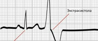
Such rhythms are considered to be sound phenomena that are formed due to pathology of the heart muscle itself or valve structures. When listening to a heart with a similar pathology, a three-part rhythm is determined, in which the second sound seems bifurcated (split). But in fact, the split sound is nothing more than an additional tone. What exactly causes the pathological tone depends on the type of rhythm. Split rhythms include the gallop rhythm and the quail rhythm.
Pathological split rhythms
Such rhythms are considered to be sound phenomena that are formed due to pathology of the heart muscle itself or valve structures. When listening to a heart with a similar pathology, a three-part rhythm is determined, in which the second sound seems bifurcated (split). But in fact, the split sound is nothing more than an additional tone. What exactly causes the pathological tone depends on the type of rhythm. Split rhythms include the gallop rhythm and the quail rhythm.
Video: description of pathological additional tones during auscultation
Diagnosis of chronic heart failure
Heart failure is not an independent disease. This is a complication of various pathologies of the cardiovascular system, in which the contractility of the heart muscle weakens, cardiac output does not meet the metabolic needs of the tissues, which leads to a decrease in venous return and an increase in total vascular resistance.
Most often, chronic heart failure develops in patients suffering from the following diseases:
- Coronary heart disease;
- Arterial hypertension;
- Diabetes mellitus;
- Cardiomyopathies;
- Systemic connective tissue diseases;
- Valvular heart defects.
Less commonly, the causes of chronic heart failure are metabolic and endocrine diseases, neuromuscular diseases, intoxications, deficiency of potassium, selenium, magnesium, selenium, and chronic respiratory diseases.
Patients suffering from chronic insufficiency present the following classic complaints:
- Inspiratory dyspnea;
- Orthopnea (shortness of breath when lying down);
- Paroxysmal nocturnal dyspnea;
- Cough;
- Hemoptysis;
- Heartbeat;
- Thirst;
- Weakness, fatigue;
- Cognitive disorders (decreased memory, mental performance).
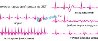
How dangerous is tachycardia, and what consequences are possible with its development?
Types of tachycardia
Before talking about why cardiac tachycardia is dangerous, you need to understand that this disease occurs due to disruptions in the functioning of its various parts. Different types of disease carry different dangers for humans.
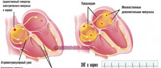
Anatomically, the heart is divided into the atrium and ventricles. Depending on where the foci of rhythm disturbances are localized, there are:
- sinus tachycardia, which develops in the atrium, that is, in the sinus node;
- atrial or ventricular paroxysmal tachycardia - it occurs in the atrium or ventricles when a distortion of the signal supplied by the sinus node occurs in these sections.
How is tachycardia dangerous for humans?
Answering the question whether cardiac tachycardia is dangerous, any doctor will definitely answer in the affirmative, because in essence it is the work of the heart for wear and tear. It works ineffectively, because due to the increased rhythm, the ventricles do not have time to fill with blood. It turns out that the organ works faster, but the speed does not allow it to do its job efficiently. The lack of blood in the ventricles does not supply oxygen to other organs and this is the main danger of rhythm disturbance.
Ventricular tachycardia can cause coronary artery disease and myocardial infarction, since it disrupts the blood supply to the heart. In especially severe cases, rhythm disturbances can lead to heart failure, and this disease develops even in those who are completely healthy, but the symptoms have remained unattended for a long time.
It also leads to pulmonary edema and impaired blood supply to the brain, as well as pulmonary embolism. The paroxysmal type of the disease leads to these consequences. Chronic heart failure, in addition to wear and tear of the heart muscle, also causes other side problems - arrhythmic shock, cardiac asthma. As almost everyone understands, tachycardia is dangerous and can result in sudden death.
But even with the most favorable prognosis, when the disease does not lead to serious consequences, it is extremely difficult for the body to tolerate. If the heart rhythm is abnormal, a person quickly begins to feel fatigue, shortness of breath, severe dizziness and chest pain. Therefore, even the symptoms themselves can pose a health risk.
How to prevent the development of dangerous consequences of this type of arrhythmia?
First of all, you need to listen carefully to your body. At the first signs of an increased heart rate at rest, seek medical help, find the cause and focus on treating it. Tachycardia can be chronic, when its symptoms are felt constantly, which is very dangerous due to the rapid development of the underlying disease. This condition requires close monitoring and constant monitoring by a doctor.
But even episodic attacks cannot be ignored, otherwise they can lead to the development of a chronic form. The main thing to remember is the signal that the heart gives us when something goes wrong in the body.
Do pathological rhythms need to be treated?
Therapy in the presence of pathological rhythms in a patient is necessary only after a thorough examination and establishment of an accurate diagnosis. The type of medical institution where treatment will be carried out depends on the underlying disease. For example, hypertension, leading to left ventricular hypertrophy, can be dynamically observed in a clinic, and more severe pathologies (heart attack, myocarditis, severe heart failure) must be treated in a hospital setting. Mitral stenosis, when diagnosed for the first time, also requires further examination and selection of therapy in a hospital, where the need for surgical correction of the defect will be determined.
2012-2020 sosudinfo.ru
Go to section:
Diseases of the heart and aorta, arrhythmology, functional. diagnostics, cardiac pharmacology and surgery
Recommendations to SosudInfo readers are given by professional doctors with higher education and specialized work experience.
What diseases cause abnormal heart rhythms?
Based on the above, during auscultation of the heart, the doctor may suspect the presence of one or another heart pathology in the patient.
- acute myocardial infarction,
- acute myocarditis (inflammation of the heart muscle),
- hypertrophy of the left ventricle (with arterial hypertension, stenosis of the aortic mouth of rheumatic or atherosclerotic origin),
- dilated cardiomyopathy (with coronary heart disease),
- acute left ventricular failure,
- chronic left ventricular failure due to post-infarction or post-myocardial origin.
2. The quail rhythm is caused by stenosis of the left atrioventricular orifice (mitral stenosis). This pathology is an acquired heart defect that develops as a result of acute rheumatic fever, scarlet fever, tonsillitis or chronic tonsillitis.
What further examination is necessary?
If during the examination the doctor was able to listen to the rhythm of a gallop or the rhythm of a quail, he should refer the patient for further examination. First of all, an ECG and chest x-ray are necessary. Any of the listed diseases can be detected by performing these methods (heart attack - on an ECG, hypertrophy or dilatation of the heart - on an x-ray, etc.).
To clarify the nature of pathological tones in the heart, phonocardiography (PCG) is used - a study in which the sounds of tones are amplified using a microphone and then converted into a graphic image using a writing device. FCG is interpreted by a specialist and helps to reliably determine what causes pathological sound phenomena. FCG is often performed in children with suspected heart disease.
How and under what pathologies is the gallop rhythm (RG) formed?
In a healthy heart, two pure tones are clearly audible - I and II. In asthenics, two more physiological ones are sometimes added - III and IV. Severe myocardial lesions, accompanied by rapid heartbeat, give rise to an auscultatory phenomenon - a diastolic gallop rhythm.
These successive sounds, reminiscent of a horse's gait, form the I and II tones, to which the third joins. It represents the third or fourth heart sound (or their fusion), which in this case are considered pathological. In various diseases, this tone appears at the beginning, middle or end of diastole (relaxation of the heart muscle).
3 types of gallop rhythm:
- Protodiastolic RG occurs immediately after the second sound. It is caused by a decrease in the tone of the heart muscle. The sound is produced due to the rapid stretching of the walls of the ventricle during filling. This is the third physiological tone. It is heard when the muscle tissue of the left ventricle is greatly relaxed and stretched.
- Presystolic (before systole) is formed by vibration of the walls of the ventricles. They are joined by the physiological IV sound - the sound of atrial contraction.
- The mesodiastolic gallop rhythm is when, against the background of tachycardia in the middle of diastole, pathological III and IV sounds merge as an additional one.
Diastolic gallop rhythm appears in myocardial infarction, cardiomyopathies and heart failure accompanying hypertension and nephritis. To accurately diagnose the rhythm of quail and gallop, the phonocardiography technique is used.
Gallop rhythm
The gallop rhythm of the heart is the threefold rhythm of the heart at a high heart rate. It got its name from its resemblance to the sound of a galloping horse. The famous French clinician Henri Huchard said: “There is no gallop without tachycardia.”
The reason for the appearance of the gallop rhythm is changes in the properties of the ventricular myocardium.
Pierre Potin (Potin Pierre Charles Edouard, 1825 -1903), a French therapist, explained the gallop rhythm by vibration of the walls of the left ventricle when it loses tone . As is known, in protodiastole the flow of blood from the atria into the ventricles begins. Normally, it occurs silently (not perceived by the ear), since the ventricular muscle, which has tone, straightens gradually and absorbs the shock of a portion of blood coming from the atrium. If the muscle has lost its tone and “hangs like a bag,” then the blood, falling to the bottom of the ventricle, causes an additional beat, heard as an additional sound - a pathological III heart sound.
The gallop rhythm can be of two types: protodiastolic and presystolic:
Protodiastolic gallop rhythm (the so-called ventricular gallop rhythm) - it is based on a pathological III tone , which has higher frequency components than normal; an additional tone is heard after II then at the apex of the heart. George Taylor (South Carolina, USA, 2004) gives the following brief description of the third tone and protodiastolic gallop: “a large flabby ventricle (volume overload).”
Presystolic gallop rhythm (atrial gallop) - it is based on a pathological IV tone - this rhythm is better recorded with a long PQ ECG interval; The appearance of a pathological IV tone depends on the value of the end-diastolic pressure in the ventricle, the rigidity of the left ventricle, its poor “straightening”, and poor relaxability.
It is very important that it can be “palpated”, i.e. feel this diastolic tone with your hand. The rhythm of the gallop indicates heart failure - this is the “cry of the heart for help”, they used to say that this is the “cry of the heart for digitalis”.
The gallop rhythm is a cry from the heart for help!
Summation gallop
Summation (or mesodiastolic) gallop is a three-term ventricular rhythm, when, as a result of a sharp shortening of the slow filling phase against the background of tachycardia, pathological III and IV sounds merge into one additional fairly loud tone.
The main conditions for the occurrence of a summation gallop are:
- a decrease in the contractility of the ventricular myocardium in patients with heart failure and acute myocardial damage, leading, on the one hand, to a decrease in its diastolic tone and rate of relaxation (“flabby ventricle, the ventricle “hangs like a bag”) - this gives the third pathological heart sound;
- an increase, of course, in diastolic pressure in the ventricle - this gives the IV pathological heart sound;
A characteristic combination for such patients is the presence of hypertension and heart failure.
Summation gallop can also occur with AV block of the first degree, when an extended PQ interval leads to a displacement of the IV sound closer to the beginning of diastole, where it merges with the III sound.
The summation gallop tone has a higher frequency, is louder and longer lasting than the III and IV tones separately.
The dual nature of the summation gallop can be revealed by slowing down the rhythm, for example, during massage of the carotid sinus. In this case, a four-part gallop rhythm with the third and fourth heart sounds heard separately.
Total
So, the protodiastolic (ventricular) gallop is associated with an audible III sound. An additional tone is heard after the second tone. It is perceived both by the ear and by palpation. Gallop is usually observed with tachycardia. Many clinicians believe that the ventricular gallop rhythm is the most reliable sign of heart failure, determined physically.
IV tone and presystolic gallop are “a hard, unyielding ventricle (pressure overload).”
The protodiastolic gallop rhythm is more unfavorable prognostically.
It should be noted once again that gallop refers to any three-term diastolic heart rhythm, the speed of which is so high that it imitates the running of a horse. The term "gallop rhythm" implies a high heart rate (often combined with dulling of the 1st and 2nd sounds) and a typical melody. The rhythm of the gallop is a more threatening sign than the simple presence of the third tone . As mentioned above, the third heart sound may be “close to physiological”, and also be a reflection of the relative volume overload of the left ventricle and not form a gallop rhythm. On the contrary, the gallop rhythm with the third tone is almost always pathological.
As the observations of V.P. Obraztsov and M.M. Gubergrits, now confirmed by many Soviet clinicians and foreign therapists, have shown, that in healthy people the heart melody heard by direct auscultation is three-membered (the so-called normal third tone) and that with a relatively small pathology, as well as under physiological circumstances, splitting and bifurcation of the first tone (? V.O.) can be observed (over 10% according to V.P. Obraztsov), it is clear that there are gradual transitions between the gallop rhythm and the normal melody of the heart, and not a sharp, inseparable edge .”
The importance of gallop rhythm was interestingly defined by George Taylor (2004):
“Traditionally, a lot of attention is paid to heart murmurs, but they, while helping to make a correct diagnosis, cannot tell us as much about the severity of the disease. It’s the gallops and the apical impulse that do this: they tell us about the size of the ventricle, its function and diastolic compliance.”
How is the rhythm of a quail formed?
In case of mitral valve stenosis, during auscultation the quail rhythm should be listened to at the top of the heart. The valve separating the left atrium from the ventricle is a bicuspid septum. These are two plates of connective tissue whose task is to prevent the backflow of blood into the atrium from the ventricle. Typically, during diastole, the valves open and press tightly against the muscle walls, allowing blood to flow freely. During contraction, the valves close tightly, preventing the backflow of blood.
One of the pathologies of the left-sided valve structure is stenosis. A healthy mitral valve covers an opening with an area of 4 to 6 square meters. see. Narrowing of the lumen between the atrium and the ventricle to 2 or less centimeters occurs when, due to inflammatory or other pathological processes, the tissue of the valves fuses with the edges of the atrioventricular opening, adhesions and scars form on it.
The valves become rigid (inelastic), so the sound of their closing is clearly audible. The quail rhythm is a clapping, loud sounding 1st heart sound and a split 2nd heart sound. The triple melody resembles the cry of a bird.
With mitral stenosis, the quail rhythm is caused by an increase in the first tone. It occurs because the left ventricle does not fill completely. Cardiomyocytes react by increasing tension (to push through an insufficient amount of blood they have to contract more), which causes an increase in sound. This is followed by the usual II sound, accentuated by the closure of the leaflets of another valve - the pulmonary valve.
Immediately after, an additional sound is produced by the mitral valve when it opens. It merges with the third normal tone and enhances the previous one, forming the phenomenon of splitting the second tone. The quail rhythm in mitral stenosis is one of the first “pre-electrocardiographic” symptoms of the disease.
The mechanism of quail rhythm formation
In addition to the gallop rhythm, the quail rhythm also has three parts. This rhythm is due to the apparent bifurcation of the second tone. In fact, the second sound does not bifurcate, it is simply accompanied by a sound called a “mitral click.” The occurrence of such an additional tone is due to the presence of adhesions and commissures between the leaflets of the mitral valve, therefore, when the valve opens, a characteristic clicking sound occurs. The quail rhythm is most clearly heard at the point of projection of the mitral valve (in the fifth intercostal space under the nipple). The additional tone begins in diastole and can be heard immediately after the second tone. It is otherwise called TOMK, or the mitral valve opening tone.
quail rhythm on FKG
Along with the additional tone, when the mitral valve ring narrows, the first two tones also become more intense. Thus, the first sound is enhanced due to the fact that the left ventricle vibrates more strongly due to less blood flow into it than normal. That is, the muscle creates a stronger sound. The second tone is enhanced by the sound of the pulmonary artery valves opening. This is due to the fact that with mitral stenosis, the blood does not completely enter the ventricle; accordingly, in the left atrium the volume of blood is greater than normal, and in the pulmonary veins that bring blood to the left atrium, pressure increases - pulmonary hypertension is formed. In conditions of pulmonary hypertension, the valves of the pulmonary trunk, on the contrary, carrying blood from the right ventricle into the arteries of the lungs, slam shut louder than usual - the second tone intensifies.
Letter designation of the heart melody with the rhythm of a quail:
– sleep – by – ra – sleep – by – ra – I tone – II tone – additional. tone – I tone – II tone – additional. tone