My favorite style which harness to choose for a cat
With the current wealth of choice, when coming to a pet store, an inexperienced “cat breeder” may be confused - which harness to choose for a cat? We give advice: dress your pet the way you would dress a child, that is, focus on three main factors: comfort, ease of putting on and practicality.
Upon closer examination, it is easy to notice that cat harnesses come in two types: more “substantial” ones, in the form of a vest, and those made of fabric or leather straps. Both have their fans.
Pros and cons of "vests"
What speaks in favor of the vests is their reliability - try jumping out of one of these! In addition, it is warm, which is especially important now, and there is room for decoration - here everything depends on the owner’s imagination.
But such harnesses also have disadvantages. Firstly, many owners, not without reason, believe that a cat in a vest is not so free to move. Secondly, in the summer you will have to change your warm winter vest to a lighter one, preferably “breathable”, mesh. And those who are not used to wasting money don’t like this. The same thing with size: the pet has grown or gained weight - the outfit will have to be changed.
Pros and cons of “straps”
Harnesses made from straps, although not as elegant, are much more practical - especially those that are adjustable in volume. As for the shape, there are two main ones: one resembles a figure eight, the second - the letter H.
In the first case, two loops (one is put on the cat’s neck like a collar, and the second is on the body behind the front paws) are fastened together on the animal’s shoulder blades. In the second - the principle is the same, only the loops are connected by a bar on the cat's back
According to the owners, both models are equally reliable and lightweight - those with mustaches quickly get used to them and stop paying attention to them
Ischemic ST segment elevation on ECG and Holter:
↓Trend of ST segment position during ischemic elevation: high “peaks” are visible during ischemic attacks.
↓The beginning of the ischemic episode: in the leads characterizing the anterolateral areas of the LV myocardium (I, V3-V5), ST elevation began. Reciprocal ST depression begins in lead AVR.
↓Development of an ischemic episode: ST segment elevation increases, changes begin in previously “quiet” leads. In the middle chest leads, the complex takes on the shape of a “cat’s back”, characteristic of acute myocardial infarction.
↓Peak of the ischemic episode: ST segment elevation is maximum, in V4-V6 the QRS complex has taken on the character of a monophasic curve, in lead AVR the curve is also monophasic, but directed downwards (reciprocal changes). Interestingly, the patient came to the outpatient clinic for holter removal on his own feet, although with a mention in his diary of handfuls of nitrates taken. After decryption, he was hospitalized by ambulance.
How to understand the language of cats
It's not difficult at all, the main thing is to have a little observation. The main group of gestures is associated with a procedure such as licking. It means not only the desire to wash, it is a kind of language of cats, with the help of which they can demonstrate:
confusion - the cat begins to lick itself when it was scolded (this is the best time for communication with the animal and reconciliation!); good mood - the cat begins to lick the owner’s hand; search for affection - the cat carefully licks the owner’s hand and tries to climb onto his lap; boredom - the cat licks itself with overly prolonged, deep and intense movements: in this way she asks to caress her or play with her; readiness for training - the cat washes rationally and carefully and is able to accept several commands from the owner.
In addition to licking, a cat's tongue also involves all kinds of movements of the body and tail. The most common among them is friction against the owner’s legs. In this way, the cat expresses love and can also ask for something. If a cat rubs its muzzle against your face and nuzzles your cheeks or lips, this expresses the extreme degree of its love and appreciation.
As for the tail, it can be in several positions:
- flies up - the cat is excited, but not angry or scared;
- slightly lowered - the cat is slightly frightened or dissatisfied (for example, with its food), in this state it is better to leave it alone;
- lowered low - the cat is very frightened or unhappy;
- actively twitching - the cat wants to be alone (if only the tip of the tail is twitching, this is an extreme degree of concern for the animal);
- fluffed up - the cat is preparing to attack;
- squeezed between the paws - the cat is afraid.
Cat Body Language: Basic Secrets
In addition, you should monitor the cat's body position, which can also express your pet's emotions. So, a straight back and a raised head indicate that a cat is relaxed, but also attentive. If the cat sits a little sideways, this indicates that he is scared. A grinning and angry cat is about to attack from the front, and if he suddenly crouches down on the ground with a gloomy look, it means he has changed his mind about doing it.
You can interpret the signs that a cat gives with its eyes in a similar way.
A cat's attention is expressed by a direct and open gaze directed directly at you. At the same time, if a cat stares at you too openly, this expresses a challenge and a requirement to keep your distance.
If the eyes are half-closed, the cat is cautious, and if they become like narrow slits, the cat is confident and wary. You should also be careful if the cat has round pupils and a crazy look - this expresses the animal’s fear, in which it cannot control itself. But if a cat looks at you, crouched to the ground, there is no danger, he is simply expressing devotion.
Finally, cat sign language also involves communication using their paws. If your cat rubs his paw against you or gently massages your hand with it, he is showing satisfaction and contentment. If a cat pulls at clothes with its paw or even fights, it means it is asking for something, including love and affection.
But the most powerful demonstration of love is a hug with paws. Also, a very strong emotion is expressed when a cat lies on its back - if it exposes its stomach to you, it means it completely trusts and loves you, so in this position you should definitely stroke and caress it - the cat will be very grateful to you!
| Newsletter Subscribe.Ru |
| Subscribe to our newsletter about cats from around the world |
| Subscribe by letter |
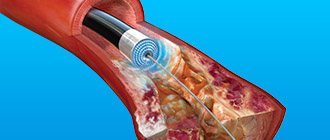
Treatment of pathology
Based on the electrocardiography examination, the specialist prescribes medications and other physiotherapeutic methods for treating the disease as a complex therapy.
The main points that must be followed for the purpose of treatment:
- limit movements and perceive emotional stress as little as possible, while continuing to move as much as necessary;
- proper nutrition, but you should not eat fatty meats or sweets; you must include lean meat, fish, low-calorie vegetables and fruits in your diet;
- taking medications, including antagonists, diuretics, beta blockers, nitrates during attacks.
Everything done in combination will help maintain the condition of the body, and the pathology will recede, but it is important to note that a slight deviation from following the diet prescribed by a specialist or failure to take medications on time entails adverse consequences.
Marjariasana
This asana can be used by anyone who is just starting to practice yoga, as it is quite simple to perform and does not require special physical training. With its help, you can successfully correct problems with the spine, strengthen weak muscles, make joints more mobile, and improve blood supply to internal organs and their normal functioning. Although the cat pose in yoga is considered one of the simplest, its benefits are very great. It is recommended to perform it every day and include it in all asana complexes used.
Performing the exercise
1. You need to get down on all fours so that your knees are under your hip joints and your palms are under your shoulders.
2. Hands and knees should be on the same line.
3. Then you need to round your back as much as possible, trying to arch it upward, as a cat does.
4. It is necessary to monitor the position of the shoulders and neck - the head should be lowered down, the neck should be relaxed, and the shoulders should not be tense. The asana is aimed at developing the flexibility of the spine, so all movements are made smoothly, truly like a cat.
5. The cat pose continues with a smooth change in the arch of the spine to maximum deflection. To do this, the back smoothly returns to its original position.
6. With a gentle movement, the head rises, the whole body is extended behind the neck, the back is arched.
7. Look up, neck extended. Don't throw your head back!
8. Maximum stretching of all muscles should be felt, but without overexertion.
This exercise has many modifications and variations. You can do it leaning on your elbows. In this case, you need to monitor not only the position of your knees, but also look at how your elbows are positioned. They should not be brought together and “squeeze” the chest.
Another option is with your arms extended forward along the floor. This pose is more difficult and requires a fairly developed spine, but it works great. To do this, from a position on all fours, you need to stretch your arms forward and try to bend so that your shoulders and chest are as close to the floor as possible, the spine is arched, and the tailbone is directed upward. In this case, you cannot lie down with your chest on the floor. The pose loads the shoulder and chest area and greatly stretches the arms. The neck should also be pulled forward without overstraining its muscles, so as not to cause discomfort.
At the first attempts, a full-fledged position may not work out, but repeating it regularly after a good warm-up will soon begin to give pleasure, and one day you will notice how easily and gracefully you “pretend to be a cat.” Bitilasana (cow pose) is similar in execution.
Benefits brought
The cat pose helps to cope with enslavement on several levels at once. The muscular frame is physically strengthened, the abdominal muscles are trained, the flexibility of the spine increases, especially its thoracic region, pain in the spine decreases, and a feeling of a light, flexible, well-trained body appears. This asana has a great effect on your figure.
After classes, a person feels a surge of energy, it becomes easier for him to move, the feeling of stiffness goes away, and the perpetually bent back straightens out. At the energy level, a person’s back also “bends” – he begins to feel much more confident and calm.
If this asana is practiced for a certain time and is accompanied by meditation, in which a person can concentrate on the image of a cat, he will begin to feel its grace, elegance, and will acquire smoothness and softness of movements.
https://youtube.com/watch?v=EL0M4tL6bzY
Highly probable and possible ECG signs of ischemic heart disease:
Highly probable signs of “pre-scar” IHD include ST segment displacements: rise (elevation) and decrease (depression). With Holter monitoring, these changes are visible as a deviation of the ST trend from the zero level of “peaks” and “beards”.
The fact of the death of all layers of the myocardium on the ECG is reflected by the pathological Q wave (it is wide and its amplitude is more than a quarter of the height of the R wave in the same lead).
ST elevation and the presence of Q are included in the diagnosis formulations: ST-segment elevation AMI and Q-forming myocardial infarction.
ST elevation can also be observed in other conditions, remember this (early repolarization syndrome - characterized by notching on the descending knee of the R wave and the duration of this condition on the holter, pericarditis - changes in it are present in all or almost all leads). ST depression can also occur with an overdose of glycosides, but the shape of the segment is very characteristic and resembles a “trough”.
The remaining options for changing the QRS complex are considered possible (i.e., a diagnosis cannot be made based on them). Most often this is a negative T wave. If you are dealing with a patient with acute chest pain and any changes on the ECG, remember a simple rule: it is better to hospitalize ten patients without a heart attack than not to hospitalize one patient with a heart attack. Don’t worry, the ambulance doctors will treat you with understanding.
Cat body language - emotions expressed without words
Cats can't talk because they don't need to. They are quite capable of expressing all their emotions through body language and facial expressions. That is why communication with a cat comes down to understanding its gestures and actions, which can be a whole spectrum.
Cat body language consists of communication using the head, ears, paws, tail, posture and, of course, the eyes. At the same time, communication with a cat should be based on understanding the complex of all gestures as a whole, besides, the animal always tries to express its emotions quite unambiguously.
Conventionally, the body language of cats can be divided into two groups - gestures that are directed directly at the owner (the cat licks his hand, hisses, rushes, rubs), or those that the cat makes as if in relation to itself, but so that the owner I could understand her easily.
Prevention
ECG signs of myocardial ischemia can be prevented, which can then be treated for a long time and tirelessly, while constantly being examined and spending money and time on eliminating the pathology.
Of course, it is good that ischemia on the ECG is recognized literally immediately, and you can begin to treat it without delay, but it is still easier to prevent it. To do this, you just need to lead the right lifestyle:
- eat well and properly;
- do physical education;
- get enough sleep;
- abstain from drinking;
- spend more time in the fresh air;
- don't be upset over trifles.
Only in this case will the heart be healthy, and the results of the electrocardiogram will only please you.
Arrhythmic form
Cardiac arrhythmia or signs of left ventricular heart failure (in the form of attacks of shortness of breath, cardiac asthma, pulmonary edema) occur as equivalents to attacks of exertional angina or spontaneous angina. Diagnosis of these forms is difficult and is finally formed on the basis of the totality of the results of an electrocardiographic study in stress tests or during monitor observation and data from selective coronary angiography.
Diagnostics
Clinical symptoms
Complaints
Irradiation of pain in ischemic heart disease. The color intensity shows the frequency of occurrence of irradiations in this area.
The most typical complaints for coronary heart disease are:
- chest pain associated with physical activity or stressful situations
- dyspnea
- Interruptions in heart function, feeling of rhythm disturbance, weakness
- Signs of heart failure, for example, swelling starting from the lower extremities, forced sitting position.
Anamnesis
From the medical history, the duration and nature of pain, shortness of breath or arrhythmia, their connection with physical activity, the amount of physical activity that the patient can withstand without an attack, the effectiveness of various medications when an attack occurs (in particular, the effectiveness of nitroglycerin) are of great importance. It is important to find out the presence of risk factors. Physical examination
Physical examination
Physical examination may reveal signs of heart failure (moist rales and crepitus in the lower parts of the lungs, “cardiac” edema, hepatomegaly - enlarged liver). There are no objective symptoms specific to coronary heart disease that do not require laboratory or instrumental examination. Any suspicion of coronary heart disease requires electrocardiography.
Electrocardiography
An ECG is an indirect research method, that is, it does not tell how many myocardial cells have died, but it allows you to evaluate some of the functions of the myocardium (automatism and, with some assumptions, conduction). To diagnose most pathological conditions of the myocardium (cardiomyopathies, myocardial hypertrophy and some other diseases), in addition to ischemic heart disease, but often combined with ischemic heart disease, the ECG has an auxiliary function, and ultrasound and other methods are also needed.
Some electrocardiographic signs of acute myocardial infarction
A characteristic sign of large-focal myocardial infarction (transmural) is the presence of a pathological Q wave on the ECG.
- in lead I:
- there is a pathological Q wave (>0.03 s, amplitude exceeds 1/3 of the amplitude of the R wave)
- there is a negative T wave.
in lead II there is a pathological Q wave (>0.03 s, amplitude exceeds 1/4 of the R wave)
in lead III there is a pathological Q wave (>0.03 s, amplitude exceeds 1/2 R wave)
in leads V1, V2, V3 there is a QS or QR wave and at the same time the T wave is negative.
in leads V4, V5, V6 there is a pathological Q wave (>0.04 s) and a negative T wave.
The T wave allows you to dynamically determine the stage of the process. For example, in lead II: in the most acute stage of myocardial infarction, it is sharply positive (Purdy curve, “cat’s back”), in the acute stage it is negative (usually with a smaller amplitude), in the subacute stage and the stage of scarring, the T-wave rises to the isoline, but more often it does not reach it (if there is a large-focal infarction). A pathological Q wave and a weakly expressed negative T wave, which do not change for several days, are an electrocardiographic sign of a scar in the myocardial tissue.
ST elevation in acute myocardial infarction.
ST depression (marked with an arrow) is a characteristic sign of myocardial ischemia. An ECG in chest leads is shown.
ECG data are an objective instrumental criterion for the presence of myocardial infarction, the duration of the damage, and its location.
Typical dynamics of the stages of infarction on the ECG
1. The most acute stage lasts 3 - 72 hours. During this time, the formation of necrosis and the Q wave may occur. Elevation of the ST segment outside the isoline is observed. The most acute stage can be recognized by the formation of a monophasic curve (cat's back).
ST segment elevation on ECG
2. The acute stage lasts 14-20 days. It is characterized by a clear localization of the ischemic zone and existing damage. In case of AMI, the ST segment comes close to the isoline and covers the T wave.
3. The subacute phase can last up to three months. The damaged area is replaced by connective tissue. The patient's condition is stabilized. When repeating the ECG, ST elevation to the isoline is recorded.
4. The scar stage is the stage of formation of a strong connective tissue scar. In other words, this is a scar that will not disappear for the rest of your life. When deciphering the ECG tape, the ST segment does not go beyond the isoline. During scarring, the amplitude of the T wave should not exceed 5 mm, and it is also important that its height does not reach the middle of Q and R in the same lead. An old scar of unknown date is recorded on the ECG tape in the form of a scar.
MI can be large-focal or small-focal. This takes into account how extensive the area of damage to the heart muscle is.
transmural Q infarction ECG - stages of development
Important details of what a proper cat harness looks like
Expensive leather harnesses are not considered the best option: they are not as easy to wash as fabric ones, and are also much heavier. Inexpensive lightweight cotton or nylon straps are quite worth it. For particularly delicate people with a mustache, it is recommended to choose harnesses with a soft lining so that they do not rub the delicate skin. The optimal width of the ribbons from which a cat harness is made is a centimeter to one and a half centimeters.
The right harness for cats is equipped with reliable fastenings that are easy for a person to fasten or unfasten. It is better if the leash is detachable - when it is no longer needed, it will not interfere. The convenience of a roulette leash was appreciated not only by dog lovers, but also by cat lovers - depending on the situation, the owner will decide how far to let his four-legged friend go.
As for the size, the optimal case is when two fingers fit freely between the harness strap and the cat’s body. If the volume is adjustable, you can do without trying on when purchasing. If not, it’s better to take your pet with you.
Many manufacturers write the volume of the cat’s “carcass” on the label, but we ourselves buy very little without trying it on. We hope that our tips will be useful to you and you will buy the ideal harness for your pet. And see you outside!
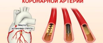
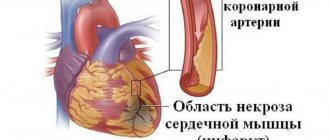
What does a cardiogram show during a heart attack?
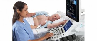
Electrocardiography (ECG) is a test performed first when a myocardial infarction is suspected. Based on the recorded cardiogram, several conclusions can be drawn:
1. The area of the dead zone located in the thickness of the heart muscle is indicated by a change in the QRS complex . Usually this part increases or a pathologically high Q wave .
2. About the place around the formed necrosis, which is affected by the displacement of the ST segment .
3. About the ischemic zone, which is located even further from the point of injury. An electrocardiogram records a violation of the myocardial restoration processes in the form of changes in the T wave .
The concept of ST segment elevation: above without atrial repolarization, below with atrial repolarization without QRS
During an ECG, each lead is recorded on the film with a new line, which helps to accurately determine the location of the cardiac lesion. In total, the electrocardiogram consists of 12 graphic lines and 5 teeth - P, Q, R, S, T. Each tooth is distinguished by its width, height, depth and goes in its own direction.
projection of ECG leads
projection of ECG leads
The dynamics of the development of a heart attack on the ECG become visible in the first minutes after the development of the disease. On the film, changes are displayed depending on the shape of the infarction, its location and stage of progression. They can be barely noticeable (with small focal lesions) or have a clear “classical” form, known to all literate people.
Post-infarction cardiosclerosis
An indication of post-infarction cardiosclerosis as a complication of coronary artery disease is included in the diagnosis no earlier than 2 months from the date of myocardial infarction. The diagnosis of post-infarction cardiosclerosis as an independent clinical form of IHD is established if the patient does not have angina pectoris and other forms of IHD provided for by the classification, but there are clinical and electrocardiographic signs of focal myocardial sclerosis (sustained rhythm disturbances, conduction, chronic heart failure, signs of scar changes in the myocardium on ECG). If in the long-term period of examination of the patient there are no electrocardiographic signs of a previous infarction, then the diagnosis can be substantiated by medical documentation data relating to the period of acute myocardial infarction. The diagnosis indicates the presence of a chronic cardiac aneurysm, internal myocardial ruptures, dysfunction of the papillary muscles of the heart, intracardiac thrombosis, determines the nature of conduction disturbances and heart rhythm, the form and stage of heart failure.
conclusions
The disease described is an extremely serious and life-threatening pathology. A person’s life depends on its timely diagnosis and treatment. Therefore, people should be very careful about their health and seek emergency help if symptoms of myocardial infarction appear. The examination of such a patient necessarily begins with an ECG. If latent forms are suspected, a cardiogram is taken in additional leads. If a patient who has previously suffered a heart attack has clinical signs of such a pathology, then a repeat episode of the disease is suspected. But often on the ECG, due to the scar from previously suffered atherothrombosis, new changes may not be seen. In such situations, additional research methods are used to determine markers of MI in the blood.
Features of exercises for pregnant women
Special exercises for pregnant women necessarily include those that help relieve the back muscles, strengthen the abdominal muscles, increase, if possible, the volume of the lungs, or teach the lungs to use their full capacity. During their implementation, the permissible load on the body is taken into account depending on the period.
What does an expectant mother need to know when starting to perform the complexes?
- Before classes, you must consult a doctor;
- It is necessary to take into account the specifics of the condition - for example, the loads of the first trimester are unacceptable in the third;
- The intensity of the training process is adjusted depending on the duration;
- If the condition worsens, palpitations and pain occur, training should be stopped immediately.
Exercises are performed at a slow pace, the loads are increased gradually. There should be no fatigue or muscle pain after exercise.
Special exercise – “cat”
One of the effective exercises for pregnant women that helps strengthen the back muscles is the cat breathing exercise.
In the classic version, it works like this:
- You need to get on all fours, leaning on a horizontal surface with your knees and palms. You should keep your head straight;
- Take a deep breath, tilting your head down, arching your back up;
- Count to yourself to 8, exhale, return to the starting position, relaxing your back muscles as much as possible.
This exercise is extremely useful for the female body. During it, all organs that will be involved during labor receive a load.
As the period increases, the cat exercise for pregnant women is adjusted. Already at the end of the first trimester, it is not recommended to bend too much, and by the 20th week it is performed only as a breathing and relaxing exercise.
Get into the starting position - on all fours. Breathe slowly and evenly, relaxing the abdominal and back muscles as much as possible. This exercise is especially recommended for women with increased body weight and, accordingly, a heavy load on the spine. Exercise cat reduces stress on the back and kidneys.
How does a heart attack manifest?
Symptoms of the dangerous disease IHD or the identified pathology may manifest differently in each person.
List of common symptoms:
- Pain in the area of the heart muscle behind the chest, which radiates to the left arm, to the area of the scapula, and with every slight movement it intensifies.
- Frequent shortness of breath that occurs when walking quickly, which cannot stop in a short time.
When similar symptoms occur frequently and last more than 30 minutes, the pain goes away after taking nitroglycerin, but again makes itself felt - this phenomenon is called angina pectoris. If such sensations intensify each time, then with any load on the body this means that the pathology is progressing.
Myocardial infarction is a very severe form of hypertensive pathology, manifested by burning sensations due to damage to the tissue of the heart muscle. If the pain continues to intensify and the patient's condition worsens, fear of death appears.
Other manifestations of coronary heart disease include arrhythmia, including atrial fibrillation, tachycardia, and disruption of intracardiac conduction by creating a block. Under such circumstances, palpitations, a feeling of fading and noticeable interruptions in the heart muscle occur.
The main enemy of ischemia is sudden death. It may result from necrosis or angina pectoris. In this case, the patient faints, cardiac and respiratory arrest occurs. This condition needs to be urgently reanimated.
If the pathology exceeds all worst-case scenarios, then when complaints are clarified, blue skin, pallor of the mucous membrane are observed, and fluid accumulates in the chest and abdominal area. The patient, as a rule, complains of chronic weakness, fatigue, shortness of breath and is forced to take a reclining and sitting position.