You are here: Blood test -
Platelets
- Structure
- Life cycle
- Functions
- Normal blood levels
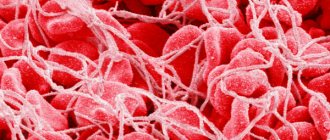
Platelets are components of blood that are oval in shape and may be slightly flattened in the center. The functions of platelets are difficult to overestimate, since these cells are responsible not only for blood clotting, but also for resistance to pathogenic microorganisms, and take an active part in the construction of blood vessels.
An increased or decreased amount of these formed elements in 1 mm3 of blood is a consequence of a certain pathological process in the body. If you have appropriate symptoms, you should immediately seek medical help, and not ignore the problem or self-medicate.
Structure
The structure of platelets is quite complex, and is not limited to just one plate with its constituent components. Each layer of the plate performs its functions:
- Outer layer or three-layer membrane. In the thickness of this membrane there is phospholipase A, which is responsible for the formation of a blood clot. Receptors are located here, which are responsible for adhesion to other plates and attachment to body tissues.
- Lipid layer. Consists of glycoproteins. The substance is responsible for gluing the components of the plate together and remaining in this state for a long time.
- Microtubules. Responsible for ensuring contraction of the structure and movement of cell contents outward.
- Organelle zone. It consists of various components that are generally responsible for wound healing.
It should be noted that microtubules are the cytoskeleton that forms the shape of platelets. The size of the “adult” body is in the range of 0.002–0.006 mm.
Cytoskeleton and shape change
The platelet cytosol is permeated by a three-dimensional network of water-insoluble protein threads (filaments), which forms the cytoskeleton. The filaments consist of polymerized actin protein and ensure that the platelet changes shape when activated. In addition, just below the plasma membrane is a membrane skeleton associated with the cytoplasmic “tails” of some receptors. It consists of short actin filaments connected to each other using special proteins. The membrane skeleton not only supports the plasma membrane, regulating the contours of the cell, and stabilizes it, preventing fragmentation, but also regulates the distribution of receptors attached to it in the plane of the membrane. It is also suggested that it plays an important role in the regulation of various intracellular events that are triggered upon activation.
Rice. 5.
Scanning electron micrographs of the process of spreading an activated platelet (
a–d
) over the surface []
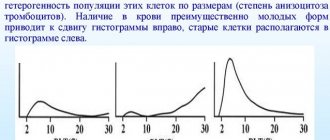
Interestingly, the cytoskeleton is a dynamic structure, thanks to which a platelet can not only change shape, but also grow “tentacles” (filopodia). With their help, it spreads over the surface of the damaged vessel (Fig. 5) and more easily adheres to other platelets (Fig. 6). Relatively recently, it was discovered that upon strong activation (by thrombin alone or together with collagen), platelets are divided into two groups (subpopulations), very different in properties and even shape, which suggests a fundamentally different organization of the cytoskeleton in them. Some of them (“regular” activated) have the appearance of amoebas - lumps with filopodia, others (procoagulant, since there is a lot of phosphatidylserine on the outer surface of their membrane) - balls without “tentacles”. The data obtained in our laboratory indicate that some membrane receptors responsible for binding cells to the surface and to each other are unequally attached to the cytoskeleton in platelets from the two subpopulations. This means that they can interact differently with the damaged vascular wall and with each other in the forming blood clot.
The sequence of processes during the restructuring of the platelet cytoskeleton has generally been studied quite little, but here is a new question: why do some cells become “amoebas” when activated, and others become “balls”?
Education and life cycle
The lifespan of platelets is significantly shorter than that of red blood cells - cell breakdown and death occurs on days 7–14; on average, these blood components live for about ten days.
Where are platelets destroyed? The process of platelet destruction occurs in the liver or spleen. The answer to the question of where platelets are destroyed is identical to the situation with red blood cells.
Where are these blood components formed? Cell production begins in the bone marrow, the place of development and maturation is non-hollow bones (vertebral area, pelvic bone).
These plates in the blood are formed as follows: the spongy mixture produces stem cells that do not have the ability to differentiate, that is, by their nature, they are not predisposed to one type or another. As a result of certain pathogenetic factors, they are converted into the necessary cells.
The resulting cell goes through several stages of formation:
- the stem cell becomes a megakaryocytic unit;
- the megakaryoblast stage begins;
- the already formed proplatelet becomes a promegakaryocyte;
- a full-fledged platelet is formed.
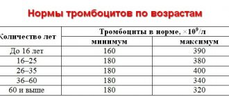
This is how the structure of a platelet occurs in several stages. The normal number of platelets in the blood for an adult is 150–375,000,000,000 per unit volume of blood. The norm of platelets in women and the norm of platelets in the blood of men differ, which is due to the peculiarities of the physiological structure of the human body.
Life cycle of platelets
Interaction between platelets and the vascular wall
The bloodstream constantly contains from 20 to 40% of activated “standby” platelets, ready to immediately begin the process of blood clotting when adhesion molecules appear. In the process of platelet-vascular interaction, the stages adhesion , their activation and aggregation .
Adhesion
When the vessel wall is damaged, the collagen of the basal membrane is exposed and a foreign “thrombogenic” contact surface is created. At the same time, adhesion proteins, primarily von Willebrand factor . The contact surface adheres to platelets and initiates the coagulation process.
The adhesion process involves the attachment of platelets caught in the damaged area to subendothelial structures. direct contact of platelets and collagen of the basement membrane occurs At the same time, von Willebrand f. isolated from damaged endothelial cells binds one part to the platelet receptor GPIb, and the other to subendothelial collagen.
Platelet adhesion
Once attached to the damaged surface, platelets become activated.
Activation
Adhesion of platelets to collagen (GPIa/IIa receptors) and interaction with von Willebrand factor (GPIb receptor) leads to their activation.
The binding of von Willebrand factor to the GPIb receptor triggers the calcium-phospholipid signal transduction mechanism and this ultimately leads to an increase in the intracellular concentration of Ca2+ ions and activation of protein kinase C. As a result:
- ATP-dependent aminophospholipid translocase, which maintains membrane asymmetry of phospholipids, is inhibited, and as a result, negatively charged phosphatidylserine .
- Together with phosphatidylserine, a special glycoprotein ( tissue factor ) comes to the surface, and a tissue factor complex is formed. The membrane becomes a surface for the interaction of plasma clotting factors, also called platelet thromboplastin.
- thrombostenin protein is reduced, as a result degranulation and factors that activate adhesion and aggregation are released,
- the shape of the platelet changes, pseudopodia appear, and it spreads out on the contact surface,
Processes that occur during platelet activation
- Phospholipase A2 is activated, which splits off polyunsaturated (for example, arachidonic) acid from the membrane phosphatidylcholine and from it thromboxane A (for example, thromboxane A2) is synthesized, a strong inducer of platelet aggregation and a vasoconstrictor. Thromboxane counteracts the effects of prostacyclins by interfering with the activation of adenylate cyclase and interrupting the effects of prostacyclins.
Antagonism of the action of prostacyclins and thromboxanes
Thromboxane further accelerates the release of active substances ( prothrombin , PAF, ADP Ca2+ ions , serotonin , thromboxane A , etc.) from the activated platelet, which maintains and enhances the activation of this and neighboring platelets. Activation is also enhanced by the action of ADP , released from damaged erythrocytes and endothelial cells of the vascular wall.
Already activated platelets have on their surface receptors for active and inactive factors V, VIII, IX, X, XI, prothrombin and thrombin.
Aggregation
The process of aggregation involves the stabilization of the thrombus by fibrin and the adhesion of activated platelets to each other.
Any triggering signal in an activated platelet leads to conformational changes in the GPIIb/IIIa receptor, which moves to the membrane. After binding to this receptor, fibrinogen acts as a bridge between adjacent platelets and a fibrinogen-reinforced platelet thrombus is formed in the damaged area. At first, the connection between platelets is not yet strong and such aggregation is reversible . Activation and aggregation is maintained by continuous secretion of granule contents from binding platelets.
Continued degranulation of platelets and their secretion of prostaglandins (PgG2 and PgH2), thromboxane A2, ADP, and the conversion of fibrinogen to fibrin (catalyzed by thrombin) make aggregation irreversible . Such a platelet is tightly bound to other cells; it has lost the contents of the granules and cannot return to its original state.
Platelet aggregation
Retraction
Retraction is the compaction of a blood clot with the release of excess serum. The stimulus for retraction is various substances released by the platelet during the stages of activation and aggregation. Retraction occurs due to the fact that the contractile protein thrombostenin (similar to actomyosin in muscle fibers) of the GPIIb/IIIa receptors
Compression of the clot causes an increase in pressure inside the platelet and causes an additional release of substances from its granules, which further enhances retraction and finally compacts the thrombus. Normally, bleeding from small vessels lasts no more than 5 minutes.
0
Functions
The shape of platelets and their structure are aimed at performing the main function - stopping blood in the event of mechanical damage to the integrity of the skin and tissues. Blood plates perform the following functions:
- serotonin metabolism;
- protective - the plates capture foreign cells and destroy them;
- release of growth factors, since after their death the components that are responsible for this are released;
- hemostatic - for its implementation, cells are grouped into large and small compounds.
Therefore, platelets in the blood are very important, which means it is necessary to maintain their optimal number. This is what annual preventive examinations at the clinic are intended for.
How red blood cells transport hemoglobin in the body
Passing through the capillaries of the lungs, where there is the greatest oxygen tension, the hemoglobin in the blood is completely saturated with oxygen.
This process occurs according to the laws of gas diffusion. Oxyhemoglobin is then transferred to the capillaries of other body tissues, where the oxygen tension is very low, making it easily separated from hemoglobin. The released oxygen is used by cells to maintain their energy metabolism.
Domestic scientist P. A. Korzhuev, using examples of individuals of the animal world at various levels of development, showed that the placement of different species of animals in the evolutionary series depends on their provision of hemoglobin (and therefore oxygen).
- For example, fish have relatively little hemoglobin per kilogram of body weight;
- Amphibians (the next stage of development) have a little more;
- Birds have even more of it, etc.
- The largest amount of it is contained in the blood of mammals.
What happens to dead red blood cells
The main task of red blood cells is to carry oxygen. They have a minimal metabolism. On average they live 100-120 days. As they age, red blood cells undergo decay: at the end of their life in the spleen and liver they stick to special cells on the walls of blood vessels.
Such cells have the ability to capture various high-molecular and foreign particles that enter the blood. This process of absorption (phagocytosis) also extends to aged red blood cells, which have already become foreign to the body.
The spleen is directly related to the process of blood destruction. This organ, a “spongy sac” of very loose tissue filled with blood, is capable of destroying red blood cells, which has long given rise to calling it the “graveyard” of these cells. (According to some data, over 70% of all red blood cells that have completed their life cycle end up in it).
It should be noted that in a healthy person, the spleen destroys only old or accidentally damaged red cells. What is the mechanism for releasing blood from those that have already become obsolete or damaged? This was discovered through interesting experiments on animals using modern electron microscopy.
Rats were injected with substances toxic to erythrocytes and their passage through the wall of splenic vessels was observed. Normal cells are easily filtered through vascular pores: when passing through them, “flexible” red blood cells change their shape and slip into the general blood flow.
But as they age or become damaged and become less elastic, they are no longer able to penetrate the capillaries, are filtered in the spleen and are absorbed (phagocytosis) by reticuloendothelial cells. When red blood cells break down in the liver, the pigment bilirubin is formed, which in the intestine, under the influence of microbes, undergoes further chemical transformation.
Blood cells had an evolutionary predecessor
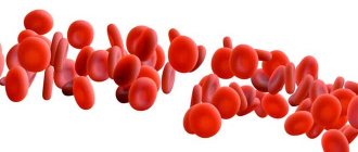
The blood of humans and other vertebrates consists of a liquid medium - plasma and cells suspended in it: red blood cells, platelets and leukocytes. Red blood cells carry oxygen, white blood cells protect the body from external and internal pathogenic agents, and platelets clog damage to the walls of blood vessels and participate in blood clotting. Although the difference between them is colossal, during the process of hematopoiesis (hematopoiesis) they are created from one precursor - hematopoietic stem cells of the bone marrow. During evolution, blood cells evolved from amoebocytes, colorless cells in the body of invertebrates.
The first organisms on Earth were single-celled. When multicellular organisms appeared, there was a need to deliver nutrients to every cell of the body. Some organisms pumped fluid through their bodies, others tried to be as flat as possible. The most effective option is a liquid internal environment, which carries nutrients and carries away waste products. But there is one problem: how to protect yourself from the leakage and invasion of foreign bodies and organisms during injuries? In response to these threats, special cells have evolved - amoebocytes, which are found in many invertebrates.
Amebocytes float in a liquid environment, attack, capture and digest foreign particles and microbes and regenerate damaged tissues. But these cells do not carry oxygen: in insects the trachea is responsible for oxygen delivery, and in many other organisms the carrier pigment is simply dissolved in the blood. Vertebrates cannot live like this: their flow rates and pressure are too high, and the protective cells themselves cannot swim to the site of damage. The immune system has evolved to require a more advanced system to counteract external and internal pathogens. In addition, vertebrates need a lot of oxygen, and simply dissolving the oxygen carrier in a liquid will not work: because the medium will be too thick. Therefore, it needs to be packaged in something. As a result, in vertebrates, amoebocytes split into several dissimilar branches and became blood cells.
Mechanism of thrombus formation
When blood vessels are damaged, changes occur in the blood. Thanks to the release reaction, enzymes and compounds are released that convert lycoprotein, an inert protein, prothrombin (produced by the liver) into thrombin (serine protease), a clotting factor. Fibrinogen (plasma protein) is transformed into fibrin. Red blood cells are retained in its threads.
Platelets secrete substances that stimulate vascular contraction and stick closely together, stretching the fibrin threads. The impulse is transmitted to the fibrin threads, they constantly tighten, compressing the blood clot. Gradually, the dense formation becomes impenetrable and decreases in size—the bleeding stops.
Description of blood cells
The structural components of blood are divided into 2 fractions - red and white. Platelets are classified as red. They do not have nuclei and are synthesized by the bone marrow from megakaryocytes, large cells with a standard structure (volumetric nuclei).
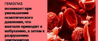
In a calm state, if there is no vascular damage, the red blood cells are flat and resemble circles or ovals in appearance. They can only be seen under a microscope; their sizes are 2-5 microns. As soon as the integrity of the capillaries, arteries or veins is compromised, the shape of the platelets changes - they increase in size and swell.
By the appearance of the structural components of physiological fluid, you can understand what is happening in the body:
- Calm state - no changes. 90% of cells are fully mature.
- With massive blood loss, under a microscope you can see a lot of large platelets on a glass slide; the bone marrow is intensively producing new cells.
- When hematopoiesis is disrupted, anucleate cells are small and degenerative.
- Slow development and uneven shapes suggest a malignant process.
- A sharp increase in size indicates diseases of the hematopoietic system.
The lifespan of platelets is 8 days. They are then destroyed in the spleen, bone marrow and liver. During normal life, renewal is continuous. Characteristics by size: megaforms, macroforms, microforms and normoforms.
Platelet Purpose
When vascular walls are damaged, the synthesis of flat anucleate blood cells increases, which are released into the bloodstream. The reserve from the spleen is also used. During the process of aggregation, a blood clot is formed on the surface of the wound, which closes the wound. The main function of platelets is hemostasis, stopping bleeding. But you shouldn’t hope that any wound, including a deep one, will heal. Blood is expelled from the arteries under pressure. Aggregation does not occur quickly enough, and if help is not provided on time, the likelihood of death is high.
But this is not the only function of nuclear-free flat blood cells. They transport nutrients through the blood vessels, normalize the permeability of the walls and increase tone. The structural features of platelets slow down age-related changes, reduce the fragility of blood ducts, and prevent internal hemorrhages.
Not only a decrease in the level of platelets in the blood is dangerous, but also an increase. Symptoms of thrombocytosis are abdominal pain localized on the left side (in the area of the spleen), frequent headaches, and the appearance of bruises even with mild physical impact. In this case, the risk of blood clots, kidney dysfunction, and the development of heart attacks or strokes increases. If such signs appear, you should consult a doctor.
Secondary thrombocytosis occurs without severe symptoms against the background of general malaise. That is why people over 45-50 years old need to take a detailed blood test at least 2 times a year.
Content
Platelets are the most important component of blood. The role of platelets in peripheral blood analysis is not clear to the average person, but this indicator can tell a doctor a lot. Blood is not a homogeneous liquid running through the vessels; erythrocytes, leukocytes, and different types, circulate in it. Platelets and other blood components are essential for the human body. Each of the elements plays an important role.