Translated from the ancient Greek language, “tachy” means fast and “cardia” means heart.
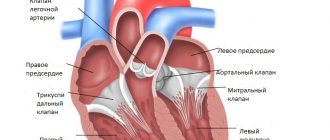
Tachycardia is rapid heartbeat. It is not an independent disease, but only indicates a violation in the regulation of heart activity. In addition, not every tachycardia is a disease; in some cases it is a normal reaction of the body. In order to understand the causes of tachycardia, you need to know the physiological norm. So, the human heart consists of four chambers - two atria, two ventricles.
The right atrium receives blood from the organs and has little oxygen. Then it enters the right ventricle, and from there straight into the vessels of the lungs. Here the blood is maximally saturated with oxygen and rushes to the left atrium and ventricle, from where it is distributed to all tissues of the body. This is how they receive the element necessary for life – oxygen.
The sinus node (a collection of special cells in the right atrium) causes the chambers of the heart to contract. It is the main one and in a healthy body suppresses all underlying foci of impulse. The node generates an electrical impulse, which first runs through the atria, then through the ventricles, and they contract.
The sinus node provides heart contraction at a rate of 60 - 90 per minute (normocardia). Anything less than this value is called bradycardia (“brady” - slow, “cardia” - heart), and more - tachycardia.
What is cardiography
The essence of cardiography is the study of electrical currents arising during the work of the heart muscle. The advantage of this method is its relative simplicity and accessibility. Strictly speaking, a cardiogram is the result of measuring the electrical parameters of the heart, displayed in the form of a time graph.
The creation of electrocardiography in its modern form is associated with the name of the Dutch physiologist of the early 20th century, Willem Einthoven, who developed the basic ECG methods and terminology used by doctors to this day.
Thanks to the cardiogram, it is possible to obtain the following information about the heart muscle:
- Heart rate,
- Physical condition of the heart
- The presence of arrhythmias,
- The presence of acute or chronic myocardial damage,
- The presence of metabolic disorders in the heart muscle,
- Presence of electrical conductivity disturbances,
- Position of the electrical axis of the heart.
Also, a cardiac electrocardiogram can be used to obtain information about certain vascular diseases not related to the heart.
An ECG is usually performed in the following cases:
- Feeling of abnormal heartbeat;
- Attacks of shortness of breath, sudden weakness, fainting;
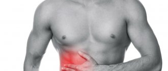
- Heartache;
- Heart murmurs;
- Deterioration of the condition of patients with cardiovascular diseases;
- Passing medical examinations;
- Medical examination of people over 45 years of age;
- Examination before surgery.
An electrocardiogram is also recommended for:
- Pregnancy;
- Endocrine pathologies;
- Nervous diseases;
- Changes in blood counts, especially with an increase in cholesterol;
- Over 40 years of age (once a year).
Diagnosis of the disease
To carry out high-quality diagnostics, it is necessary to complete all successive steps:
- studying the anamnesis by interviewing the patient and studying the history of his diseases. What matters is the strength and frequency of manifestation of arrhythmia symptoms, how long ago the first signs of abnormalities appeared, even the time of day characteristic of the manifestations of the disease is important,
- making a primary diagnosis is possible based on the results of auscultation - listening to the rhythm and heart rate using a stethoscope,
- laboratory tests of blood and urine: general and biochemical analysis, determination of hormonal levels,
- conducting electrocardiography. The procedure helps determine the type of arrhythmia. In some cases it is performed with a load,
- ultrasound examination to determine the affected areas of the myocardium and other heart pathologies,
- MRI allows you to clarify the stage of the disease, identify complications in other organs, pathologies affecting the heart rhythm,
- diagnosing tachycardia using carotid sinus stimulation. Changes in heart rate are monitored when pressing on the right carotid artery. The cells in this area perform a regulatory function in heart rate.
Methodology of the procedure
ECG recording is usually performed in a supine position. To take a cardiogram, a stationary or portable device is used - an electrocardiograph. Stationary devices are installed in medical institutions, and portable ones are used by emergency teams. The device receives information about electrical potentials on the surface of the skin. For this purpose, electrodes are used that are attached to the chest area and limbs.
These electrodes are called leads. There are usually 6 leads installed on the chest and limbs. The chest leads are designated V1-V6, the leads on the limbs are called basic (I, II, III) and reinforced (aVL, aVR, aVF). All leads give a slightly different picture of oscillations, but by summing up the information from all electrodes, you can find out the details of the functioning of the heart as a whole. Sometimes additional leads are used (D, A, I).
Typically, the cardiogram is displayed in the form of a graph on paper containing millimeter markings. Each electrode lead has its own schedule. The standard speed of the belt is 5 cm/s; other speeds may be used. The cardiogram displayed on the tape can also indicate the main parameters, normal indicators and a conclusion generated automatically. Data can also be recorded in memory and on electronic media.
After the procedure, the cardiogram is usually deciphered by an experienced cardiologist.
What does sinus rhythm look like on an ECG, normally and with pathologies?
During pregnancy, the heart experiences significant stress. The organ begins to function at an accelerated rate, enriching the maternal and child’s body with oxygen. As a result, arrhythmic manifestations during pregnancy are quite common conditions.
Abnormal heart rhythm can result from various diseases or high cardiac load. Pregnant women experience sinus rhythm with a heart rate that exceeds the normal values by 10 beats over a one-minute period. If sinus rhythm disturbances occur as a result of gestation, they disappear on their own at the end of the birth process.
Conclusion The ECG is called an electrocardiogram. It allows you to record the rhythmic contractions of the heart on paper in the form of a special graph. An ECG records information from a person’s limbs and cardiac zone. The sinus rhythm of the heart is determined using standard leads, which are designated by Roman numerals I, II, III.
Doctors analyze the following components of the electrocardiogram:
- P wave;
- PQ distance;
- QRS complex;
- P-wave spacing;
- distance between R teeth;
- number of heart beats.
Holter monitoring
In addition to stationary devices, there are also portable devices for daily (Holter) monitoring. They are attached to the patient's body along with electrodes and record all information received over a long period of time (usually within 24 hours). This method provides much more complete information about processes in the heart compared to a conventional cardiogram. For example, when taking a cardiogram in a hospital setting, the patient must be at rest. Meanwhile, some deviations from the norm may appear during physical activity, sleep, etc. Holter monitoring provides information about such phenomena.
Photo: kostastudio/Shutterstock.com
Irregular sinus rhythm: appearance on the ECG, treatment, symptoms
Additional education:
"Cardiology"
State educational institution "Institute for Advanced Medical Studies" of the Ministry of Health and Social Development of Chuvashia
Contacts
Physiological processes in the body are determined not only by internal biochemistry - they are also influenced by external factors. Changes in body temperature, breathing rate and heart rate are a natural reaction to the external environment, stress, and psycho-emotional state. Cardiac activity directly depends on the momentary state of a person.
Sports, physical work, and stress make the heart beat faster, as muscles and nerve tissues require more oxygen. At rest, the heart returns to its normal rhythm. Irregular heart rhythm can also be a response. When should you be concerned and contact a cardiologist?
What is sinus rhythm
Sinus heart rhythm means normal heart function.
The electrical impulses that cause the heart to contract and pump blood evenly come from the right place - the sinus node, located in the upper part of the right atrium.
Pulses are generated at regular intervals. The heart rate (HR) for a healthy adult at rest is 60–90 beats per minute. This is called regular sinus rhythm.
Deviations from the genetically determined mode of cardiac activity are called arrhythmias. Rhythm disturbances occur in different parts of the heart muscle - in the atria, ventricles and septa. If the cause is a malfunction of the sinoatrial node, then the arrhythmia is called sinus.
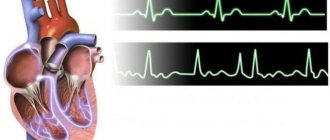
This is what sinus rhythm looks like on an ECG
Sinus tachycardia
Sinus tachycardia is an increase in heart rate to 100 beats per minute or more. In this case, the interval between contractions remains the same. During and immediately after physical or nervous stress, this condition is considered normal. At rest, the heartbeat returns to normal. If an increase in heart rate begins for no apparent reason, this indicates pathology.
Sinus bradycardia
A slow, uniform heartbeat occurs in physically trained people. With each beat, the heart pumps enough blood to fully supply the body with oxygen and nutrients. Therefore, a heart rate of 59–50 beats is the norm for them. The cause of pathological slow heartbeat is certain diseases that affect the functioning of the sinus node.
Sinus arrhythmia
The condition when the frequency of contractions remains normal (increased or decreased), but the intervals between them differ, is called sinus arrhythmia. The reason for its appearance in children and adolescents is uneven growth and development of organs or respiratory arrhythmia. Sinus arrhythmia as an isolated disease is more common in older people.
Sinus arrhythmia - view on the ECG
Changes in the sinus rhythm of the heart sometimes do not manifest significant symptoms. But they are clearly visible on the ECG. Electrocardiography shows the parameters of the heart muscle - the uniformity and frequency of contractions, the conductivity of muscle fibers, the work of the cells that generate the impulse.
To do this, the doctor evaluates the teeth, intervals and segments on the displayed graph. What matters is their presence or absence, sequence, height, location and direction. Each parameter has a numerical value. Teeth are areas located above or below the isoline. They show moments of excitation and relaxation of the myocardium of different parts of the heart:
- P - wave, reflecting contraction and relaxation of the atria;
- Q, S - waves showing excitation of the septum between the ventricles;
- R—ventricular excitation parameter;
- T – the process of their relaxation.
The P-Q interval is the time it takes for an impulse to travel from the atrium to the ventricles. The QRS segment is the ventricular complex (reflects the excitation of the ventricles), T–P is the period of diastole (relaxation of the heart muscle).
Heart rate is determined by RR intervals - their number in 3 seconds is multiplied by 20. Normally, this figure ranges from 60 to 90 contractions.
If sinus rhythm is disturbed, the following changes are observed on the ECG:
ECG indicatorsNormalTachycardiaBradycardiaSinus arrhythmia
| Heart rate | 60–90 | 100 and above | Less than 59 | – norm – tachycardia – bradycardia |
| P waves and QRS complexes | Correct alternation in all cycles | Correct alternation in all cycles | Correct alternation in all cycles | Correct alternation in all cycles |
| P waves | 1.5–2.5 mm | Amplitude increased | Amplitude reduced | Norm |
| PQ interval | 0.12–0.2 s | Shortened | Increased | Minor interval changes |
| RR intervals | Uniform, 0.15 s | Uniform | Uniform | Changes abruptly, lasting more than 0.15 s |
Sinus arrhythmia - physiological and pathological
Irregular sinus rhythm can be physiological and pathological. The cause of physiological arrhythmia is often the breathing process, which is inextricably linked with the work of the heart. When you inhale, it beats faster, and when you exhale, it slows down.
This irregular rhythm in some people is a feature of the body and occurs constantly. Physiological sinus arrhythmia does not interfere with blood circulation and does not affect cardiac activity in any way. Respiratory arrhythmia manifests itself:
- when overworked or in moments of stress;
- in childhood and adolescence;
- with vegetative-vascular dystonia;
- after severe infectious diseases.
Pathological sinus arrhythmia has cardiac and extracardiac causes. Cardiac - diseases or defects of the heart.
Extracardiac origin is associated with other pathologies that disrupt the functioning of the heart - hypertension, viral infections, diseases of the lungs and thyroid gland. In older people, heart rhythm is affected by age-related changes.
Bad habits, lack of potassium and magnesium, obesity are also possible causes of sinus arrhythmia.
Symptoms
Heart problems sometimes cause panic attacks. They worsen existing symptoms:
- shortness of breath;
- inability to take a full breath;
- weakness;
- dizziness.
During an attack, the feet and hands become cold, the person feels blood pulsating in the temples, and pain in the heart area may appear, radiating to the left arm. One of the indirect signs of pathological sinus arrhythmia is mood swings and irritability.
Features of sinus arrhythmia in children
A child's heart rate is significantly different from an adult's. In newborns, the normal heart rate is between 120 and 170 beats per minute. With age, the heart rate decreases and reaches adult values by adolescence.
Doctors distinguish three types of arrhythmia - mild, moderate and severe. In moderate form, it occurs in children under 5 years of age and in adolescents. Severe bradyarrhythmia occurs after rheumatism or in children involved in sports.
The main causes of arrhythmias are congenital heart defects, endocrine pathologies, metabolic disorders (lack of microelements and impaired water-electrolyte metabolism). In some children, arrhythmia appears during periods of rapid growth - at 5–7 and at 9 years. In adolescence, it is caused by vegetative-vascular dystonia.
Each type of arrhythmia is characterized by certain threshold values of heart rate. Severe bradycardia in children under one year of age is a pulse less than 100 beats. In children from 2 to 7 years old, it is diagnosed with a heart rate of less than 75, from 8 to 18 years old - with a heart rate of less than 62 beats per minute. Severe tachycardia occurs when normal pulse values are exceeded by 40–60 beats.
Common symptoms for all types of sinus arrhythmias in children:
- fast fatiguability;
- intolerance to stuffiness;
- restless behavior (typical of children under one year old);
- poor appetite;
- pale skin;
- When counting the pulse, interruptions are heard (uneven rhythm).
Older children complain of headaches and morning dizziness. If these symptoms are observed continuously, the child should be examined by a cardiologist.
How to treat arrhythmia in a child
First of all, it is necessary to organize the child’s life in such a way as to minimize the risk of further development of the disease and complications. It is necessary to protect him from any conflicts in the family and reduce his time in front of the TV or computer. Daily long walks, balanced nutrition and physical activity will benefit your health.
Sinus arrhythmia in children is not treated with special medications. Usually it is symptomatic, and the efforts of doctors are aimed at correcting the underlying disease.
Treatment
Not all types of sinus arrhythmia need to be treated. Doctors distinguish two types - cyclic arrhythmia associated with breathing and non-cyclic or pathological. Cyclic arrhythmia is not treated, considering it physiological. In case of pathological arrhythmia, diagnostics are carried out to determine the cause of the rhythm disturbance. The choice of treatment depends on the type of pathology detected and the cause of its occurrence.
If heart failure is caused by chronic stress, the doctor prescribes sedatives. They improve sleep, relieve irritability and increased excitability. For nervous disorders, tranquilizers are used.
If the heart rhythm disturbance is organic and associated with changes in the heart muscle, antiarrhythmic drugs are prescribed. In addition to medications, the doctor gives recommendations:
- make changes to your diet - give up junk food, strong tea and coffee, sweets and alcohol;
- normalize weight (for obesity);
- use traditional medicine.
A patient with sinus bradyarrhythmia less than 40 beats per minute requires implantation of a pacemaker. Often arrhythmia is symptomatic, that is, it is a consequence of another disease. After it is cured, cardiac activity returns to normal, and the risk of complications tends to zero.
Source: https://CardioPlanet.ru/zabolevaniya/aritmiya/ritm-sinusovyj-neregulyarnyj
Other types of procedures
There are several other methods for carrying out the procedure. For example, this is monitoring with physical activity. Abnormalities are usually more pronounced on the stress ECG. The most common way to provide the body with the necessary physical activity is a treadmill. This method is useful in cases where pathologies can only manifest themselves in the case of increased heart function, for example, if coronary artery disease is suspected.
During phonocardiography, not only the electrical potentials of the heart are recorded, but also the sounds that arise in the heart. The procedure is prescribed when it is necessary to clarify the occurrence of a heart murmur. This method is often used when heart defects are suspected.
How to treat an increased heart rate of 80 beats: preventive measures
If a heart rate of 80 beats is too high for you or is at a constant level, but there are no sudden jumps, then you can simply reconsider your regime.
- Exercise more and take a walk outside every day. Your heart will beat faster during activity. Regular exercise gradually slows your resting heart rate.
- Stress should be reduced —try meditation, relaxation, or other stress-relieving techniques to help lower your resting heart rate
- Avoid smoking, as tobacco products increase your heart rate. Therefore, reducing it may help improve oxygen absorption and your heart health.
- Lose weight if necessary. The larger the body, the harder the heart must work to supply it with blood. Losing weight may help slow your heart rate
- Run at least 3 times a week for at least 30 minutes. This is one of the most effective methods of training your heart. But you don’t need to push yourself to the point of exhaustion - do the load gradually!
- Drink at least 1-1.5 liters of water per day. Often dehydration of the body gives such a signal
- Rest and sleep at least 7-8 hours. Don’t forget that overwork is far from the only cause of increased heart rate.
A few facts
- Watch your diet and take proper vitamins. By the way, they can be obtained from food. But we include in our diet: potassium, which protects against high blood pressure and heart attack
- magnesium, which helps maintain potassium levels and regulates heart rhythms
- ascorbic acid or vitamin C for elasticity and strengthening of blood vessels
- selenium, which prevents blockage of blood vessels
- chromium to reduce coronary heart risk
- Coenzyme Q10 to energize the heart
- Omega 3, as an indispensable storehouse for the functioning of the heart and great vessels
Even if a pulse of 80 beats per minute is normal for you and your body, you should not ignore the body’s signals to visit a doctor. Even a small adjustment in your routine will help improve not only your heart rate, but also your overall well-being. Giving up bad habits and food will only add strength and energy! Take care of yourself and your body!
Types of teeth
First we should talk a little about how the heart works. It has 4 chambers - two atria and two ventricles (left and right). The electrical impulse, due to which it contracts, is formed, as a rule, in the upper part of the myocardium - in the sinus pacemaker - the sinoatrial (sinus) node. The impulse spreads down the heart, first affecting the atria and causing them to contract, then passes through the atrioventricular nerve node and another nerve node, the bundle of His, and reaches the ventricles. The main burden of pumping blood is taken on by the ventricles, especially the left one, which is involved in the systemic circulation. This stage is called heart contraction or systole.
After contraction of all parts of the heart, the time comes for their relaxation - diastole. The cycle then repeats again and again - this process is called heartbeat.
The condition of the heart, in which there are no changes in the propagation of impulses, is reflected on the ECG in the form of a straight horizontal line, called an isoline. The deviation of the graph from the isoline is called a spike.
One heartbeat on the ECG contains six waves: P, Q, R, S, T, U. The waves can be directed both up and down. In the first case they are considered positive, in the second - negative. The Q and S waves are always positive, and the R wave is always negative.
Photo: Infmedserv.ru
The teeth reflect the different phases of heart contraction. P reflects the moment of contraction and relaxation of the atria, R – excitation of the ventricles, T – relaxation of the ventricles. Special designations are also used for segments (spaces between adjacent teeth) and intervals (sections of the graph that include segments and teeth), for example, PQ, QRST.
Correspondence between the stages of heart contraction and some elements of cardiograms:
- P – atrial contraction;
- PQ – horizontal line, the transition of the discharge from the atria through the atrioventricular node to the ventricles. The Q wave may be absent normally;
- QRS – ventricular complex, the element most often used in diagnostics;
- R – ventricular excitation;
- S – myocardial relaxation;
- T – ventricular relaxation;
- ST – horizontal line, myocardial recovery;
- U – may be absent normally. The reasons for the appearance of the prong are not clearly understood, but the prong is valuable for diagnosing certain diseases.
Below are some abnormal ECG findings and their possible explanations. This information, of course, does not negate the fact that it is more advisable to entrust the decoding to a professional cardiologist who better knows all the nuances of deviations from the norm and associated pathologies.
Main deviations from the norm and diagnosis
| Description | Diagnosis |
| The distance between the R teeth is not the same | atrial fibrillation, heart block, sinus node weakness, extrasystole |
| P wave is too tall (more than 5 mm), too wide (more than 5 mm), has two halves | atrial thickening |
| The P wave is absent in all leads except V1 | the rhythm does not come from the sinus node |
| PQ interval extended | atrioventricular block |
| QRS extension | ventricular hypertrophy, bundle branch block |
| No gaps between QRS | paroxysmal tachycardia, ventricular fibrillation |
| QRS as a flag | heart attack |
| Deep and wide Q | heart attack |
| Wide R (more than 15 mm) in leads I, V5, V6 | left ventricular hypertrophy, bundle branch block |
| Deep S in III, V1,V2 | left ventricular hypertrophy |
| ST is more than 2 mm above or below the isoline | ischemia or heart attack |
| Tall, double-humped, pointed T | cardiac overload, ischemia |
| T merging with R | acute heart attack |
Table of cardiogram parameters in adults
| Index | Value,c |
| QRS | 0,06-0,1 |
| P | 0,07-0,11 |
| Q | 0,07-0,11 |
| T | 0,12-0,28 |
| PQ | 0,12-0,2 |
Normal duration of cardiogram elements in children
| Index | Value,c |
| QRS | 0,06-0,1 |
| P | <0,1 |
| PQ | 0,2 |
| QT | <0,4 |
The norms indicated in the table may also depend on age.
Main forms of arrhythmia and their characteristics
Heart rate is the basis for classifying heart rhythm deviations into the following forms:
- normocardia: 60-90 heart beats per minute, heart rate is normal, rhythm is disturbed,
- bradycardia: less than 60 heart beats per minute, characterized by a slow heart rate, blood supply to organs and tissues is disrupted,
- tachycardia: more than 100 heart beats per minute, accelerated heartbeat, weak heart filling and blood ejection, resulting in tissue hypoxia.
If we classify pathology according to the cause of its occurrence, we distinguish the following groups of diseases:
- Normotropic disorders of automaticity. The sequence and correctness of the phases of the cardiac cycle do not change, the mode of generation of impulses by the sinus-atrial node is changed. The heart speeds up or slows down its rhythm.
- Heterotropic disorders of automaticity arise due to the influence of the lower nodes of the conduction system on heart rate. The impulse control center is displaced, the synchrony of contractions is disrupted.
- Extrasystole occurs when the ability of the myocardium to perform a response function to incoming impulses is impaired. Incomplete contractions are formed due to the generation of signals by other parts of the heart.
- The mixed type is distinguished by an increase in the supply of exciting impulses and a simultaneous decrease in their conductivity. This type includes atrial fibrillation and cardiac flutter.
Rhythm of contractions
Violation of the rhythm of contractions is called arrhythmia. The irregularity of the rhythm during arrhythmia is measured as a percentage. An irregular rhythm is indicated by a deviation in the distance between similar teeth by more than 10%. Sinus arrhythmia, that is, arrhythmia combined with sinus rhythm, may be normal for adolescents and young adults, but in most cases it indicates the onset of a pathological process.
A type of arrhythmia is extrasystole. They say it in the case when extraordinary contractions are observed. Single extrasystoles (no more than 200 per day with Holter monitoring) can also be observed in healthy people. Frequent extrasystoles that appear on the cardiogram in the amount of several pieces may indicate ischemia, myocarditis, or heart defects.
What types of sinus rhythm disorders can occur?
The ECG conclusion may have various errors.
Even if the electrocardiogram demonstrates features of sinus rhythm, a person may develop pathological processes. It happens that despite the fact that electrical impulses are generated in the sinus node, the rhythmic cardiac pulsation does not satisfy the norm. What pathologies of sinus rhythm are most common:
- An increased number of heart contractions may indicate that the patient has sinus tachycardia;
- On the contrary, a reduced number of heart beats may signal the development of sinus bradycardia;
- Irregularity of heart contractions, in other words arrhythmia, is characterized by the same frequency of beats that do not occur regularly. The specialist may also suspect that the patient has extrasystoles - unexpectedly occurring impulses in the intervals between normal heart beats. Another pathology that irregular beats may indicate is sick sinus syndrome. This pathology is characterized by a stable, rare heartbeat, moments of “stopping” of cardiac activity, and in addition, the alternate occurrence of an accelerated and slowed down rhythm;
- Disturbed regularity of sinus rhythm indicates the absence of response reflexes in the muscular lining of the heart to stimuli from the environment.
In some cases, the sinus rhythm of the heart undergoes certain disruptions, which can manifest themselves in:
- Arrhythmia and its varieties. This pathology indicates an irregular heart rhythm when there is an unstable sinus rhythm.
- Sinus tachycardia. Represents an accelerated heartbeat. Tachycardia of a physiological nature occurs in athletes during training sessions. The cause of this disorder is rapid blood circulation, which occurs as a result of stress, provoking a more intense contraction of the heart. Accelerated sinus rhythm can also be caused by increased adrenaline levels due to severe emotional turmoil. The pathological form of tachycardia becomes a consequence of the use of certain substances in the form of alcohol or medications. Diseases of the blood and heart can also cause a rapid heartbeat.
- Bradycardia. It is a condition when there is a disturbance in the repolarization process with a simultaneous decrease in heart rate. Most often, the causes of this pathology are infectious diseases.
- Coronary sinus rhythm. It is a type of cardiac contraction of a passive nature, when the myocardial region, localized close to the coronary cardiac minus, acts as the rhythm controller. The coronary sinus rhythm is detected only by ECG.
Even if the ECG shows signs of sinus rhythm, this does not mean that there cannot be any abnormalities in the body. It is possible that pulses occur in the main node, but do not correspond to normal characteristics. The most common violations are shown in the table.
| Nature of changes | Possible medical names and diseases |
| Too frequent contractions (more than 90/min) | Sinus tachycardia |
| Contractions too infrequent (less than 60/min) | Sinus bradycardia |
| Irregular heartbeats | Arrhythmia – irregular contractions with normal frequency |
| Extrasystole - spontaneous extraordinary impulses between regular contractions | |
Sick sinus syndrome:
| |
| Too regular rhythm | Rigidity of heartbeats - lack of response of the myocardium to any external influences |
Sinus rhythm disturbances can be both a variant of the norm and a signal of a serious pathology!
Heart rate
This option is the simplest and most understandable. It determines the number of contractions in one minute. The number of contractions may be higher than normal (tachycardia) or lower than normal (bradycardia). The normal heart rate in adults can range from 60 to 80 beats. However, the norm in this case is a relative concept, so bradycardia and tachycardia may not always be evidence of pathology. Bradycardia can occur during sleep or in trained people, and tachycardia can occur during stress, after exercise or at elevated temperatures.
Heart rate norms for children of different ages
| Age | Heart rate, beats/min |
| Newborns | 140-160 |
| 6 months | 130-135 |
| 1 year | 120-125 |
| 2 years | 110-115 |
| 3 years | 105-110 |
| 5 years | 100-105 |
| 8 years | 90-100 |
| 10 years | 80-85 |
| 12 years and older | 70-75 |
Photo: Africa Studio/Shutterstock.com
Pulse 80 beats per minute - is this normal: how to check what is normal?
It’s immediately worth noting why a pulse of 80 beats is cause for concern. Let us remind you that the heart rate (heart rate) should be similar to the number of pulse beats. And the higher the number of these contractions, the higher the load on the heart. But this does not necessarily require more blood to be pumped.
Important: The heart grows with a person, and also increases from regular physical activity. And a large heart makes significantly fewer contractions to pump the same amount of blood as a person without physical training and with an increased heart rate. For this reason, children's standards are slightly higher than those of adults! Also, do not forget that with age the frequency increases due to wear and tear of the heart. Therefore, the verdict is this: the lower the pulse, the better!
You can calculate the number of arterial beats or pulse rate (HR) at home yourself:
- on the wrist of the left or right hand, in the area of the radial artery. In this case, choose a place under the bend of the hand in the conditionally divided half on the left. That is, under the thumb
- can be measured along the ulnar artery also on the wrist, but on the right, conditionally under the little finger
- immediately at the beginning of the axilla on the axillary artery, in the area of the lateral wall
- a reliable and simple way to measure the pulse in the carotid artery, the so-called carotid pulse. A stronger blow is felt on the left side, but can also be measured on the right side of the neck. To do this, place the pads of your fingers just under the cheekbone.
Where and how to measure
It is worth noting that many people take such measurements at home using their thumb. But this is not entirely correct - the chance of incorrect calculation increases. Therefore, to determine the pulse, we use the pads of the thumb and middle fingers, and even better, together with the ring finger.
Important: Ideally, counting should be done for the entire minute, that is, 60 seconds. But methods are allowed when they count for 30 seconds and multiply by 2, or when they count only for 15 seconds and then multiply by 4.
What are the normal indicators?
Even when we rest, our heart continues to work. It's important to understand that there are many factors that affect your heart rate, such as age, weight, and even life situations. And what is good for one can be deadly for another.
Table of standards
As we can see from the table, the average value for an adult should not exceed the limit of 70-75. At the same time, the pulse rate can be used to judge the proper functioning of the cardiovascular system. And also a healthy person with a low heart rate lives significantly longer than those who have a high frequency. After all, the heart wears out to a lesser extent. Therefore, if your pulse is 80 beats per minute at rest, then this is already considered an overestimated reading! But we should not forget about other factors that influence such a norm. By the way, do not forget that the pulse should not be low either - the minimum acceptable limit is up to 50 beats!
Heart Rate Types
There are several types of heart rhythm, depending on where the nerve impulse begins to spread, causing the heart to contract:
- Sinus,
- Atrial,
- Atrioventricular,
- Ventricular.
Normally, the rhythm is always sinus. In this case, sinus rhythm can be combined with both a heart rate above normal and a heart rate below normal. All other types of rhythms are evidence of problems with the heart muscle.
Atrial rhythm
Atrial rhythm also often appears on the cardiogram. Is atrial rhythm normal or is it a type of pathology? In most cases, the atrial rhythm on the ECG is not normal. However, this is a relatively mild degree of heart rhythm disturbance. It occurs when the sinus node is suppressed or disrupted. Possible causes are ischemia, hypertension, sick sinus syndrome, endocrine disorders. However, isolated episodes of atrial contractions can also be observed in healthy people. This type of rhythm can take on both the character of bradycardia and the character of tachycardia.
Atrioventricular rhythm
Rhythm emanating from the atrioventricular node. With atrioventricular rhythm, the pulse rate usually drops to less than 60 beats per minute. Causes: weakness of the sinus node, atrioventricular block, taking certain medications. Atrioventricular rhythm, combined with tachycardia, can occur during heart surgery, rheumatism, and heart attack.
Ventricular rhythm
With ventricular rhythm, contractile impulses propagate from the ventricles. The contraction frequency drops to below 40 beats per minute. The most severe form of rhythm disturbance. Occurs in acute infarction, heart defects, cardiosclerosis, cardiac circulatory failure, and in a preagonal state.
Causes of sinus bradycardia
All factors influencing the development of pathology can be divided into two large groups: internal and external. In some cases, there is a joint influence of causes from one and another group. Their correct determination allows you to accurately establish a diagnosis and subsequently prescribe effective treatment.
Internal causes of sinus bradycardia
First of all, physiological
aging of the body
. Many processes slow down, resulting in age-related degeneration of organs and systems, including the heart muscle.
Cardiac diseases
are in second place in the list of external factors provoking the development of bradycardia. We are talking about inflammatory diseases of the myocardium, coronary heart disease, and cardiomyopathies. Some infiltrative pathologies such as amyloidosis and sarcoidosis also cause a slow heart rate.
Intracardiac causes:
- arterial hypertension;
- atrial fibrillation;
- myocardial infarction;
- heart defects;
- myocarditis;
- cardiac ischemia;
- degenerative disorders of the sinus node;
- atherosclerotic changes in the coronary arteries.
The development of sinus bradycardia is often observed in non-cardiac diseases. These include pathologies of various organs and systems of the body.
External factors for the appearance of sinus bradycardia
These include:
- disorders of the nervous system (stroke, trauma and infectious lesions of the central nervous system) and peripheral system (neuritis and neuralgia, tumor processes);
- improper use of medications, resulting in the development of toxic and poisonous conditions;
- alcohol abuse;
- hormonal imbalance (hypothyroidism, aldosteronism);
- physiological impact factors associated with fasting, aging of the body, low body temperature;
- hereditary predisposition.
The disease is often associated with various electrolyte disturbances
. With an increased concentration of calcium and potassium in the blood, as well as with a deficiency of the latter element, the heart rhythm may be disturbed towards slowing down.
Metabolic pathology
, associated with the thyroid gland or gallbladder, also negatively affects the performance of the heart muscle. First of all, diseases such as cholestasis (stagnation of bile) and hypothyroidism (insufficient functioning of the thyroid gland) affect.
Nervous system diseases
may be complicated by sinus bradycardia, which is often one of the first signs of a brain tumor. Neurotic conditions and increased intracranial pressure directly affect the heart rhythm, as well as vegetative-vascular dystonia, which is common at a young age.
In some cases, there may be not one, but several causes, then the symptoms become unclear and the course becomes protracted. But sometimes it is not possible to accurately determine the factor of influence, then they speak of an idiopathic manifestation of rhythm disturbance.