The essence of this analysis
Not every patient knows what a platelet count according to Fonio is. It is carried out on 1000 red blood cells in a material mixed with a special dye reagent. After this, the resulting number is recalculated in relation to 1 μl or 1 liter of blood.
The advantage of this method is a fairly accurate determination of the number of these blood cells, which naturally leads to a more accurate diagnosis.
Let's figure out exactly how platelets are counted according to Fonio.
What kind of analysis
The analysis makes it possible to obtain information about the number of particles. They participate in the process of blood clotting, create blood clots, form a substance necessary for tissue repair, promote cell division, normalize the permeability of cell walls and restore the elasticity of blood vessels.
The analysis is used in the diagnosis of cardiovascular diseases. An elevated platelet count is called thrombocytosis, and a low platelet count is called thrombocytopenia. The color of the particles changes according to age.
The study of platelets using the Fonio method allows you to calculate the exact number of cells and identify many pathologies. During the study, the proportion of microparticles in a blood smear per 1000 red blood cells is calculated. This indicator is converted to 1 ml or l. This method is effective for diagnosing diseases.
During the analysis, the blood is colored. For testing, blood is taken from a finger. The number of cells in the blood is a coagulogram. This test is performed on many people. It makes it possible to diagnose various pathologies.
Advantages and disadvantages
The Fonio method has many advantages and has been used for many years.
Advantages:
- Under a microscope, the studied material, size, color and number of particles are clearly visible.
- The study is carried out at any time of the day.
- The technique is easy to use, has a simple formula, and the analysis is highly informative.
- Now other research methods have appeared, but the Folio method is more informative.
Important information: What is ESR in a blood test and the norm in adults
Flaws:
- While other methods allow you to immediately obtain a ready result using instruments, the laboratory assistant must visually calculate the indicators under a microscope, which takes time and requires special qualifications.
- The collection of material - red blood cells - is carried out manually.
Preparation for the procedure
The last meal should be 8 hours before the test, since some foods affect the color of the particles being tested.
The day before the test, you should not play sports and avoid physical activity. You need to come to the laboratory in the morning. Taking a repeat test makes it possible to monitor the number of cells over time. The patient must inform the doctor in advance about the medications he is taking.
How is the analysis carried out?
The total number of platelets in a particular blood smear represents a coagulation profile. To carry out this analysis, capillary blood is taken from a finger. It is necessary for every patient being examined to take this test, since its results can help the attending physician determine the presence or absence of many dangerous diseases.
When taking a blood platelet level test, consider the following:
- The concentration of these blood cells is maximum after eating and physical activity. For this reason, it is necessary to submit the material at least eight hours after the last meal.
- This requirement also applies to physical activity: if a patient donates blood in a state of excitement or after heavy physical activity, the result may be false.
- Blood must be donated early in the morning.
- To achieve maximum accuracy, the analysis must be carried out twice more with an interval of 3-5 days.
What is the Fonio method?
Platelet count according to Fonio is an analysis that helps to identify the exact number of these cells in the blood and diagnose many diseases in a person. In this case, platelets are counted in a blood smear per thousand red blood cells, after which this number is recalculated in the ratio of 1 microliter or liter. The technique is considered very accurate and is used to diagnose many pathologies.
During the analysis, the blood is stained using the Romanovsky-Giemsa method
To carry out the analysis, blood is taken from a finger. The total number of platelets in a unit of blood is called a coagulogram. This type of study is prescribed to every patient, because the analysis helps to suspect and identify many diseases.
Calculation of results
To count the platelet count according to Fonio, a 2.6% solution of sodium ethylenediaminetetraacetate (EDTA) and a 14% solution of magnesium sulfate are used. Reagents are drawn up using a pipette to an ESR meter (or a Panchenkov capillary).
- First, one of the reagents is introduced into the test tube, which in the capillary should be at division level 75. Blood taken with a pipette is also added there (up to division level K).
- The components are thoroughly mixed, and then thin strokes are prepared.
- After this, the smears are stained using the Romanovsky-Giemsa method for 35-45 minutes if EDTA was used, or for two to three hours if magnesium sulfate was used. During this time period, platelets turn purple-pink.
How is platelet counting performed using the Fonio method? To do this, platelets and red blood cells up to 1000 red blood cells are counted in the field of view of the microscope. The number of platelets in the blood plasma is determined by multiplying their number in the smear by the number of red blood cells in 1 μl, and then the resulting value is divided by 1000. The specific color helps the doctor more accurately count the number of cells being examined.
The study is highly accurate, so a large number of doctors use the results obtained by this method.
The platelet rate according to Fonio is of interest to many.
How to count platelets
Laboratory workers perform platelet counting according to Fonio using several reagents. For analysis, magnesium sulfate or the drug ethylenediaminetetraacetate is used. The specialist draws these substances from the container with a pipette.
First of all, blood and 1 reagent are dripped into the container. The amount of the substance is brought to 75, the plasma is brought to the K mark. The resulting solution is stirred; it is needed to create a smear. When using magnesium sulfate, the smear changes color in 120 minutes. When ethylenediaminetetraacetate is used, the smear is stained within 40 minutes. The particles take on a purple hue.
Cells are counted in a smear using a microscope. When counting, to determine the level of platelets, they are multiplied by the content of red blood cells in 1 ml, then divide the value by 1000. To make the result more accurate, a particle staining technique is used.
Important information: What does it mean to have elevated eosinophilic cationic protein (protein) in a blood test?
Features of the method
This technique is by far the most popular and widespread. The main difference of this technique is that the formed elements are counted in a stained blood smear.
This method has the following advantages compared to others:
- the laboratory technician can very well examine all the blood cells under a microscope;
- blood can be examined at any convenient time, without being tied to the moment of receiving the material;
- cells are counted using a fairly simple formula; in addition, the number depends on 1000 red blood cells per microliter of blood.
What does the Fonio platelet test reveal?
What do the lower numbers indicate?
If the level of platelets in the examined patient is low, this indicates the presence of thrombocytopenia, which develops from:
- exposure to ionizing radiation (radiation);
- poisoning with lead and other substances;
- various diseases of the blood system;
- from the presence of collagenosis in the patient;
- developing chronic nephritis.
People with low platelet levels often experience hematomas, bruising and internal bleeding, which is a sign of reduced vascular elasticity and resistance. Petechiae, which are pinpoint red or purple spots on the skin, can also often be observed.
A platelet count using the Fonio method may reveal an increase in platelet count.
What does an elevated platelet level indicate?
Thrombophilia is a high level of platelets in the blood, observed in the following conditions:
- taking certain medications;
- after surgery;
- after removal of a patient’s spleen;
- due to injuries accompanied by large loss of blood;
- when a person develops iron deficiency anemia;
- if the patient has developing malignant neoplasms.
An increased concentration of platelets in the blood is very dangerous for the life and health of the patient, since in this case the risk of thrombosis - the formation of a blood clot inside a vessel that prevents normal blood flow - increases significantly.
An elevated platelet count can be very dangerous for pregnant women. This phenomenon can lead to the following extremely unpleasant consequences:
- severe form of toxicosis in pregnant women;
- intrauterine growth retardation syndrome;
- spontaneous termination of pregnancy;
- varicose veins during pregnancy;
- development of leg thrombosis during pregnancy;
- an increase in the size of a blood clot formed inside the vessel;
- heart attack
Also, in the presence of inflammation of the lungs, liver, meninges, toxoplasmosis, the level of platelets in the blood may increase. In this condition, the level of leukocytes also increases.
Role of platelets
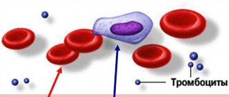
Platelets are the smallest non-nuclear spherical blood elements, formed in the process of synthesis from large cells (megakaryocytes) of the bone marrow.
They play a critical role in the mechanisms of primary and secondary hemostasis in case of vascular damage.
Platelets are the first line of defense when bleeding occurs.
Moreover, they are a source of enzymes (regeneration and growth factors) that contribute to the following processes:
- stimulate cellular synthesis;
- regenerate tissues;
- promote the elasticity of artery walls;
- participate in the regulation of the level of permeability of cell membranes.
Like leukocytes, platelets have their own life cycle (no more than 7 days) and division according to the maturation phase - young, mature, old. However, the aging process is most pronounced in them.
Normally, in a healthy person, the level of mature cells should fluctuate within 95%.
Deviations from the norm indicate the following:
- high concentration of young cells - the development of numerous disorders in the circulatory system;
- the presence of degenerative (old) cells in the blood - destabilization of the production and processing of blood cells.
The state of a person's coagulation system can be determined by the number of platelets. An excess of the norm is called thrombocytosis, a decrease is called thrombocytopenia.
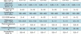
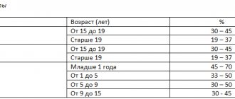
Normal values in children and adults
The normal platelet count according to Fonio ranges from 120 to 400 thousand per 1 cubic meter. millimeter of blood. But we should not forget that for certain groups of the population the obtained indicators will vary significantly. Such differences are considered variations of the norm.
The norm for children over 1 year old is 180-320 thousand in 1 cubic meter. millimeter of blood. For newborn children, the platelet count limit is somewhat wider - from 100 to 420 thousand per 1 cubic meter. millimeter.
For men, the normal platelet count ranges from 180 to 400 thousand. The maximum concentration of these blood cells is observed at the age of 40 years. Subsequently, the platelet count decreases slightly, but does not fall below 320 thousand.
The normal level of the blood elements in question for women ranges from 180-340 thousand. The maximum concentration is observed at the age of 16 years. During menstruation, the norm drops to 150 thousand. In adolescence, this number can even drop to 75 thousand.
The Fonio platelet blood test is very informative.
What affects concentration?
Many factors are implicated in increasing platelet levels. Thus, all infectious diseases of the body in acute form lead to an increase in the number of these blood cells in the blood. The same is observed in the presence of disorders of the hematopoietic system and cancer.
Also, the number of platelets in the blood often changes in people experiencing constant stressful situations. In addition, some may find it strange that during large blood losses the number of these blood cells increases, but this suggests that the human body compensates for blood loss in this way.
Increased platelet concentrations are also observed in people who drink alcohol for a long time.
With prolonged and uncontrolled use of drugs that have a thromocytopenic effect, the blood picture will change significantly. The same happens with diseases of certain organs, for example, the liver, thyroid gland, etc. Sometimes cuts or nosebleeds can negatively affect the number of platelets in the blood, lowering their number.
Evaluation of platelets and leukocytes in blood smears
N. Puletti
In addition to the evaluation of red blood cells, examination of blood smears involves the presence of platelets and leukocytes, mainly to determine immature neutrophils, detect toxic and lymphocytic reactions. The study of platelets and leukocytes in blood smears is the most important step in the diagnosis of inflammatory disorders and causes of bleeding.
Platelet assessment
Assessment of platelets by counting is indicated, in particular, when the clinical picture indicates possible thrombocytopenia (petechiae, ecchymoses - extensive hemorrhages under the skin or mucosa; melena; epistachys - nosebleeds; hematuria, etc.). If platelet function is not impaired, hemorrhagic manifestations usually occur when platelets decrease to 25–50 × 109/L. However, even such a decrease in some animals does not lead to hemorrhagic syndrome. This can be explained by the presence of larger platelets or an increase in their activity. The first stage involves checking for the presence of clots, which form mainly at the end of the smear. In a healthy animal, platelet counts using a hematological analyzer sometimes erroneously give low results due to the formation of large clots, so it becomes necessary to use blood smears for recounting. The formation of platelet clots may be a consequence of difficult blood sampling and insufficiently rapid transfer of the material into the EDTA tube. They may also be due to poor homogenization of the material placed in the test tube (KE Russel, CB Grindem, 2000). But in cats, platelets tend to form clots, which often provokes the misconception that they decrease during counting (C. Tasker, PJ Gripps, AJ Maskin, 1999). Finally, during hardware counting, aggregated platelets are sometimes mistakenly recognized as leukocytes, which distorts the results of the study.
In dogs and cats, the number of platelets observed in the field of view of a microscope with high magnification (× 1000) ranges on average from 10 to 30 cells. When multiplied by 15x109 or 20x109, the number of cells corresponds to the content of 1 liter (JW Harvey, 2001). In an animal with immune thrombocytopenia, the number of platelets in the microscopic field of view is usually reduced (Photos 1a and 1b). A decrease in the number of platelets in a blood smear in the absence of clots alerts the doctor to excessive peripheral uptake (hemorrhages, disseminated intravascular coagulation - DIC, etc.), impaired formation in the bone marrow, or destruction mediated by an immune reaction (figure) (SL Stockman, MA Scott, 2008).
| 1a | 1b |
| Photos 1a and 1b. Counting blood smears using microscopy (x 1000). Lack of platelets in a dog with immune-mediated thrombocytopenia 1b compared with a healthy dog 1a. | |
Drawing. Main causes of thrombocytopenia considered after final confirmation by blood smears
Leukocyte assessment
Studying the morphology of leukocytes can give us serious information. A large number of hematology analyzers give the total number of leukocytes and differentiate (percentage in relation to the total number of leukocytes) into three or five parts (neutrophils, monocytes, lymphocytes, eosinophils and basophils). However, as for red blood cells and platelets, these modern diagnostic methods often provide distorted or incomplete information on the morphology of leukocytes, which also requires additional examination of the blood smear.
Neutrophils
Immature neutrophils
With a pronounced inflammatory focus (pyometra, bronchopneumonia, etc.), immature forms of leukocytes often appear in the peripheral blood. This is indicated by a shift in the leukocyte formula to the left. This picture is associated with the expressed need of body tissues for neutrophils, despite the fact that the pool of mature neutrophils in the bone marrow is depleted.
The degree of deviation to the left is manifested by the number of immature neutrophils and their level of maturity. Most often, a shift to the left is manifested in an increase in the number of non-segmented (band) neutrophils (cells with stripes) (photo 2). In the presence of a more pronounced focus of inflammation, the presence of meta- and myelocytes is sometimes observed (photo 3). Long-term persistence of a pronounced deviation to the left, especially in combination with neutropenia (degenerative deviation to the left) is considered an indicator of an unfavorable prognosis. Sometimes unsegmented neutrophils can be observed in healthy animals.
| Photo 2. Reading blood smears using microscopy (x 1000) in a dog susceptible to acute bronchopneumonia. Presence of an immature unsegmented neutrophil (cell with a streak) with signs of toxicity (heterogeneous and basophilic cytoplasm) | Photo 3. Reading a blood smear under a microscope (×1000) in a dog with pyometra. Presence of a myelocyte with signs of toxicity (heterogeneous and basophilic cytoplasm) |
Toxic changes
Toxic changes in neutrophils include the appearance of Doyle bodies, as well as cytoplasmic basophilia, diffuse cytoplasmic vacuolization and nuclear lysis (JW Harvey, 2001). The presence of toxic granulation (pink granules are usually absent in mature neutrophils) is also an indicator of toxicity.
Doyle bodies are clusters of rough endoplasmic reticulum that form bluish granules. The detection of several neutrophils with Doyle bodies in a cat is considered normal (photo 4).
Basophilia of the cytoplasm is associated with a large amount of rough and polyribosomal reticulum. Diffuse vacuolization and nuclear lysis appear last, with severe toxemia. Typically, a change in the degree of toxicity accompanies a shift in the leukocyte count to the left.
| Photo 4. Reading a blood smear under a microscope (×1000) from a healthy cat. A mature (or segmented) neutrophil with Doyle bodies is visualized (arrow) |
| Photo 5. Reading a blood smear under a microscope (×1000) from a healthy cat. The presence of a reaction neutrophil is noted |
Lymphocytes
Reactive lymphocytes
Incidentally appearing reactive (or reactive) leukocytes can be observed upon stimulation with antigen (for example, during vaccination). These cells are often larger in size with basophilic and more abundant cytoplasm relative to small normal lymphocytes. The chromatin is more or less well condensed and nucleoli are generally not visible (photo 5).
Large granulosa lymphocytes
Large granulomatous lymphocytes, unlike small normal lymphocytes, have pale, more abundant cytoplasm and contain purple granules (Figure 6). These cells belong to T-lymphocytes or natural killer cells (CD8+ immunological markers). Their number increases with infection with Ehrlichia canis and chronic lymphocytic leukemia (JW Harvey, 2001).
| Photo 6. Reading a blood smear under a microscope (×1000) from a healthy dog. Presence of a large granular lymphocyte |
Atypical lymphocytes and lymphoblasts
Atypical lymphocytes and lymphoblasts are much larger than normal lymphocytes. Chromatin in their nuclei with clearly defined and weakly concentrated granulation. Lymphoblasts, in addition to the above, contain protruding nucleoli (photo 7). In small quantities, their presence can be caused by inflammation. An increase in their number may indicate acute lymphoblastic leukemia or stage 5 lymphoma (lymphoma with bone marrow infiltration through neoplastic lymphocytes). Morphologically, lymphoblasts can be difficult to differentiate from blasts appearing from other cell lines, such as myeloblasts. For final identification, modern immunocytochemical methods and specific cytochemical stains (directly on smears) are used (JA Ramos-Vara, AC Avery, PR Avery, 2010).
| Photo 7. Reading blood smears under a microscope (×1000) from a dog susceptible to acute lymphoid leukemia. The presence of lymphoblasts with protruding nucleoli and granular chromatin with weak condensation is noted | Photo 8. Reading a blood smear under a microscope (×1000) in a dog. Morula Ehrlichia spp (arrow) observed in a mature neutrophil is visualized |
Inclusions
Microorganisms can be found in leukocytes, such as Ehrlichia spp, represented by morula in monocytes or granulocytes (photo 8).
Mast cells (mast cells)
Mast cells are usually identified at the end and edge of the smear. They can be distinguished from basophils by a round nucleus and a large number of dark inclusions in the form of granules that can mask the nucleus. Mastocytes are found during inflammation (hypersensitization), as well as during neoplastic processes (systemic mastocytoma).
Examination of white blood cells and platelets in a blood smear allows rapid diagnosis of a number of diseases, such as thrombocytopenia or hematopoietic neoplasia (for example, acute lymphoblastic leukemia). In some cases, timely making a final diagnosis significantly improves the prognosis and quickly guides the choice of therapy. In addition, this allows you to assess the severity of the inflammation or clarify the prognosis for an animal in critical condition.
Basic provisions
— In the absence of platelet dysfunction, hemorrhagic syndrome manifests itself when platelets decrease to 25–50 × 109/l.
— With severe inflammation, immature neutrophils are always found in blood smears.
— In cats, the presence of several neutrophils with Doyle bodies is normal.
— Atypical lymphocytes and lymphoblasts are found in large numbers in acute lymphoblastic leukemia and lymphoma in the fifth stage.
— For canine ehrlichiosis, morula Ehrlichia spp. may persist in monocytes or granulocytes.
SVM No. 5/2010
What should be done to normalize the platelet count?
It is clear that measures to normalize platelet concentration will be somewhat different for different versions of the results obtained. But all of them will be useless if harmful factors continue to affect the body. First of all, it is necessary to avoid physical and emotional overload.
So, you can increase your platelet level in the following ways:
- Eliminate alcoholic beverages, seaweed, pickles and red grapes from your diet.
- Include fresh fish, bell peppers, liver, buckwheat and apples in your diet.
- Sometimes your doctor may prescribe special medications to raise platelet levels. You should absolutely not self-medicate!
- Do not take medications that may cause thrombocytopenia as a side effect.
- Consume vitamins A, B and C.
To lower the platelet count, you must first establish a healthy lifestyle and eliminate stressful situations. It is strictly forbidden to engage in traumatic sports. The following will help lower platelet levels:
- use of acetylsalicylic acid drugs;
- refusal of bananas, pomegranate, mango, rose hips;
- inclusion of beets, blueberries, cranberries, and sea buckthorn in the diet;
- taking multivitamins and magnesium supplements.
How to normalize the number of platelets in the blood
Obviously, in order to normalize PLT in the blood, it is necessary to conduct a further examination, find out and cure (eliminate) the reason for their decrease or increase.
In case of secondary thrombocytosis or thrombocytopenia, cure of the primary disease leads to normality and PLT.
Thrombocytosis - how to reduce the number of platelets in the blood
| Thrombocytosis | How to reduce PLT |
| Secondary (reactive) | 1. Treatment (or elimination) of a primary disease (or condition), one of the symptoms of which was an increase in PLT in the blood 2. Stop smoking 3. Normalization of body weight 4. Sufficient consumption of clean water (at least 30-40 ml per 1 kg of body weight per day) |
| Primary (essential) | 1. Aspirin at a dose of 81 mg per day (to reduce the risk of blood clots) 2. Medicines that reduce the level of platelets in the blood (hydroxyurea, anagrelide, etc.) 3. Thrombocytopheresis (according to indications) 4.Interferon alpha-2b (according to indications) 5. Cytotoxic antitumor agents (according to indications) 6. Bone marrow transplant (if indicated) |
Products useful for thrombocytosis
Unrefined vegetable oil (flaxseed, olive, pumpkin, etc.) Fish oil, seafood Tomatoes, tomato paste Lemons Sea buckthorn Ginger Garlic Green tea Drinking water 40-50 ml per 1 kg of body weight per day or more
Thrombocytopenia - how to increase the number of platelets in the blood
When platelets decrease to 100-50 x10^9/l, special treatment, as a rule, is not required. This condition is not dangerous, but requires mandatory monitoring and clarification of the cause of the decrease in PLT.
Important! Patients with thrombocytopenia are recommended to completely abstain from alcohol.
Foods useful for thrombocytopenia
Pomegranate Beetroot Nuts Green leafy vegetables, herbs Liver, red meat Buckwheat Oatmeal Rosehip decoction Chokeberry Stinging nettle (tea, decoction)
It is important to note that there is no special diet to truly effectively increase blood platelets.
The patient's diet should be hypoallergenic and complete and varied: rich in proteins, vitamins and microelements. Dishes are steamed, boiled or baked. Spicy, mechanically rough, too hot or cold foods should be avoided.
How to increase platelets in the blood
- Treatment of thrombocytopenia begins when PLT is below 50 x10^9/l
| How to increase PLT in the blood | Note |
| Prednisolone (corticosteroid hormone) | Used for the treatment of immune (autoimmune, drug) thrombocytopenia (ITP). The individual daily dose and duration of taking prednisolone is prescribed by the doctor |
| Human immunoglobulin (Intraglobin, Imbiogam) | It is administered intravenously, drip-wise, slowly, in a hospital setting. Used for the treatment of ITP, chronic lymphocytic leukemia, multiple myeloma |
| Stimulator of thrombopoiesis in the bone marrow (thrombopoietin receptor agonist): Eltrombopag (Revolade) | Indicated for chronic ITP and unspecified thrombocytopenia when treatment with prednisolone, immunoglobulin is ineffective, and with insufficient effect of splenectomy |
| Stimulators of hematopoiesis in the bone marrow (recombinant erythropoietins): Recormon, Eralfon, Erythrostim | Used strictly according to indications under the supervision of an oncologist-hematologist for the complex treatment of symptomatic thrombocytopenia and anemia in patients receiving chemotherapy |
| Transfusion of donor platelets (thrombomass) | Performed in the hospital for life-threatening conditions associated with severe thrombocytopenia, including in patients receiving chemotherapy |
| Vincristine (antitumor drug) | Used to treat ITP when prednisolone is ineffective |
| Vitamin B12 | Indicated for the treatment of thrombocytopenia in megaloblastic anemia |
| Splenectomy (surgical removal of the spleen) | Performed when treatment with ITP with prednisone is ineffective |
| Bone marrow transplantation | Use strictly according to indications |
Which doctor should you contact if you have low or high PLT in your blood test: general practitioner, hematologist
- Tags:
Save the article for yourself!
3 comments
Tatyana08/14/2018 at 20:20
Everything is very detailed, just chewed on the shelves. Thank you very much for the article.
Christina 08/17/2018 at 16:19
Please help me decipher the smear analysis! Leukocytes uterus>100, vagina>100. Epithelium. cells of the cervix um/n, vagina b/n. Cocci cervix b/p, vagina vol.?
Tatyana21.10.2018 at 09:16
Hello! Please help me with decoding. Description b/m sticks +++, squamous epithelium in the form of layers of mature cells. Description of c/c mucus, erythrocytes, fibrin, groups of flat cells, numerous glandular structures proliferating, including hyperplastic type glandular cells, their endometrial origin cannot be excluded; groups of cylindrical cells. NILM. Signs of hyperplasia of the glandular epithelium. Thank you