0
Author of the article: Marina Dmitrievna
2017.08.03
5 108
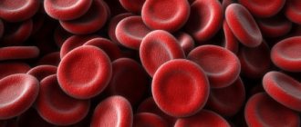
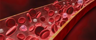
Leukocytes
Eosinophils and monocytes are types of blood cells that belong to the class of leukocytes (white bodies). Their main task is to protect the body from microbes and malignant cells, removing foreign and dead particles. Therefore, it is so important to monitor the level of monocytes and eosinophils in the blood of both children and adults, since an increase or decrease in their number can have serious causes.
Why are eosinophils and monocytes needed?
Let's talk a little about eosinophils and monocytes. They, of course, perform different functions, and when their numbers are increased, it is necessary to find out which subpopulation is increased the most. And the most important thing is whether this increase is related to each other, or whether the patient is experiencing two independent pathological processes. The doctor, when he interprets the analysis, must understand that any changes in the leukocyte formula do not clearly indicate any disease, that is, they do not have specificity.
They can be caused by a variety of diseases, and the blood can react with different populations of cells in the same way. On the other hand, with the same disease in different patients, the blood can react in different ways.
It should not be forgotten that the assessment of a subpopulation of leukocytes has certain characteristics. The age of the patient must be taken into account. It is very important to remember that any deviation can be absolute or relative. With an absolute deviation, the number of monocytes and eosinophils increases, since more of them are simply actually produced. If this deviation is relative, then in reality there are exactly the same number of them, but in a given blood sample there are more of them, since there are fewer of them elsewhere. Therefore, in order to get a complete picture of the real number of leukocyte subpopulations, it is necessary to know not only their number per liter, but also as a percentage.
Monocytes
Monocytes in a typical blood smear always look like large cells, their nucleus resembles a bean or legume, without any constrictions and without segments. Monocytes do not live long in the blood, no more than a day, and then the pool of released monocytes rushes into the tissue. It enters the tissue through the endothelium, or the inner wall of capillary vessels, through active movement. In the tissue, the monocyte turns into special cells - macrophages, and are forever lost from the systemic circulation. They live in tissues for a very long time, up to several years, which is several times longer than the lifespan of any blood cells in blood vessels.
Tissue macrophages are active defenders, and in tissues they do the same thing as neutrophils in blood plasma. They recognize, capture and destroy all foreign microorganisms, including fungi; tissue macrophages have a high degree of phagocytosis. They actively appear in foci of tissue inflammation, where they also destroy all microbes, damaged cells, as well as their dead brothers - leukocytes. Macrophages are the most active cells capable of phagocytosis in our body, and their task in the area of inflammation is complete cleansing, elimination of the pathogen, destruction of tissue detritus and pus and preparation of the area for restoration or tissue regeneration. However, monocytes can not only be beneficial, but sometimes they also support pathological, autoimmune inflammation, such as in rheumatoid arthritis.
Therefore, the number of monocytes in the blood is the tip of the iceberg, a huge part of them is in the tissues, and therefore it is, of course, impossible to judge the state of macrophages by the monocytes that are in the bloodstream.
Read more about monocytes in our article Monocytes in the blood: functions, normal, causes of abnormalities.
Eosinophils
Eosinophils are cells of which there are very few in the blood, fewer of them, perhaps, only basophils. Yes, eosinophils are also capable of phagocytosis, but still their role is different. They are particularly good at recognizing parasitic antigens and engaging in various allergic cellular reactions.
Not a single neutrophil can compare with eosinophils in their ability to recognize and bind antigens of multicellular parasites. These primarily include various flatworms, roundworms and tapeworms. These are pinworms and roundworms, the cat fluke - the causative agent of opisthorchiasis and the liver fluke, the causative agents of diphyllobothriasis and taeniasis, that is, the broad tapeworm, and the pork tapeworm. These cells also remain in the blood for a very short period of time, literally a few hours, and then are transported to the tissues in order to restore order there, just like monocytes.
For more complete information, read the articles - Eosinophils: age norm and causes of deviations and Eosinophilia in adults.
Procedure for determining the level of monocytes and eosinophils
Determining the level of eosinophils and monocytes in the blood is considered an important study that can indicate serious infectious and viral pathological processes occurring in the body of a child or adult. Based on the results of this analysis, the doctor prescribes further diagnostics for the patient and draws up an approximate clinical picture of his current state of health.
Taking blood from a child
To determine the levels of monocytes and eosinophils in the blood of an adult or child, the doctor takes fluid for analysis exclusively from capillaries (from a finger or heel), although in rare cases a sample is also taken from a vein. However, before this procedure, the patient must undergo training consisting of the following steps:
- Refusal to eat food (except water) 12 hours before the procedure (in the case of infants, blood is taken at least two hours after feeding).
- Calming the nervous system. Before the blood collection procedure, it is recommended not to be exposed to stress, as an excited state can affect test results.
- Correct filling of forms. Before starting the procedure, the patient must fill out a special form indicating information about his body (age, weight, height, presence of chronic pathologies, etc.).
- Refusal of physical activity. If the patient is a professional athlete, or his work involves serious physical activity, then 1–2 days before the procedure, he is recommended to refrain from his usual schedule and give the body a rest.
Important! If the patient is undergoing a course of drug treatment, then 2-3 days before taking blood for analysis, it should be temporarily interrupted. However, in the case of children, before stopping medications, parents should definitely consult a doctor about this issue.
Normal blood cells
As mentioned above, you need to know not only the absolute, but also the relative value. Normally, monocytes in boys, girls and adults make up from 3 to 11% of all leukocyte populations. Some laboratories give from 3 to 12%. This means that in a fixed blood smear, out of all 100 leukocytes that catch the eye of a laboratory assistant, monocytes will make up no more than 12%. If we determine the absolute value, then it will be 0.05 - 0.82 * 109 / l.
However, there are no significant changes from the neonatal period to old age. So, for babies less than 1 year old, the reference values range from 4 to 10%. Agree, this is very close to adult values. When can we talk about monocytosis, or an increase in these cells in the plasma? This is when there are more than 1000 of them in one microliter, and when examining a smear, their number exceeds 15%.
The situation is approximately the same with eosinophils. In children over 6 years of age and in adults, their blood levels are 0.02 - 0.5 *109/l, in absolute values. If we talk about the relative content, then in adults, as well as in children over 5 years old, the number of eosinophils should not exceed 5 representatives out of every 100 leukocytes in the smear, which was counted by the laboratory technician, or 5%.
As with monocytes, a significant increase in the eosinophil population, or eosinophilia, is an increase in their absolute number of more than 700 per microliter. There is a clinical phenomenon of eosinophilic infiltrate, when there are too many eosinophils and they are elevated for a long time. Such infiltration and damage to internal organs develops when the absolute number of eosinophils is at least 1500 per microliter. This toxic effect on tissue is due to the fact that eosinophils rushing into the lesion develop high chemical activity, and tissues are affected by toxic products of oxygen breakdown and degradation products of granular eosinophilic proteins.
What do eosinophils show in a blood test?
Tests do not always show normal blood cell counts. Less commonly, a decrease or absence is observed. This condition is called eosinopenia, and is usually caused by congenital characteristics of the body and weak immunity.
Eosinophilic cells are sometimes absent in children affected by viral or bacterial infections. A decrease is also observed after emotional and mental stress, excessive physical exertion. Cells are completely absent after serious injuries, operations, burns.
Deviations from reference values
Let us name those conditions in which the population of monocytes in the blood plasma significantly increases. These are all acute and chronic inflammatory diseases. These are furunculosis, streptoderma, arthritis, purulent inflammation of the subcutaneous tissue and phlegmon.
Since monocytes are tissue protectors, they surround various specific granulomas, which are infectious and inflammatory in nature. The most well-known specific granulomatous inflammation is tuberculosis infection and syphilis. Therefore, monocytes can be elevated in the blood for a long time in these diseases, as well as in the subacute course of bacterial endocarditis.
They can be increased in various forms of sarcoidosis, which also occurs as a type of granulomatous inflammatory process. It is interesting that sarcoidosis is a non-contagious disease, and granulomas, although inflammatory, are non-infectious in nature, but tissue macrophages are actively involved in its formation.
This is an autoimmune pathology, which is also inflammatory in nature, it is a state of convalescence after an acute infectious pathology, for example, after typhoid fever or dysentery. These are various malignant neoplasms and growing tumor tissues. Of the malignant neoplasms, the growth of monocytes is most typical for cancer of the stomach, mammary glands, and ovaries, as well as lymph nodes.
The cause of high monocytes can be oncohematological diseases, when the hematopoietic germ in the red bone marrow that produces granulocytes is affected. Finally, there is acute poisoning with organophosphates and tetrachloroethane.
Eosinophils, in addition to various allergic diseases and parasites, also increase upon the administration of many medications. They react with an increase in allopurinol, beta-blockers, heparin and many antibiotics.
There is a sharp increase in the number of eosinophils in various skin diseases. Their growth increases with eczema, atopic dermatitis, systemic pathology, for example, autoimmune diseases. Their growth also increases with malignant neoplasms in tissues and with damage to the red bone marrow. Rare causes include volatile eosinophilic infiltrates, or Loeffler's disease. The number of eosinophils increases in the initial period of various acute infections: scarlet fever, gonorrhea, tuberculosis and infectious mononucleosis.
What are the common reasons for both monocytes and eosinophils to increase at the same time? This:
- autoimmune pathology, or systemic connective tissue diseases;
- malignant neoplasm of lymph nodes;
- sarcoidosis;
- the oncohematological process and myeloblastic leukemia also lead to a joint increase in these populations.
Therefore, if a patient’s blood has repeatedly and consistently elevated monocytes and eosinophils, then it is necessary to be extremely wary of the risk of tuberculosis and sarcoidosis, be examined by an oncologist, and also pass all the necessary tests for autoimmune pathology, and consult a rheumatologist.
These steps will be correct, and depending on the clinical symptoms, epidemiological history, and data from instrumental and laboratory studies, the specialist can certainly make the correct diagnosis.
Reasons for the increase in monocytes and eosinophils in the blood of a child
An increase in the level of monocytes and eosinophils in a child never occurs just like that; there may be serious reasons for this:
- Autoimmune diseases (rheumatism, lupus erythematosus, etc.). With such pathologies, the body begins to produce leukocytes more intensively, as a result of which the level of monocytes and eosinophils increases significantly.
- Infectious mononucleosis. This pathology affects the patient’s liver, spleen and tonsils, as a result of which the composition of the blood changes significantly. With this phenomenon, not only monocytes and eosinophils increase, but also other cells related to leukocytes.
- Tuberculosis. In the first stages of the development of this pathology, the level of leukocytes in the patient’s blood drops, but a few days after the child’s body is infected with tuberculosis, the doctor will be able to observe the opposite picture.
- Malaria. With this pathology, as a rule, there is an increase in the number of not only monocytes and eosinophils, but also all other leukocytes in the blood.
- Congenital syphilis. If a child has elevated eosinophils and monocytes from birth, then the reasons for this deviation may lie in congenital syphilis, which is transmitted from mother to child during pregnancy or childbirth.
- Toxoplasma. These parasites, like any other similar organisms that enter a child’s body, can provoke significant changes in the composition of the blood. As a result, the patient's overall white blood cell level will also be quite high.
- Poisoning with toxic substances (phosphorus, chlorine or tetrachloroethane). All these substances have a very detrimental effect on neutrophils, which are also part of the blood, due to which the level of monocytes and eosinophils increases noticeably.
- Oncological and malignant hematological diseases (lymphoma, leukemia, etc.).
- The level of eosinophils increases with parasitic diseases and helminthic infestations (giardiasis, opisthorchiasis, ascariasis, etc.). These cells are precisely responsible for antiparasitic immunity. Another reason for the increase is allergies and diseases that are associated with them (bronchial asthma, Quincke's edema, atopic dermatitis, allergic rhinitis, etc.).
Important! If monocytes and eosinophils are elevated in an adult, then the reasons for this deviation may differ from those listed above. This is due, first of all, to the fact that children’s immunity has not yet had time to strengthen, and many diseases occurring in the children’s body are clearly manifested in test results (especially on the blood test). It is worth noting that with monocytosis, the child may not have any accompanying symptoms, and an increase in monocytes or eosinophils in the blood is detected only during a routine examination.
Reasons for the sharp increase in basophils in the body
As mentioned earlier, the normal level of basophils in the blood is 1-5%.
If it is greatly enlarged, this indicates to the doctor the development of various diseases, such as:
- allergies (medicines, products);
- cancer developing in the respiratory system;
- the appearance of a viral infection in the body;
- blood flow leukemia;
- diabetes of any form;
- disorders of the thyroid gland;
- hepatitis, which causes jaundice;
- anemia caused by iron deficiency;
- disorders of the gastrointestinal tract.
These signs most often appear as a result of taking medications that contain estrogen.
Also, an increase in basophils usually occurs at the onset of menstruation or during ovulation.
An increase in the element occurs when a reaction to an allergen develops due to the fact that it begins to fight this phenomenon. This leads to the redirection of basophils into human tissue, which causes the development of red spots on the face, the appearance of swelling, and itching on the skin.
Basophil function
If the level of this element in the bloodstream is greatly increased, it means that inflammation is occurring in the body, and basophils are considered the main criterion for diagnosis, allowing for a more accurate result.
Penetrating into the inflamed area, these substances open up, forming such elements as:
- histamine;
- heparin;
- seratonin.
These elements are able to quickly respond to the development of the disease, especially if a person is in anaphylactic shock. Together with leukocytes, these elements are also involved in slow reactions, and are also considered an excellent aid in diagnostics.
Often in people the number of basophils is greatly increased - this phenomenon is called basophilia. In an adult’s body, the normal amount of these substances is 2-3%, which is not considered dangerous to health, but if their level constantly increases, a person urgently needs to undergo a full examination of the body to identify the cause of this phenomenon.
It is important to remember that basophils become active in the bloodstream only when an allergic reaction develops in the body. Also, an increase in their number indicates the obvious development of pathologies in the circulatory system, which also requires prompt treatment.
Indicators in childhood
According to the norm, there are few eosinophils in the blood test in children. Acceptable indicators are given in the table
| Age | Index |
| Eosinophils in infants | 1-4 units |
| Infancy up to 12 months | 1-5 units |
| After 1-2 years | 1-4 units |
Any changes in children indicate problems in the functioning of the body, requiring consultation with a pediatrician.
Eosinophils are normal
Neutrophils, basophils and eosinophils
Concept and types of immunity
Immunity concept
comes from the Latin "immunis". Even before our era, doctors understood its meaning as “well protected, resistant to infectious disease.”
The immune system (immunity) is the natural defense mechanism of our body. Immunity maintains the constancy of the internal environment, eliminates the foreign influence of infectious pathogens, chemicals, abnormal cells, etc.
The immune system is responsible for two important processes in the body:
1) replacement of spent or damaged, aged cells of various organs of our body;
2) protecting the body from the penetration of various types of infections - viruses, bacteria, fungi.
When an infection invades the human body, the body’s defense systems come into play, the task of which is to ensure the integrity and functionality of all organs and systems. Macrophages, phagocytes, lymphocytes are cells of the immune system, immunoglobulins are proteins that are produced by cells of the immune system and also fight foreign particles.
In other words, immunity is:
- the body's immunity to infections;
- the ability to remove any foreign material from the body (bacteria, viruses, atypical (tumor) cells).
There are two types of immunity:
1. specific immunity
acquired after an infection (for example, after influenza, measles, rubella) or vaccination.
It is individual in nature and is formed throughout a person’s life as a result of contact of his immune system with various microbes and antigens.
Specific immunity preserves the memory of the infection and prevents its recurrence. Sometimes specific immunity can last a lifetime, sometimes for several weeks, months or years;
2. nonspecific (innate) immunity
– the innate ability to destroy everything alien to the body. This is the ability of cells formed in intrauterine life to synthesize membrane receptors for antigens of other organisms, other tissues and some microorganisms, as well as to synthesize the corresponding antibodies and remove them into body fluids.
During intrauterine ontogenesis, all cells, including blood cells, commit to the antigens of another organism and to the antigens of other tissues.
Therefore, the main part of innate immunity is the acquisition by cell membranes of receptor molecules capable of binding to certain molecules fixed or produced by cells of other organisms (maternal), as well as the own cells of other organs and tissues. This part of the innate immunity (immune status) is called the major histocompatibility complex.
Immune system organs
Two factors play a major role in the immune system: antibodies and leukocytes (white blood cells). The immune system of the human body consists of different organs.
Thymus
The thymus is the thymus gland. This organ is located in the upper part of the chest in front. It is like a breeding ground for T-lymphocytes.
Spleen
This is a very fragile organ, which is located in the abdominal cavity in the left hypochondrium. It plays the role of a filter for the entire blood of the body, removing old red blood cells and platelets from its bloodstream. In addition, it is also a reservoir of blood, and also some cells of the immune system are formed in it. Of course, a person can live without a spleen, but he will be more susceptible to infections.
Bone marrow
Most of the bone marrow is contained in large tubular bones (femurs), as well as vertebrae and pelvic bones. Bone marrow is a source of not only erythrocytes - red blood cells, but also lymphocytes, as well as macrophages.
Lymph nodes
Lymph nodes can be compared to roadblocks along the path of lymphatic vessels. They are lymph filters, purifying it from various antigens: bacteria, viruses, cancer cells. They retain antigens, after which antibodies, macrophages and T-lymphocytes come to fight them.
Antibodies
Antibodies are special proteins (proteins) that are produced by cells of the immune system. Antibodies are able to fight specific antigens. Antibodies are usually only protein molecules, but sometimes they can also be non-protein molecules.
Protein molecules as antigens are, for example, pathogenic bacteria, viruses, tumor cells, foreign cells that entered the body artificially (blood transfusion, organ and tissue transplantation), as well as other protein substances.
Antibodies have their own specificity. That is, certain antibodies can only act on certain antigens. For example, a serum containing antibodies is introduced into the body for a certain disease and does not have any effect for other diseases, since in this case there is a different antigen in the body.
Antibodies can fight antigens in several ways:
- They stick antigens (cells) together in piles so that they cannot move, after which they are absorbed by macrophages.
- They form “holes” in the wall of antigen cells, as a result of which their contents leak out and the cells die.
- They block antigens, which allows cells of the immune system (especially macrophages) to devour these cells.
White blood cells
White blood cells are leukocytes. There are a lot of them in the blood and they circulate throughout the body, as if on guard, in order to repel the attack of antigens at any moment. Normally, the number of leukocytes ranges from 4 to 9 billion in one liter of blood.
Leukocytes, in turn, are divided into 5 types:
Lymphocytes. This type of white blood cell is a key element of the immune system. Lymphocytes have a unique property - they can remember any antigen that they once encountered.
Thanks to this property, in particular, there is immunity from various infectious diseases. This means that when any antigen enters the body, lymphocytes “remember” how to fight them.
Lymphocytes are divided into two large classes:
- T lymphocytes. These lymphocytes interact with the antigen only after special cells “let them know” about it. After interacting with the antigen, T lymphocytes begin to produce substances that attract other immune cells - macrophages, which attack the antigen, devouring it. Sometimes the immune system is not able to completely destroy the antigen, but only seems to isolate it, enveloping it as if in a net. Therefore, the function of T-lymphocytes is to collect cells of the immune system to fight the antigen.
- B lymphocytes. These cells of the immune system play a very important role - they produce antibodies. B lymphocytes also have memory and can remember for a long time which antibodies need to be produced against a particular antigen. The principle of vaccination is based on this. In this case, an antigen is introduced into the body, but not an ordinary one, but a much weakened one or even a dead one. Sometimes the vaccine does not contain the entire antigen, but only a part of it, the one that the immune system “remembers”. As soon as such a weakened or killed antigen appears in the body, the immune system produces antibodies to it and thus a “memory” is formed - this is immunity. The next time a real antigen enters your body, your immune system will already know how best to fight this antigen, as a result of which the disease is very mild or does not even have time to enter the clinical stage.
Macrophages
The next type of immune system cell is macrophages. The word macrophage itself is formed from two words: macro - large and phage - devour. These cells are the white blood cells that devour the antigen.
Neutrophils, basophils and eosinophils
The remaining three types of cells: neutrophils, basophils and eosinophils are responsible for the development and course of inflammation.
Source: //megaobuchalka.ru/13/54183.html
Eosinophils
Eosinophils - responsible for protecting humans from parasites
Norm of eosinophils by age
The normal percentage in the structure of leukocytes is from 0.5% to 5%
Why are eosinophils elevated?
An increase may occur due to the development of:
- leukemia;
- tuberculosis;
- rheumatism;
- vegetative-vascular dystonia;
- hypothyroidism.
Why are eosinophils reduced?
Main reasons:
- heavy metal poisoning;
- development of purulent processes;
- development of inflammation.
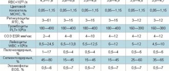
Leukocyte formula
Leukocyte formula is the percentage of different types of leukocytes in the peripheral blood.
There are five populations of leukocytes found in peripheral blood. Neutrophils, basophils, eosinophils belonging to the granulocytic series (their cytoplasm is granular and contains a large number of granules, including, for example, myeloperoxidase, elastase, lysozyme); monocytes and lymphocytes (B cells, T cells).
When testing blood on hematological analyzers, the leukocyte formula is automatically calculated with the determination of the five main populations of leukocytes. Technologies for calculating the leukocyte formula vary among different manufacturers.
Reference interval, %
Neutrophils s/o Neutrophils s/o Eosinophils Basophils Lymphocytes Monocytes
| 1–5 | 47–72 | 1–5 | 0–1 | 19–37 | 3–11 |
NEUTROPHILS make up the bulk of all leukocytes (up to 95%). The main function of neutrophils is phagocytosis. The lifespan of neutrophils is short – 2–3 days.
From the bloodstream, neutrophils actively move to areas of inflammation and tissue decay, to foci of bacterial and viral infections, where they perform their main function - phagocytose microbes and tissue breakdown products, and then destroy them with their granular inclusions, for example, lysosomal enzymes
Increased values
- inflammation;
Reduced values
– reduced immune status.
MONOCYTES are the precursors of macrophages. They make up 4–8% of all leukocytes. Circulating in the blood for up to 20 hours, monocytes migrate into tissues, where they differentiate into macrophages. Their main function is phagocytosis.
Quickly accumulating at the site of inflammation and tissue destruction, they eliminate microorganisms, lifeless cells and cellular fragments.
Macrophages, unlike neutrophils, actively function in an acidic environment and have a longer lifespan.
Increased values
associated with the presence of an infectious process.
Reduced values
, neutropenia - drug-induced, autoimmune, lymphogranulocytic leukemia, genetically determined syndrome, etc.
EOSINOPHILES are cells that phagocytose antigen-antibody complexes, including mainly IgE. After maturation in the bone marrow, eosinophils remain in the circulating blood for about 3–4 hours and then migrate to tissues, where their life expectancy is 8–12 days.
In a healthy person, eosinophils account for 2–5% of all leukocytes. Eosinophils are characterized by a daily rhythm of fluctuations in the blood, the highest levels are observed at night, the lowest during the day. The effect of eosinophils is manifested in sensitized tissues.
They are involved in immediate and delayed hypersensitivity reactions.
Eosinophils participate in the body's reactions to parasitic (helminthic and protozoal), allergic, infectious and oncological diseases, when an allergic component is included in the pathogenesis of the disease, which is accompanied by hyperproduction of IgE.
Elevated values, eosinophilia
- Allergic diseases;
- parasitic infestations;
- taking medications (antibiotics, antimicrobials, cytostatics, psychotropic drugs, etc.);
- eosinophilic pneumonia;
- hereditary diseases.
Reduced values
, eosinopenia – long-term steroid therapy.
BASOPHILES are the smallest representatives of leukocytes, accounting for less than 1% of the total number of leukocytes. Large cytoplasmic granules of basophils contain sulfated or carboxylated acidic proteins. The lifespan of basophils is 8–12 days, the circulation time in the peripheral blood is several hours.
the function of basophils is to participate in immediate hypersensitivity reactions. They are also involved in delayed-type hypersensitivity reactions, inflammatory and allergic reactions. Basophils secrete heparin, histamine, and serotonin.
The last two substances affect vascular permeability and smooth muscle tone, determining an allergic reaction of the “urticaria” type.
Increased values
, basophilia – rarely occurs in isolation. Basophilia in chronic granulocytic leukemia indicates the transition of the process to a malignant form.
LYMPHOCYTES play an important role in the processes of cellular (T-lymphocytes) and humoral (B-lymphocytes) immunity. Lymphocytes are actively involved in the pathogenesis of immunodeficiency conditions, infectious, allergic, lymphoproliferative, oncological diseases, transplantation conflicts, as well as autoimmune processes.
Increased values
, lymphocytosis - for childhood infections, infectious mononucleosis, cytomegalovirus infection, viral hepatitis, tuberculosis, brucellosis, lymphoproliferative diseases.
Reduced values
, lymphopenia, less than 1000 cells per microliter - severe bone marrow failure, for example, after irradiation or immunosuppression.
Source: //www.cmd-online.ru/vracham/spravochnik-vracha/leykotsitarnaya-formula/