The device used to measure intraocular pressure (IOP) is called a tonometer. The procedure is prescribed to patients who are suspected of developing glaucoma or other disorders of the visual organs. In ophthalmology, there are contact and non-contact tonometry, which are prescribed by the doctor individually for each patient. Intraocular pressure is measured in adults and children in a medical facility, and after the results obtained, treatment is prescribed as necessary.
Intraocular pressure
How to measure eye pressure?
Source: o-glazah.ru Without correction of pressure in the tissues of the eye, irreversible changes can occur. Intraocular pressure increases due to increased production of intraocular fluid, congenital structural features of the eye, and cardiovascular diseases can also be the cause. Less common is low intraocular pressure - this condition develops due to injury or complications after surgery, as well as due to underdevelopment of the eyeball.
Low intraocular pressure is dangerous because the nutrition of the eye is disrupted, and this can lead to the death of eye tissue. Eye pressure is increased due to increased production of intraocular fluid, congenital structural features of the eye, and cardiovascular diseases can also be the cause.
Increased intraocular pressure is often the cause of the development of such a serious pathology as glaucoma. An imbalance in the production and outflow of intraocular fluid leads to its accumulation, which causes an increase in eye pressure in glaucoma.
First, visual acuity decreases, then peripheral vision is impaired, the visibility zone is limited, and, as a result, complete blindness occurs. These changes are irreversible, so timely diagnosis and treatment of the disease is important.
Glaucoma can occur with normal intraocular pressure, but in this case there is a sharp deterioration in blood circulation in the optic nerve and disruption of its functions.
Glaucoma and eye pressure
Men and women over 40 years of age who suffer from diabetes and atherosclerosis are more susceptible to the development of glaucoma. The occurrence of the disease is often associated with a hereditary predisposition (if close relatives of the patient had cases of glaucoma).
This disease is insidious in that in some cases it may not manifest itself, i.e. be asymptomatic. Vision loss can occur gradually over many years, which patients often mistake for natural age-related changes. The early form of glaucoma is diagnosed only during an ophthalmological examination.
In other cases, the development of the disease may be accompanied by pain and severe disturbances in visual function: the appearance of periodic blurred vision, a halo around the light source, pain in the temples and above the eyebrows.
These forms of glaucoma can be accompanied by periodic acute attacks: a sharp increase in intraocular pressure. An acute attack is characterized by nausea and a general deterioration of the condition.
In these cases, in order to avoid complete blindness, you must immediately seek medical help.
The main goal of glaucoma treatment is to reduce intraocular pressure and bring it back to normal. In some cases, this is achieved through conservative therapy, in others, surgical treatment is used.
If glaucoma treatment is not started in a timely manner, it can lead to blindness, so if you detect the slightest suspicion of glaucoma, you should immediately consult an ophthalmologist.
Modern treatment methods make it possible to achieve satisfactory results and ensure that patients maintain visual function, despite the fact that a complete cure for glaucoma is still not possible.
Drug treatment for glaucoma begins with eye drops that help reduce intraocular pressure. If the drugs are selected correctly and constant monitoring by a specialist is carried out, the progression of the disease slows down significantly.
Important
This disease is insidious in that in some cases it may not manifest itself, i.e. be asymptomatic. Vision loss can occur gradually over many years, which patients often mistake for natural age-related changes. The early form of glaucoma is diagnosed only during an ophthalmological examination.
Carrying out classical microsurgical operations for glaucoma can reduce and normalize intraocular pressure for many years. However, even with a persistent decrease in intraocular pressure, visual function in some patients continues to deteriorate.
Therefore, the treatment process for glaucoma must be comprehensive, aimed at reducing intraocular pressure, improving blood supply and normalizing metabolic processes in the eye tissues. Traditional medicine offers a huge number of means to alleviate the condition of a patient with glaucoma:
- mix crushed plants in equal proportions: licorice root, ginger, buckwheat flowers, lemon balm and cinnamon, mix the mixture thoroughly. 1 tbsp. pour boiling water (1 tbsp.), then leave for half an hour. Drink ½ glass 3 times a day 20 minutes before meals;
- ½ tbsp. dry nettle mixed with 1 tsp. l. lily of the valley flowers and 1 tbsp. hot water, leave for 7-8 hours in a dark place, then add ½ tsp. baking soda. Use the infusion for lotions;
- grate raw potatoes (2-3 pcs.) on a fine grater, mix with 1 tsp. apple cider vinegar, let sit for 15 minutes. Then apply the mixture to a gauze cloth and apply to the eyes for 30 minutes, apply a terry towel over the compress.
If eye pressure is increased, it is recommended to use bee honey, always liquid, which should be placed into the retracted eyelid using a glass rod. Then close your eyes and make rotational movements with your eyeballs (without opening your eyes) down and up, left and right.
This procedure is recommended to be carried out twice a day. Herbal teas to reduce eye pressure:
- Mix birch leaves, string, tansy, horsetail and knotweed in equal proportions, add plantain and coltsfoot (2 times more), chop thoroughly. Pour boiling water (500ml) over 2 tablespoons of the herbal mixture and let it brew for 9-10 hours;
- Mix lingonberry, birch, plantain, nettle, flax, horsetail, string and knotweed leaves (one part each) with St. John's wort (three parts), rosehip and rowan (two parts each), mix thoroughly. Prepare the infusion in the same way as the first recipe.
Take infusions three times a day, 1 tbsp. daily, for 30-40 days. The course of treatment can be repeated after a break.
How to prevent complications from developing
The earlier you seek help from a medical specialist, the more successful the treatment of glaucoma. The effectiveness of treatment also depends on how pedantically the patient follows the recommendations of the ophthalmologist.
Instillation of eye drops prescribed by a medical specialist should be carried out regularly, without interruption. The importance of self-motivation is important: the patient must be interested in treatment, believe in its effectiveness and not start the therapeutic process.
A patient with glaucoma should reconsider their usual lifestyle: limit physical and emotional stress, lifting heavy objects (the maximum weight that can be lifted should be less than 10 kg).
You should try to avoid being in a dark space, since an increase in intraocular pressure can provoke dilation of the pupils in the dark.
Particular attention must also be paid to the diet. Preference should be given to products of plant origin; the consumption of meat, fatty and fried foods should be significantly reduced.
It is also necessary to exclude the use of spices, pickles, hot seasonings, strong tea and alcoholic beverages. It is useful to include in your diet a large amount of fermented milk products, especially fresh kefir, vegetables, bread and wholemeal flour products, white and cauliflower, prunes, dried apricots and other dried fruits.
You should drink no more than one and a half liters of purified water per day. Heavy smokers need to get rid of this addiction as soon as possible.
For preventive purposes, men and women over 40 years of age are recommended to be examined by an ophthalmologist at least once a year (especially patients suffering from pathologies such as farsightedness and a hereditary predisposition to the development of glaucoma).
Currently, there are no methods to prevent the development of this disease. Only timely contact with a qualified medical specialist can prevent possible serious complications, the most dangerous of which is complete loss of vision.
At the age of over 40 years, it is recommended to measure intraocular pressure once every 3 years for preventive purposes and to identify pathologies.
Thanks to modern diagnostics, regular measurement of intraocular pressure can prevent the development of optic atrophy, glaucoma and other consequences of high or low intraocular pressure.
How is intraocular pressure measured?
Measuring intraocular pressure (tonometry) - determining the pressure inside the eye using special devices (Maklakov tonometer, pneumotonometer, electrotonography). This is a very important procedure, which is performed under local anesthesia and causes slight discomfort, but no pain.
Maklakov tonometer is the most famous device for determining intraocular pressure. During the measurement process, specially colored weights are placed on the central part of the cornea of the eye and leave an imprint, which is subsequently measured and deciphered.
When measuring intraocular pressure using a Maklakov tonometer, the norm of eye pressure in women and men is less than 24 mm Hg. Art.
If intraocular pressure is measured with a pneumotonometer, the norm may not be higher than 15-16 mm Hg. Art., in general, the limits of the norm are determined by the device with which tonometry is performed.
The electrotonography method makes it possible to determine an increase in intraocular pressure by the increased production of intraocular fluid and its accelerated outflow.
Symptoms of increased intraocular pressure
Increased intraocular pressure may not cause any symptoms. In some cases, signs of high eye pressure in adults include pain in the eyes or temples. Rarely, increased intraocular pressure is indicated by decreased clarity of vision, redness of the eyes, and increased fatigue of the visual organ.
A sharp increase in intraocular pressure is an acute attack of glaucoma, which occurs with severe pain, clouding of the cornea, swelling of the eyelids, nausea and vomiting.
It is important to regularly measure intraocular pressure and consult a doctor if there are any visual disturbances or discomfort in the eye area.
We are accustomed to the fact that human health problems often arise due to high blood pressure. But there is another type of it - intraocular. And it is no less important in order to feel comfortable and enjoy all the colors of life.
For this, intraocular pressure must be normal. How do you know if it is elevated? How do ophthalmologists measure it?
What is IOP
Intraocular pressure is the effect of liquid media on the structures of the eye. The process is formed in the presence of two factors. The first is the production of aqueous humor. The second is the drainage of condensate through the trabecular meshwork (a spongy formation between the edge of the iris and the posterior surface of the transparent membrane - the cornea), which is located in the anterior chamber of the organ of vision.
IOP is the ratio of the following indicators:
rate of formation of intraocular fluid (F)/flow rate (C) + venous pressure in the space above the sclera (PV).
During the day, IOP can change, which is a physiological norm. This depends on the rate of release of aqueous humor.
The indicators are influenced by the following factors:
- physical activity, fitness classes;
- loud sounds of wind instruments;
- increased heart and respiratory rhythms;
- amount of fluid consumed;
- medications;
- caffeine, alcoholic drinks, narcotic drugs.
Measurement methods
All of them are divided into contact and non-contact. The first involves contact of the instrument with the cornea of the eye. So, let's learn about common ways to measure pressure inside the eye. Here they are:
- Palpation method. It is considered obsolete. However, in the absence of tools and outside a medical facility, the method is used. The patient is asked to sit in a chair, relax, look down, closing his eyelids. The diagnostician gently presses his finger on the eyeball. This is how you get an idea of its density. Palpation determines intraocular pressure approximately. If the apple is very dense and hard, it means that intraocular pressure is increased, and significantly. Soft indicates normal blood pressure. When it is slightly increased, the eyeball does not feel very dense to the touch. Of course, the degree of density can be determined by a knowledgeable person with experience and practice, for example, an eye doctor outside a hospital or a person with a medical education. Although people suffering from glaucoma (increased eye pressure) over time themselves acquire the skills of such self-diagnosis.
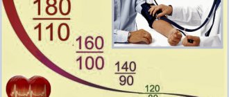
- Tonometric measurement method. It belongs to the instrumental category and is carried out using a special device - a tonometer. His indications are based on determining the degree of flattening of the eyeball. Let us note that in the practice of domestic ophthalmologists, the Maklakov tonometer has received authority and general recognition. It is a metal cylinder with flat bases and a set of glass plates of different weights. Before use, the cylinder is disinfected and then painted with a special substance. The patient is asked to lie down on the couch. An anesthetic is instilled into his eyes (most often a solution of dicaine is used for this). The plates are then lowered exactly onto the center of the cornea. Under their weight, the cornea bends. At the point of contact on the base of the cylinder, the paint is completely washed off. A circular imprint is left on the painted area. Its diameter is measured with a ruler graduated in millimeters of mercury. The larger the contact area, the higher the pressure. For this method, the generally accepted norm is a pressure reading of 18-27 mmHg. Art. As we see, this method cannot be called modern and perfect.
- Non-contact blood pressure monitors. Today, eye doctors use these in most cases. They are complex electronic devices. The patient needs to sit down in front of the tonometer and focus his gaze on a special target. Pressure is measured using a stream of air. It affects the cornea and allows you to get a very accurate result. The patient does not experience any discomfort; he only feels fast and light air movement. This method is also called pneumotonometry.
Why is intraocular pressure measured? Normally, it should not exceed 27 millimeters of mercury. This is the maximum indicator, and measurement results can range from 10 to 21. Why measure intraocular pressure?
First of all, for the timely detection of glaucoma. It is a dangerous ophthalmological disease, which is accompanied by an increase in this pressure and a deterioration in visual acuity.
Eye doctors strongly recommend that all men and women over 40 years of age systematically undergo preventive eye examinations with such a diagnostic procedure. You should see an eye doctor twice a year, especially if the patient has a genetic predisposition to diseases of the visual system.
This makes it possible to detect not only glaucoma, but also other pathologies before they begin their destructive effects. Note that tonometry is mandatory before and after eye surgery.
The following patient complaints may serve as a reason for measuring IOP:
- heaviness in the eyes;
- redness;
- fast fatiguability;
- headache;
- discomfort in the orbit;
- deterioration of twilight vision;
- the appearance of haze before the eyes;
- narrowing of visual fields;
- dry eye syndrome;
- deterioration of visual acuity.
Eye pressure is measured using instrumental techniques and palpation diagnostics through a closed eyelid. Palpation through the eyelids makes it possible to only roughly assess the pressure and understand whether there are any deviations from the norm at this time.
The interpretation of the results is based on the degree of eye hardness. Instrumental methods are more accurate. Currently, specialists use contact and non-contact methods to measure IOP. Next, we’ll talk in more detail about these verification methods.
Finger
In this case, there is no need to use special devices. An ophthalmologist conducts diagnostics using his fingertips. Using light pressure on the upper eyelids of closed eyes, you can judge the IOP values. The result is determined depending on the ophthalmotonus by the degree of hardness.
This is an indicative method, which is most often performed after surgical interventions. This method is also used in cases where instrumental examination cannot be performed, in case of changes or injuries to the cornea.
Let's consider the algorithm for measuring IOP:
- The patient closes his eyes, while his gaze should be directed downward.
- The ophthalmologist places the finger of one hand on the upper eyelid just above the cartilage and presses lightly against the eye.
- At the same time, with the index finger of the second hand, the doctor presses on the eyeball from the opposite side.
- Normally, the fingers feel shocks when pressing on the other side. With ocular hypertension, more effort is required to press on the sclera.
Contactless
This is a computer change, the essence of which is to diagnose the reaction of the cornea under the influence of air flow. This is a gentle and non-traumatic measurement method. No direct contact with the eye. There is no risk of infection.
Patients do not experience any pain during the procedure and there is no need for anesthesia. It only takes a few seconds to measure IOP.
The patient's head is fixed using a special device. The subject must open his eyes wide and fix his gaze on the luminous point. The shape of the cornea changes when air flow is applied. The results are recorded by the computer itself.
Pneumotonometry is often used in pediatric practice. However, the technique does not have high accuracy titres. Indications for pneumotonometry may include the following conditions:
- retinal disinsertion;
- endocrine disorders;
- vascular disorders;
- developmental anomalies of the visual apparatus;
- heart pathologies;
- glaucoma;
- complications after ophthalmological operations.
Experts recommend conducting examinations for patients after forty years of age, even in the absence of obvious diseases. With age, the risk of atrophic changes in the visual apparatus increases sharply.
Despite the safety and high efficiency of pneumotonometry, in some cases it is prohibited:
- diseases of the cornea and previous operations on it using a laser;
- bacterial and viral ophthalmological pathologies;
- severe myopia;
- injuries accompanied by a violation of the integrity of the eye membrane;
- state of alcohol or drug intoxication.
Contact
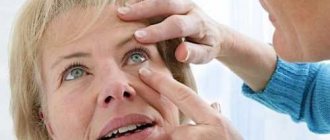
The name itself speaks for itself - the method involves direct contact of the device with the patient’s eyeball. Before the procedure, anesthesia is administered using eye drops. Contact tonometry can be applanation, dynamic and impression.
Applanation tonometry
This technique has become widespread in modern ophthalmology due to its high accuracy. The patient is instilled into the eyes with anesthetic drops and then placed on a couch. The measurement is carried out with Maklakov weights or using a Goldman tonometer.
According to Maklakov
The essence of this method is that the Maklakov tonometer displaces a large amount of moisture from the chamber of the eye. The paint is intended for painting weights. Let's consider the IOP measurement technique step by step:
- The device is wiped with alcohol or hydrogen peroxide solution twice, then wiped dry with a sterile swab.
- Specially prepared paint is applied in a thin layer to the disinfected plates.
- Anesthetic drops containing lidocaine are placed into the eyes.
- The patient is placed face up on the couch.
- Then he is asked to extend his hand up and fix his gaze on the index finger.
- The specialist widens the palpebral fissure with one hand, and with the other installs the device in the center of the cornea. The weight of the cylinder flattens the cornea.
- The weight holder is carefully lowered and quickly removed.
- At the point of contact, the paint is washed off with tear fluid. A circle remains in this place and is printed on paper.
- Finally, a disinfectant is dropped into the patient's eyes.
- The diameter of the resulting sample is measured using a ruler. The higher the IOP, the less paint is washed off from the records.
According to Goldman
To determine fundus pressure, a small special probe is used, which creates pressure on the cornea. This is a highly accurate measurement based on the force required to flatten the cornea.
The Goldmann tonometer is installed at the slit lamp. The device has a prism installed, which is applied to the cornea. When developing his device, the scientist was guided by the fact that the cornea resists deformation. And this directly affects the measurement of intraocular pressure.
Goldmann tonometry is considered the gold standard for testing. The procedure is carried out in several stages:
- The tonometer is installed at the slit lamp.
- An anesthetic and fluorescein solution is instilled into the eyes.
- The patient sits on a chair that is placed behind the slit lamp.
- He should place his head on the stand, rest his forehead on the plate and look straight into the microscope.
- Next, the prism is applied to the cornea.
- IOP is determined using the instrument scale.
You cannot save on coloring agents, as this will affect the accuracy of the results. Likewise, it is forbidden to instill too much fluorescein solution into the eye.
Non-contact methods
Non-contact tonometry uses targeted airflow to smooth the surface of the cornea. The degree of flattening is determined using an optical electronic system. Intraocular pressure indicators depend on the force of the air stream required to press the transparent membrane.
The devices do not always determine the pressure in the eyes with high accuracy. At the same time, it is an accessible, easy-to-use, quick diagnostic method. It is ideal for stubborn people and children.
Air flow blood pressure monitors
Non-contact tonometers differ in technical characteristics. On average, the range of pressure determination varies from 5 to 50 mmHg. Art. The error is 0.1 mmHg. Art.
The devices can be fully automatic or with the ability to independently project air flow into a specific area. Some models combine both functions.
Intraocular pressure is measured at a distance of 2 cm from the cornea. You can bring the device closer. If you move the tonometer away, this will give false research results.
Depending on the modification, the devices can be stationary, installed on a table or mounted on a wall, or portable (hand-held).
The average weight of an ophthalmological stationary tonometer is 18 kg. The device is equipped with a monitor and a thermal printer with high printing speed.
Optical coherence tomography
OCT is a non-invasive technique for layer-by-layer examination of various parts of the eye. Infrared optical radiation is used to probe tissue. Indications for use: diagnosis of increased intraocular pressure, glaucoma at an early stage.
This is a non-contact way to detect anatomical and physiological disorders when the patient has not yet shown clinical signs of visual impairment.
The technique is similar to ultrasound. The light beam is directed onto the tissue. Then the time of its delay before the start of reflection from the surface is recorded. The procedure is performed in a lying position. A sensor with a static flashing dot is placed on the eye. The patient should look at it without moving the pupils. After the camera is fixed in the desired position, the doctor performs a scan. The condition of the eye structures is visualized in great detail on the tomograph monitor. For a comprehensive assessment, information is displayed on a printer in the form of tables with reference values and deviations from the norm, and graphic images.
Measuring eye pressure at home
When you decide to purchase a tonometer for home use, you will be able to find out what is happening to your eyes at any time and take action. Portable devices have a forehead rest. The person touches the tonometer with his forehead and looks into the meter. Diagnostics are carried out at the touch of a button.
A transpalpebral tonometer is a device for measuring IOP without direct contact with the eyeball. Such devices allow measurements to be taken in cases of corneal pathologies and after surgical interventions. They are convenient for diagnosing children, disabled people and the elderly.
The portable tonometer is lightweight and compact. Diagnostics with their help does not cause any painful sensations.
You can also measure eye pressure at home using an ICARE tonometer, which is used to diagnose and monitor glaucoma. It has the following features:
- high accuracy;
- instant measurements;
- lack of corneal reflex;
- reliability and harmlessness;
- lack of need for pain relief;
- minimal risk of infection.
The device stores the data of the last ten measurements and operates on rechargeable batteries. Operating the device is very simple; no special training is required.
The following recommendations will help bring intraocular pressure levels back to normal:
- When sleeping, your head should be slightly elevated, so use large pillows;
- while reading a book or working at the computer, make sure there is a sufficient level of illumination;
- periodically perform gymnastic exercises to strengthen the eye muscles;
- avoid tight collars, as they impair blood flow;
- try not to bend down too much during physical activity;
- follow a daily routine and rest schedule, try not to overload your eyes;
- give up bad habits;
- regulate the amount of fluid consumed;
- try not to be nervous;
- the diet should be fortified;
- light physical activity will only be beneficial;
- do a light massage of the eyes and collar area.
So, how to measure the fundus correctly? Currently, palpation, non-contact and contact methods are used. Palpation provides only indicative information, and it is used after operations, as well as when it is impossible to carry out instrumental techniques.
Important
Non-contact equipment allows you to painlessly measure IOP, while the device itself does not come into contact with the eyeball. Nevertheless, contact methods are considered the gold standard in measuring intraocular pressure.
Ophthalmologists recommend measuring intraocular pressure according to Maklakov or Goldman. These are painful procedures that require anesthesia.
Every person should be regularly examined by an ophthalmologist. When warning signs appear, it may be very late. Patients over forty, even in the absence of any complaints, are recommended to periodically measure intraocular pressure.
If you have complaints about blurred vision, flickering before your eyes, dryness, discomfort, be sure to consult an ophthalmologist. Early diagnosis will help prevent the development of dangerous complications.
Indications and measurement results
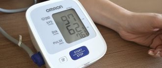
Normal true intraocular pressure is approx. 16.2 mmHg Art. Readings from 10 to 21 are not considered a deviation from the norm. During the day, its level changes slightly, after waking up it is higher, and then begins to decrease slightly. Daily fluctuations can range from 1 to 5 mmHg.
Maklakov’s tonometers do not show true V.D., but the so-called tonometric one. The readings are somewhat inflated due to the squeezing out of a certain amount of fluid from the eye chambers. Therefore, the norm for tonometric pressure according to the Maklakov method was adopted in the range of 12-25 mmHg.
It is also necessary to take into account when measuring V.d. the fact that each method and type of instrument produces slightly different data. It makes no sense to compare readings - this is a feature of each measurement method.
Therefore, if it is necessary to monitor the dynamics of the patient’s eye condition, then the check must be carried out using the same method. Only in this case can we compare the results and draw a conclusion. In particular, this is very important for patients with glaucoma.
The results of measurements using non-invasive methods are influenced by the thickness of the cornea. With a dense and thick cornea, the likelihood increases that the data obtained will be slightly higher than the true V.D. With a thin cornea, the opposite picture is observed.
What is IOP
Intraocular pressure (IOP) is the force with which the contents of the eyeball press on its walls. It maintains the shape of the eye and regulates a constant level of nutrients. The IOP value depends on the following indicators:
- production and outflow of internal fluid;
- pupil width;
- level of tone of the outer membranes of the eye (sclera and cornea);
- sensitivity and degree of filling of the choroid and capillaries of the ciliary body;
A healthy person has a clear mutual regulation of all elements. The level of intraocular pressure fluctuates throughout the day, this is normal. Typically, muscle and vascular tone is higher in the morning. But these fluctuations are insignificant and do not affect the condition of the eyes in any way.
If changes in IOP under the influence of negative factors cause anatomical or functional disorders of the eye, then serious diseases are possible. Fluctuations in pressure can be associated with eye pathologies, as well as with disturbances in the functioning of other organs and systems.
The IOP norm does not depend on age, and its readings are approximately the same in adults and children. On average, it ranges from 10 to 25 mmHg and depends on the method chosen for measurement.
Types of ophthalmic tonometers
Non-contact tonometers are modern automatic devices that allow you to quickly and accurately measure intraocular pressure. They have a number of advantages over contact ones. In particular, pneumotonometers do not require the use of eye anesthesia and dyeing solutions.
This is the most gentle and non-traumatic research method for the patient, eliminating the risk of corneal damage and infection. Also, the advantage of non-contact tonometers is that the procedure is not labor-intensive and does not require calculations, since the measurement and output of results are carried out automatically.
Using a non-contact tonometer, you can measure intraocular pressure in children and patients with hypersensitivity, as well as allergy sufferers and those who have individual intolerance to drugs.
Applanation tonometers – measurement according to the Maklakov and Goldman method. To measure according to Maklakov, a cylindrical tonometer with glass plates is used. A dye is applied to the disinfected surface of the glass plates.
The measurement is carried out under local anesthesia. After sensitivity disappears, a colored tonometer plate is lowered onto the central part of the cornea. The paint gets onto the surface of the cornea, and an unpainted spot appears on the plate.
Its size depends on the degree of flattening of the cornea, which, in turn, shows the amount of pressure. The larger the contact area, the softer the eye and the less pressure. Then repeat the measurement with the opposite plate of the tonometer. The readings are transferred to paper - an imprint is made and measured using a ruler.
The Goldmann tonometer is a more modern version of the applanation tonometry method. This is a tonometer mounted on a slit lamp. A prism is installed on the tonometer, which, after anesthesia of the eye and instillation of a fluorescein solution, is applied to the cornea.
The prism is illuminated with blue light and makes it possible to clearly see the tear menisci, which are due to the refraction of light passing through the prism into the upper and lower half ring.
Next, use the knob to slowly adjust the pressure of the prism on the cornea, flattening it until the sodium fluorescein-stained half rings converge at one point. V.d. pressure is determined by the scale of the device.
An impression tonometer is a tonometer that uses the Schiotz method (the most successful and accurate impression tonometer, although the author of the technology belongs to Graefe). According to Schiotz, pressure is measured by pressing a rod of a certain mass on the cornea.
Before measurement, local anesthesia is administered. Then a tonometer rod (piston) with a load of a certain mass is placed on the eye, which can move freely along the rod. Under the influence of the force of intraocular pressure, the piston moves and deflects the arrow on the scale.
To determine the value of V.d. using a Schiotz tonometer, it is necessary to compare the readings with calibration tables (taking into account the mass of the load on the piston).
Dynamic contour tonometer – “Pascal” tonometer. Its advantage is that it is possible to carry out measurements practically ignoring the properties of the cornea. "Pascal" is somewhat similar to the Goldmann tonometer, since the device is also mounted on a lamp.
The contact plane of the tip is concave in shape and degree of curvature of the cornea of the eye. In this case, the cornea does not flatten, as in other contact tonometers. The device records a series of data and calculates on their basis a certain average value, which is accepted as true.