Method in the 18th century. suggested by the Italian doctor Antonio Maria Valsalva. Initially it was used to remove purulent masses from ear disease.
The basic idea is to deliberately try to exhale sharply while the mouth and nose are closed. Today, the test is used in the diagnosis of cardiovascular pathologies and otorhinolaryngological diseases.
Valsalva maneuver - measurement of various conditions during forced expiration with a closed airway
General concept
To understand the indicated diagnostic technique in more detail, it is worth defining its concept.
The procedure is widely used in modern medicine by specialists in various fields.
It makes it possible to establish as accurately as possible the pathological processes of the ENT organs, vascular system, heart, etc.
The essence of the described testing is straining. In which the patient consciously closes his mouth and nose and makes a forced exhalation. This procedure is extremely simple and does not take much time. In this case, the patient performs the following actions:
- takes a deep breath;
- strains to the maximum limit, while maintaining the state of a closed mouth and nose;
- Then the person does not breathe into a special device for a certain period of time.
The procedure is usually performed sequentially in 4 phases, which begin from the moment of exhalation.
Each of the indicated stages is accompanied by certain transformations in the body:
- Inhalation is accompanied by an increase in pressure in the abdominal cavity and chest, lasting approximately 3 seconds.
- Increased tension during breathing, while the maximum permissible blood supply in the heart muscle is observed. The transparency in the lungs increases, this phase lasts from 5 to 7 seconds.
- Next, the beginning of relaxation is noted. Which is accompanied by an increase in blood flow to the heart muscle, while minimizing pressure in the chest. The contractile function of the heart returns to normal, levels out and becomes deep. As for the transparency of the lungs and the overall size of the heart, they return to their previous values.
- The relaxation period in phase 4 ends. It is accompanied by restoration of normal cardiac output and stabilization of venous reversal.
Diagnostic measures performed in this way are always accompanied by monitoring of blood pressure and pulse.
The results obtained are taken by specialists as a basis. When assessing a person’s general condition and determining a diagnosis.
Indications for the Valsalva test
The sample is used if it is necessary to evaluate:
- risk of death after myocardial infarction;
- functionality of the valve veins of the lower extremities;
- degree of development of varicocele;
- patency of the auditory canals.
In the modern world, this test is actively used not only in medicine. Divers and airline passengers use the Valsalva maneuver to relieve discomfort caused by pressure surges in the cavity of the ear canals and the upper sinus of the jaw. The breathing technique is also practiced in situations that require independent elimination of symptoms of rapid heartbeat during sinus (supraventricular) tachycardia.
It should be remembered that the use of breathing techniques is strictly contraindicated in case of tachycardia with the concomitant development of pain in the heart, low blood pressure, the development of suffocation, as well as in a condition preceding loss of consciousness. In these cases, the most justified action would be to call an ambulance.
Diagnostics using the Valsalva maneuver is not carried out in the following situations:
- a patient in the stage of acute heart attack, stroke;
- the patient has thromboembolism of large arteries;
- the patient suffers from retinal diseases;
- an acute infectious disease has been diagnosed, accompanied by a high jump in body temperature;
- for pathologies requiring urgent surgical intervention;
- with blockage of the veins of the lower extremities;
- a patient in a state of exacerbation of chronic diseases.
- any disease in the acute phase;
- stroke;
- heart attack;
- heart failure stage 2 or higher;
- thromboembolism;
- vein thrombosis;
- cerebral atherosclerosis;
- sepsis;
- retinopathy;
- fever;
- acute infectious diseases.
It is also prohibited to conduct such a test in the presence of tachycardia, in which chest pain, a sharp decrease in blood pressure, and loss of consciousness are observed.
In cardiology and phlebology, this method helps in studying the patient’s condition in the following pathologies:
- tachycardia;
- assessment of the probability of death with the risk of developing myocardial infarction;
- activity of venous valves in various types of varicose veins;
- determination of the baroreflex of the vessels of hollow organs;
- varicocele in men;
- with improper functioning of the autonomic nervous system.
The Valsalva maneuver should be performed exclusively in the presence of medical personnel, since there is a very serious risk of worsening the patient’s health condition.
The diagnostic technique is not recommended for use in persons with the following pathologies:
- fainting;
- cardialgia;
- hypertension;
- periodic attacks of suffocation.
It is prohibited to use it if there is:
- sepsis;
- blood clots in large vessels;
- advanced thrombophlebitis of the lower extremities;
- disorders of cerebral hemocirculation, as well as in atherosclerosis;
- stroke or heart attack;
- heart failure;
- in a state of fever.
Carrying out this functional technique provokes an increase in intrathoracic and intra-abdominal pressure, which triggers a number of natural mechanisms in the body. By observing their course and the patient’s reaction, the specialist draws conclusions about the presence or absence of any pathology.
Carrying out the Valsalva maneuver allows you to identify the following diseases:
- Varicocele.
- Varicose veins
- Lumbar osteochondrosis.
- Hernia of the lumbar spine.
- Iatrogenic pneumothorax (neurosurgery).
- Chiari malformation.
For varicocele, the method allows us to detect dilation of the seminal vessels at an early stage, when there are no symptoms of the disease. The Valsalva maneuver looks like this: the patient should exhale, inhale deeply and try to exhale with his mouth closed, that is, strain as much as possible. An increase in pressure in the abdominal cavity stimulates blood flow to the inferior vena cava and its branches, one of which is the spermatic vein, which is responsible for the development of varicocele.
When the vascular valves or venous wall are weak, which is the cause of the development of varicocele, the seminal vessels dilate - a specialist detects this by palpation. The absence of dilation of the seminal vessels during the Valsalva maneuver indicates that the test is negative and the patient is fine. Similar to this test is to conduct a reception when the doctor asks the patient to cough.
The negative point is that, due to the anatomical features of the seminal vessels, the Valsalva maneuver in most cases allows identifying varicocele on the left, but it is not always possible to determine the pathology on the right in this way. It is logical that in diagnosing varicose veins, the method does not have any cost, but is included in the price of the initial examination by a urologist - if we are talking about a private clinic. But most often the analysis is carried out in conjunction with urography for a more accurate diagnosis.
DETAILS: Urology and gynecology at profit
As an example, we will give the cost of such a service as excretory urography with a Valsalva maneuver in various clinics and hospitals. centers of the capital.
| Clinic | Service cost |
| Patero Clinic | 4800 rub. |
| MedpayN-Service | 4500 rub. |
| Medswiss | 4090 rub. |
| K 31 Pokrovsky Gate | 3000 rub. |
The price of the Valsalva maneuver during the diagnosis of varicose veins is determined by the need for an ultrasound examination, and since not all public medical institutions can afford such equipment, the examination will have to be carried out in a private medical center.
The Valsalva maneuver for varicose veins uses the same mechanisms as during the diagnosis of varicocele. Only the methodology for carrying it out is somewhat different.
Diagnosis begins with performing an ultrasound examination of the vessels of the lower extremities with Doppler (USDG). Afterwards, the patient exhales deeply, then inhales deeply and tries to exhale with his mouth closed, thereby increasing the pressure in the inferior vena cava. At this point, the specialist performs a repeated ultrasound scan of suspicious areas and records the presence of weakness of the venous valves, which is manifested by excessive blood filling of the vessels.
Carrying out the Valsalva maneuver to diagnose varicose veins is used at the stage of swollen and tired legs, when dilated vessels are not visually visible, that is, with the aim of diagnosing the early stage of varicose veins. The Valsalva maneuver can also be performed to evaluate the effectiveness of therapy for dilated veins.
In neurosurgery, diagnostics is very important, as it allows one to identify pleural defects during operations on the thoracic spine. Naturally, the Valsalva maneuver can only be used when the operation is performed under general anesthesia and the patient remains conscious.
When straining after a deep breath, the pressure increases not only in the abdominal cavity, but also in the thoracic cavity. If during the operation the pleural membrane was accidentally touched, then a positive Valsalva maneuver will lead to the appearance of a characteristic sound of air entering the pleural cavity.
Previously, the test was used in neurosurgery and to assess the reactivity of cerebral vessels, but today the manipulation has been replaced by more advanced techniques. However, one diagnostic step still remains valuable. During the test, a headache appears or sharply intensifies in patients with a congenital anomaly of cerebral vessels - Chiari malformation.
With an increase in intra-abdominal pressure, the lower back also experiences an increase in load, which made it possible to use the test in the diagnosis of lumbar osteochondrosis. Due to the fact that with severe osteochondrosis, the neurovascular bundles of one or several segments of the spinal cord are compressed, an increase in intra-abdominal pressure leads to irradiation of pain to the corresponding limb. If the left root of the spinal cord is compressed, the pain radiates to the left leg, and vice versa.
Also, a hernia of the lumbar spine can respond to the Valsalva maneuver. Only additional examinations, in particular x-rays of the lower back, will help to make a differential diagnosis. When pain from the lumbar region radiates to the buttock or intensifies locally, there is a possibility that the patient has a rupture or sprain of the ligaments of the intervertebral joint. This may be due to injury, irrational physical activity, or lifting a large load at once.
Performing the Valsalva maneuver for any reason is not possible for every patient. The use of a diagnostic test is contraindicated in the following cases:
- Myocardial infarction in the acute stage.
- Acute stroke.
- Thrombosis and inflammation of blood vessels of any location.
- Risk of retinal detachment.
The Valsalva maneuver is an excellent diagnostic technique that has a long history. A justified prescription and correct interpretation of the results of this test can provide the doctor with a sufficient amount of information to make or refute a diagnosis in a particular patient.
The Valsalva maneuver when scuba diving at depth helps to cope with the unpleasant sensations that arise when pressure in the middle ear increases. This technique also reduces intra-ear pressure during airplane takeoff and landing.
Carrying out a test for varicocele
When starting to examine a person in the manner described, the doctor first conducts a consultation with a detailed interview of the patient.
Clarifies the nuances regarding the general condition and possible existing contraindications.
The test, in fact, is as safe as possible, but if there are individual characteristics, it can provoke a sharp deterioration in well-being.
The effect develops with proper breathing. But doing it at home or on your own is not recommended.
All actions must be carried out under the strict supervision of a specialist. The description of the Valsalva maneuver for varicocele involves performing the following sequential actions:
- The person takes a comfortable sitting or lying position.
- Next, take a deep breath while simultaneously closing your nose and mouth.
- The resulting position is fixed within a few seconds.
- After this, the person breathes through the proposed tube, previously connected to the barometer.
This device is designed to control and correctly determine the breathing rate.
The indicated actions, which are performed by the patient, are accompanied by an examination of the testicles by a specialist through palpation. The area being diagnosed is located in the area just above the scrotum, where damaged blood vessels are found.
The described activity is performed outside the walls of a medical institution. In most cases, it can cause a decrease in cardiac blood pressure.
If suspected pathological changes are detected, the doctor immediately refers the patient to an ultrasound scan. Where the exact diagnosis of the developing pathology is already determined.
Prevention of ear barotrauma
To prevent ear barotrauma during diving, the barofunction of the auditory tube must be normal. If it is violated, as well as with a runny nose, sore throat, inflammation or swelling of the nasopharyngeal mucosa, it is better to refrain from diving.
Apparently, the simplest and most effective way to increase the patency of the auditory tubes and train the tubar muscles is to regularly blow out the ears.
Beginner divers are advised to gently increase the load on the ears, avoiding pain, so as not to injure the miniature joints of the auditory ossicles. Injury to these joints can reduce their mobility, which will cause a decrease in tolerance to pressure and loud sounds, as well as a decrease in hearing acuity.
For some novice divers who have no experience in diving at sea, salty sea water entering the mouth or nose causes swelling of the nasopharyngeal mucosa. It may take several days to get used to salt water. Since trips to the sea are usually limited in time, novice divers often strive to speed up their exploration of the depths despite difficulties with airflow. This increases the risk of barotrauma. Barotrauma further impairs barofunction. Therefore, the desire to accelerate the development of depth can give the opposite result. Gargling with sea water helps you get used to salt water. 1-2 weeks before leaving for the sea, you can start gargling with a hypertonic salt solution (2 level teaspoons per 250 ml - approximately the same concentration of salt in the Red Sea). Rinsing your nose with sea water can also be helpful, but to reduce the risk of infection, it is better to use a salt solution in clean water or boil sea water and then pass it through a paper filter. It is better to rinse the nose in the first days with a normotonic solution (0.9 g of salt per 100 ml) and gradually move to higher concentrations. However, it is hardly advisable to use more concentrated solutions than 2 g per 100 ml. Too frequent and long rinsing (daily for months) is hardly beneficial for the mucous membrane of the nasopharynx and sinuses. In particular, it is unclear whether this can lead to its hypertrophy or atrophy of the ciliated epithelium.
Swelling of the nasopharyngeal mucosa can be caused by psychosomatic factors, including mental anxiety. Excitement decreases with experience, knowledge of diving theory and a friendly atmosphere.
Swelling of the nasal mucosa occurs as a reflex when the body cools, especially the feet, so using a good wetsuit reduces the risk of barotrauma to the ear.
When diving in a helmet to depths of over 20 m in a thin suit and over 10 m in a thick suit, you must not forget about the need to equalize the pressure in the external auditory canal (see above).
The strength of the eardrum is determined by the strength of the collagen fibers of its fibrous layer. The body needs protein and vitamin C to produce collagen.[13] Therefore, to prevent barotrauma, adequate nutrition is necessary. Changes in tissues become noticeable 1-2 weeks after changing the diet.
If hearing loss is associated with conduction disturbances in the middle ear, heavy loads on the eardrum should be avoided, since abnormalities in the auditory ossicles, annular stapes ligament and round window membrane can make them intolerant to static loads. On the other hand, moderate loads on the eardrum can probably have a positive effect on the state of the sound-conducting system of the middle ear, increasing the mobility of its elements.
Decoding the results
During the test, a negative or positive result may be obtained. Moreover, borderline states can be defined, clearly indicating the patient’s predisposition to a certain disease. Its progression is only in the initial stages.
As for deciphering the results achieved, this is done only by a qualified doctor.
Taking into account existing pathological diseases and the general well-being of the patient. You should clearly understand what a negative and positive reaction is.
About the positive test result
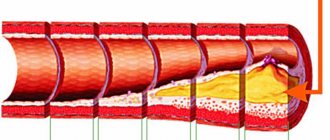
A more detailed examination of the anatomical venous structure will help to understand that this is a positive Valsalva maneuver. It is known that the vein system is equipped with numerous special valves. Thanks to them, normal blood movement is ensured.
If the valves begin to work chaotically, inconsistently, blood masses begin to be thrown in the opposite direction. As a result, stagnation appears and develops.
When a positive test is determined, we can confidently say that the entire valvular system of the blood flow is disrupted.
Also problems occurring inside the vessels and represented by pathological reverse blood flow. They are detected by the Valsalva test performed during an ultrasound.
The results obtained from the diagnosis are the basis for subsequent study of the course of the disease and the appointment of the most effective treatment.
Negative answer
This test result determines the lack of connection between the pathology progressing in the body and the vascular system.
During the diagnosis, bloated, swollen veins are not detected. Accordingly, the functioning of the valves in the veins and the circulation of blood masses are within normal limits.
The essence of the method
The differences between such concepts as the Valsalva maneuver and the Valsalva maneuver are as follows:
- The test is a diagnostic method that helps identify pathological processes.
- Reception is certain human actions that create high pressure in the middle ear, as well as in the abdominal and thoracic areas.
Operating principle
The pressure caused in the chest and abdominal area due to deep inhalation, subsequent breath holding and straining leads to changes in blood flow. This state is accompanied by the following stages – tension and relaxation.
In the process of tension, the flow of blood into the vessels of the heart decreases, which leads to a decrease in cardiac pressure and a simultaneous increase in intracranial pressure. When you relax, blood flow is restored and, accordingly, blood pressure increases.
At this stage, the receptors of the carotid artery detect changes in blood pressure, as a result of which a signal is sent to the vagus nerve of the brain, which further contributes to a reduction in heart rate. A healthy body reacts this way to the Valsalva maneuver.
The inability of the myocardium to change the frequency of contractions indicates the likelihood of pathological conditions developing in the cardiovascular system.
Holding your breath and pushing also affects the pressure in the lower extremities. Pathological changes in the venous valves of the legs and thighs lead to a reverse movement of blood flow, which indicates a violation of their functioning .
The Valsalva maneuver simultaneously increases pressure in the nasopharynx and middle ear, which, in the presence of accumulations of pus in it, causes perforation of the membrane, followed by its expulsion from the auricle.
Indications
Diagnostics using the Valsalva technique is currently used in various fields of medicine.
With its help, the following actions are performed:
- diagnosing tachycardia;
- determination of the quality of patency in the auditory tubes;
- detection of varicocele, determination of the severity of the baroreflex in the reproductive system;
- assessing the possibility of death due to a heart attack;
- diagnostics of the valve system of the lower extremities, examination for varicose veins;
- establishment of dysfunction progressing in the nervous venous system;
- The Valsalva maneuver is widely used in obstetrics.
This technique also neutralizes the unpleasant sensations that plague divers when diving to great depths. This time-tested technique effectively reduces the pressure that builds up in the middle of the hearing aid during landing/takeoff of airplanes. Which is also typical for sudden changes in atmospheric pressure.
It is necessary to correctly understand what the test and maneuver according to the principles of Valsalva are.
The test is prescribed by a doctor, it is a diagnostic measure and is performed within the walls of a hospital. As for the reception, its main task is to eliminate unpleasant sensations for a person. Which appear at certain moments in life. The reception is carried out independently.
Indications for the Valsalva test
Hardware diagnostics allows you to obtain comprehensive data on the condition of the testicles, spermatic cords and appendages with varicocele. First of all, they rely on ultrasound diagnosis of varicocele, but there are other methods.
- Ultrasound diagnosis of varicocele. The method of non-invasive examination of the scrotal organs is used to obtain information about the condition of the spermatic veins using ultrasonic waves.
Ultrasound signs of varicocele are easily noticeable in the initial stage, when there are no visual signs of the disease.The thickness of the veins of the spermatic cord is determined in a relaxed and tense state (a man is recommended to tense his abdominal muscles). Preparation for ultrasound of varicocele is not required; it is performed in a standing position.
It is possible to detect not only varicocele on ultrasound, but also additional testicular diseases - orchitis, hydrocele - and even cancerous changes.
- Valsalva test for varicocele. It is often carried out during any urological examinations and involves studying the state of the testicular vein during inhalation and exhalation. The test is also carried out during an ultrasound examination, and if the patient experiences a significant increase in the diameter of the veins in an upright position, the healthcare professional will diagnose a varicocele.
- Phlebography.
The method is designed to determine the presence of impaired blood circulation in the vein leading to the testicle. A contrast agent is injected into the vessel, after which an X-ray examination is performed, which records the speed of distribution of the solution through the veins. Phlebography also allows you to determine the structure of the spermatic vein and its diameter. This method is considered outdated due to its traumatic nature (in addition, local anesthesia and catheter insertion are required), as well as the negative impact of X-rays on the genital area. - MRI. The most advanced method for studying the smallest structural changes in the genital area.
The magnetic resonance method provides excellent tissue contrast and resolution, with the help of which initial functional transformations are easily determined.
The final results are recorded on information media that allows you to successfully enlarge any areas on the monitor.
An MRI is recommended for men with suspected first-degree varicocele and those with varicose complications.
Considering the high cost of MRI services, doctors refer men to study the testicles using this method only if other methods turn out to be uninformative.
Venography does not involve carrying out these manipulations if the image is taken without the introduction of a substance.
Reference. The diameter of the testicular vein in healthy men is 4.3 mm, and in patients with varicocele - 6.8 mm or more.
When identifying various pathologies, the methodology differs:
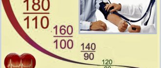
- When diagnosing cardiovascular diseases, the patient acts under the supervision of a doctor. While lying down or sitting, the patient is asked to inhale slowly and then exhale smoothly into a narrow mouthpiece. During the procedure, the physician's assistant monitors blood pressure, and electrocardiography (ECG) is performed at the same time. Thus, with the help of a test, a specialist can identify myocardial tone and deviations in changes in the size of the heart muscle during surges in intrathoracic pressure.
- In otolaryngology, the test is aimed at expelling purulent exudate from the middle ear cavity under the influence of a forced air flow. The opening of the auditory canals is facilitated by an increase in pressure in the patient's closed nasopharynx. Blood pressure monitoring is not necessary in this case.
- In urology, a breath test is performed on male patients to identify the initial stage of varicocele, a dangerous disease that causes dilation of the veins of the testicles and spermatic cord. The patient is asked to take as deep a breath as possible and hold his breath. While holding your breath, the urologist conducts a palpation examination of the man’s genital organs. The Valsalva maneuver for varicocele, performed in conjunction with ultrasound, allows an experienced specialist to accurately identify the disease at the very beginning. In the future, this will make it possible to choose the right treatment regimen.
- In vascular surgery, the Valsalva maneuver is one of the most effective methods for diagnosing varicose veins. At the same time, Doppler ultrasound (USDG) is performed, which makes it possible to give an objective assessment of the functioning of the valve apparatus of the veins of the lower extremities. At rest, the leaflets of the vein do not touch each other, but the Valsalva maneuver forces the valves to meet in the path of the return wave of blood, preventing retrograde flow.
Before carrying out the procedure, the specialist must conduct a thorough survey of the patient to determine possible contraindications.
The fact is that despite the safety of the test, if there are contraindications, it can threaten a significant deterioration of the condition.
DETAILS: Clinic of Urology of the First Moscow State Medical University named after. THEM. Sechenov
If there are no contraindications, then the doctor performs a Valsalva maneuver for varicocele, which looks like this:
- The patient lies down or sits down.
- Takes a deep breath, closing your nose and mouth.
- Fixes the position for 1-2 seconds.
- Next, he breathes into a special tube, which is connected to a barometer to control the strength and frequency of breathing.
At this time, the specialist performs manual palpation of the testicles in the area above the scrotum and determines the presence of damaged vessels.
If the doctor finds certain pathological changes, then an ultrasound is prescribed to clarify the diagnosis.
- determination of the patency of the auditory tubes;
- diagnosis of tachycardia;
- diagnosis of varicocele and severity of the genital baroreflex;
- assessment of the functioning of the venous valve system of the legs and determination of varicose veins;
- determination of dysfunction of the autonomic nervous system;
- assessment of the risk of death after a heart attack.
In addition, the Valsalva maneuver helps eliminate discomfort in divers when diving to depth.
Also, this technique will help reduce pressure in the middle section of the auditory analyzer during takeoff/landing of an airplane or during a sharp jump in atmospheric pressure.
The first is prescribed by a specialist for examination and carried out in a hospital, while the second is designed to eliminate discomfort and can be performed independently.
The Valsalva maneuver for diagnosing varicocele is as follows: first, the patient is interviewed to inform about the presence or absence of contraindications. Despite the relative safety of the Valsalva method, this test can lead to a significant deterioration in well-being in the presence of pain in the heart and a predisposition to sudden changes in pressure under negative external influences.
If no contraindications are found, the doctor proceeds with the actual procedure. To do this, the patient must take a straight position, lying or standing. Then he is asked to draw air into his chest and tense, closing his mouth and nose. After a few seconds, this tension leads to an increase in intra-abdominal pressure and vasospasm, which creates good conditions for studying the condition of the arteries in the groin area.
If the phlebologist finds “stars” on the veins and swelling of the arteries, then an ultrasound examination is prescribed to clarify the diagnosis. During an ultrasound, the Valsalva maneuver is also taken, but this method has some specifics. Only a standing position is allowed; manual palpation is not performed; instead, characteristics such as heart rate and blood pressure are examined with an ultrasound device.
Urologists perform a Valsalva maneuver during each medical examination of men, which allows them to identify varicocele at the initial stage and avoid the harm caused by this pathology to the body. In the process of diagnosing varicocele, the patient takes a deep breath and holds his breath. At this time, the doctor palpably examines the patient’s scrotum and testicles.
The Valsalva maneuver, performed during an ultrasound, can detect changes in blood flow in the affected veins. The patient is asked to take a deep breath, inflate the stomach as much as possible and tense the muscles of the lower abdomen. The spermatic cords enlarge and elastic nodules are visible on them. These signs indicate the development of varicocele.
The Valsalva maneuver is widely used in the diagnosis of varicocele in the early stages, when there are no pronounced clinical symptoms yet. The technique is used as an independent method of examination or in combination with ultrasound.
In patients with suspected varicocele, the Valsalva maneuver is performed at the first examination. The man takes a deep breath and holds his breath. At this time, the urologist palpates the scrotum and testicles. In the presence of pathological changes, even slight dilation of the veins becomes noticeable.
To assess blood flow in the affected vessels, the Valsalva maneuver is also performed during ultrasound. The specialist asks the man to take a deep breath and strongly tense his abdominal muscles. Signs of the disease are expansion of the spermatic cords and the appearance of elastic nodules.
DETAILS: Women's urology clinic center
Of the contraindications
There are many prohibitions associated with performing the described test:
- Strokes or heart attacks;
- the disease is in the acute phase;
- thromboembolism, vascular thrombosis;
- heart failure;
- fever;
- sepsis;
- infectious lesions.
The test is not permitted in the following cases:
- Severe tachycardia, accompanied by sharp chest pain;
- semi-fainting;
- a sharp drop in blood pressure.
The Valsalva technique is an accessible and informative method. In which disturbances in the functioning of the blood flow system are determined.
The procedure makes it possible to identify pathologies at the very beginning of development without identifying obvious visual signs. Early diagnosis entails timely treatment of the pathology and prevention of its progression.