Stages of transition of normoblasts into erythrocytes
It will take a little time for the described blood cells to transform into red blood cells. Initially, the development of a basophilic erythroblast is observed, in the central part of which there is a nucleus. It is characterized by the presence of a round shape and a size of 18 microns.
These cells have a rich blue color. Subsequently, polychromatophilic erythroblasts are formed from them, which are smaller in size than basophilic ones. These cells are characterized by the presence of a wheel type of chromatin, and the cytoplasm acquires a pink-blue color.
Subsequently, the present erythroblast is converted into an oxyphilic form. Such cells are characterized by the presence of a fuzzy purple nucleus. The cell becomes even smaller and more closely resembles an erythrocyte.
Over time, the cell nucleus becomes pyknotic, and the cytoplasm becomes light blue. This indicates the transition of the erythroblast to the polychromatophilic form. The cell then transforms into reticulocytes. And only at this stage are red blood cells formed in the blood without a nucleus.
What are normoblasts?
Normoblasts are cells that are formed at the initial stage of red blood cell formation. They are distinguished from fully mature red blood cells by the presence of a nucleus.
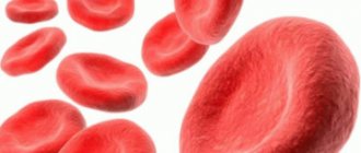
But while normoblasts grow, they are filled with hemoglobin and lose their nucleus. After its disappearance, mature erythrocytes are obtained from normoblasts.
The process of converting normoblasts into red blood cells takes a certain time. First, a basophilic erythroblast with a nucleus in the center appears. Its shape is round and its size is about 18 microns.
This cell is painted in a rich blue color. Soon a polychromatophilic erythroblast is formed from it, which becomes smaller than the basophilic one.
Its chromatin has a wheel-shaped structure, and the cytoplasm appears pink-blue in color.
Later, the oxyphilic erythroblast is formed from the polychromatophilic one. Its purple core is already losing its clear structure. The cell itself decreases in size and becomes somewhat similar to a red blood cell.
After some time, the nucleus becomes pyknotic, and the cytoplasm turns gray-blue, causing the oxyphilic erythroblast to turn into polychromatophilic.
After this, transformation occurs into reticulocytes, and then into mature red blood cells without a nucleus.
The number of nucleated red blood cells is determined in the blood using a special analyzer. Usually they count how many there are per 100 leukocytes.
Sometimes normoblasts are mistaken for small white blood cells (leukocytes), which results in a false test.
Therefore, when counting normoblasts and leukocytes, a correction factor must be entered to obtain the correct result.
There should be no normoblasts in the peripheral blood at all. They are formed in the bone marrow, where they are degenerated.
They can enter the bloodstream due to bone marrow lesions or the appearance of various diseases associated with hematopoietic disorders.
Causes of normoblasts
Normoblasts are formed and transformed in the human bone marrow. As a result, the number of 0.01 normoblasts in a general blood test is considered a deviation from the normal value. These cells should not enter the peripheral blood. Their detection on a hemogram is a sign indicating the possible formation of serious diseases associated with hematopoiesis or brain structure.
The causes of normoblasts in a general blood test include the following:
- Hemolytic type of anemia.
- Various forms of leukemia or erythroleukemia.
- Brain cancer.
- Tumors of a malignant nature.
- Problems with blood circulation.
- Severe blood loss.
- Formation of metastases in the bone marrow.
An increase in the number of normoblasts in the blood is considered especially dangerous after surgery. The presence of these cells indicates a serious condition of the patient.
In this case, the presence or absence of blood cells, and not their number, acts as a diagnostic element. Even the slightest deviation from zero is a sign of a pathological process. But do not get upset ahead of time, because the appearance of normoblasts may be associated with the presence of prolonged inflammation or hypoxia.
Why might they appear in analysis?
The reasons for the appearance of normoblasts in blood tests include pathological or acute conditions of the body - a deviation from the norm (and the norm is when they are absent) is always a sign of a disorder in hematopoiesis:
- Anemia, especially thalassemia.
- Acute or chronic leukemia. They are often manifested not simply by the presence of normoblasts in a blood test, but also by an increased number of them (there are a lot of cells). Men are affected more often than women.
- Erythroleukemia (Di Guglielmo's disease). An acute malignant form of blood cancer, the diagnosis of which begins with determining the presence in the blood of cells with non-discarded nuclei. This pathology is also distinguished by the appearance of additional foci of blood cell construction, for example, in the spleen, so that the number of normocytes can go off scale.
- Blood loss for any reason (trauma, wound, surgery, etc.). The body is forced to intensively restore the number of blood cells in this state.
- Metastases in the bone marrow requiring treatment with chemotherapy: both in the normal state and after a course of treatment, the number of normoblasts in the blood will be increased, since the body needs to repair the damage and compensate for the destructive process.
Many of these diseases are diagnosed precisely by a blood test and the presence of normoblasts.
Blood pathologies, however, are the main reason for the increase in the level of normoblasts.
Prevention of the appearance of normoblasts cannot be universal - it consists of preventive measures suitable for each pathological condition separately.
General recommendations apply to cases of anemia, since this is the main reason for the increase in the level of normoblasts in the blood:
- Do not expose yourself to radiation from radioactive substances (important for those working in production or science).
- Avoid poisoning with chemicals and poisons of chemical origin.
- Do not abuse medications – especially in case of serious illnesses and without a doctor’s prescription.
- In general, take care of yourself and lead a healthy lifestyle.
- Get blood tests done regularly.
Normoblasts in a child's body
Hematopoiesis in a child’s body is significantly different from an adult’s, so normoblasts in a general blood test are considered a completely normal process, which is explained by the fact that during birth the bone marrow, which is responsible for the production of blood cells, is located in all bones, both flat and tubular types. A heavy load, as well as increased synthesis of erythropoietin by the child’s kidneys and liver, leads to physiological changes. And they, in turn, entail the release of a small number of normoblasts into the blood.
The maximum number of normoblasts in the analysis is found in infants aged 2 to 3 months. A small number of normoblasts may be periodically detected throughout the early developmental period.
It is worth noting that normoblasts in a child’s general blood test may also indicate pathology, in particular, the development of a disease such as an acute form of lymphoid leukemia. This disease requires immediate treatment, since in advanced stages it can even cause death.
Is an elevated level always a sign of pathology?
Not at all. For example, normoblasts in a general blood test in absolutely healthy infants up to 3–4 months are a completely acceptable phenomenon associated with the active physiological formation of a small organism. In older children, blastocytes should be observed only in the bone marrow and not in the blood.
Under such circumstances, blast cells will indicate the presence of functional defense mechanisms. Moreover, people sometimes get disappointing results that are overshadowed by repeat diagnostic rates. Therefore, you should not worry when you see alarming numbers on the form; you need to calmly visit a specialist to confirm or refute them.
Decreased number of normoblasts
Since the norm of normoblasts in the blood is 0, then their number cannot be reduced.
Only the number of red blood cells that are formed from normoblasts can be reduced. Red blood cells can be diluted with a large volume of liquid, or their normoblasts can actually form in smaller numbers.
The latter problem is observed in the presence of various bone marrow diseases as a consequence of radiation exposure. But the main cause of this condition is recognized to be a deficiency of iron, which is required to create hemoglobin.
What is the normal rate of blast cells?
Blast cells in the blood test are not normal. In a good state of the body, when there is no stress or disease, the standard amount of such elements in the bone marrow will be 1%. They gradually replace their “used up” elements, and the hematopoietic organ makes up for this deficiency without exceeding the norm.
In case of stress, viral or bacterial infection, the bone marrow will increase the number of blast cells by up to 10%. In exceptional cases the quantity may be slightly higher. Any other indicators indicate deviations in the operation of the system.
If the number of blast cells reaches 20%, this means that a disease such as acute leukemia is developing. They come in different types and forms, occur quickly and are classified as cancer. Such a deviation can appear completely unexpectedly, even in those who have never had anything serious in their life. This happens to children and young people. In the older generation, leukemia is secondary, caused by diseases or treatments, for example: chemotherapy.
What do blast cells mean in a blood test, the norm of which is exceeded? In fact, such bodies in their “immature” form should not leave the bone marrow. Elements that are fully formed for their intended purpose and are ready to perform their function are sent there. If blasts are in the blood, this indicates advanced cancer.
From the above we can draw conclusions:
- Blast cells should be absent from the blood; their normal habitat is the bone marrow.
- Under normal conditions, such elements should not exceed 1% of the amount of blood.
- Up to 10% of blast bodies are found in the bone marrow during times of acute need (disease, infection).
- More than 20% in the bone marrow and the presence of deformed blasts in the blood mean the presence of acute leukemia.
An accurate diagnosis determining the presence of leukemia can be made even before blasts enter the blood. For this, a general clinical blood test is sufficient, which shows the level of red blood cells and other related indicators.
A person, cat, dog or other living creature with the appropriate body structure can become ill with acute leukemia. In any case, this is a serious cancer that requires immediate treatment. The chances of a successful outcome depend on the stage of the disease, the condition and health of the patient, and the type of leukemia.
Blasts are immature forms of blood cells. In general, they mature to completion in the bone marrow. And their appearance in a blood test in most cases indicates serious health problems. The patient needs urgent consultation with an oncologist. This will be determined by a corresponding blood test - whether there are blasts in the fluid or not.
These cells can be normal or cancerous. Normal ones are always present in the bone marrow, although there are very few of them there, the amount does not exceed 5 percent.
But with leukemia in various forms, their number increases so much that they can get not only into various organs and tissues (especially the kidneys, brain, spleen, lymph nodes and liver), but also sometimes appear in the blood.
Sometimes when making a diagnosis, the doctor indicates “leukemia”. This is because the cells under discussion are very similar to leukocytes. Blasts in the blood are young blood cells that differ from others in the fluid in the structure of their nucleus. Their main feature is the content of up to five nuclei in one cell.
In rare cases, they appear in the fluid after aplasia. Indeed, during this period, the bone marrow does its best to restore its function. But basically these are not malignant blasts, and they appear in an amount of no more than two percent.
Under any other conditions, a healthy person should not contain blasts in the blood; their norm should be zero.
Symptoms of leukemia
Early diagnosis of leukemia significantly increases the chances of cure, so it is recommended to consult a doctor immediately after the following symptoms appear:
- pale skin;
- feeling of weakness;
- dizziness;
- problems with blood clotting;
- deterioration in the functioning of the immune system;
- excessive fatigue.
If, against this background, a normoblast result of 1:100 is detected in a general blood test, then this may indicate the occurrence of leukemia (high blood cell counts may also indicate the presence of this pathology).
What are normoblasts?
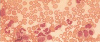
Blast cells are an intermediate form of erythrocytes (red blood cells), responsible for saturating body tissues with oxygen, as well as removing carbon dioxide from the body. These substances are formed, like all formed elements of blood, in the bone marrow. Blue-violet cells at this stage of development are actively enriched with hemoglobin, which provokes a decrease in the nucleus. When it is completely dissolved, normoblasts will acquire smaller sizes and transform into pinkish reticulocytes.
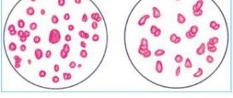
Blast cells in a blood test under a microscope
They, in turn, leave their abode, entering directly into the bloodstream, where over the course of 1–3 days they get rid of unnecessary organelles (mitochondria, endoplasmic reticulum). Reticulocytes can already carry out oxygen transport, but their functionality increases significantly after they are fully transformed into biconcave erythrocytes.
Often, when deciphering indicators, experts mention normocytes or blastocytes. It must be remembered that these terms are the medical interpretation of normoblasts.
Diagnosis of leukemia
If there are symptoms characteristic of leukemia, the doctor first sends the patient for a hemogram and a blood test to detect blast cells. Thanks to the analysis, an accurate indicator of all atypical blood elements will be obtained, which will reveal the extent of the spread of the disease. In the case of leukemia, a general blood test reveals a decrease in the number of platelets. In parallel with this, there is an increase in ESR and the number of normoblasts in the blood.
In addition, the following diagnostic studies may be prescribed:
- blood chemistry;
- study of immunoenzymes;
- myelogram (bone marrow biopsy).
Only after studying all the data obtained as a result of the presented research methods, the doctor is able to make a diagnosis.
Features of the analysis
To diagnose an acute form of leukemia, you need to take a general blood test and from it a specialist will determine what diseases you have. If the number of red blood cells is reduced, then the anemia is said to be normochromic. Most likely, such an analysis of blast cells will also reveal a reduced platelet count. In rare cases, their number is greatly increased, and they have oddly shaped nuclei. ESR (erythrocyte sedimentation rate) is increased in acute forms of leukemia, but there are cases when it is normal.
Automatic Leukocyte Analysis System
You should know that normally there are no blast cells in a blood test, since they are located in the bone marrow and do not extend beyond it. The first symptom of blood leukemia, however, is a high level of white blood cells, which are found in the blood along with blast cells.
In any case, one general blood test does not diagnose leukemia. If cytopenia is not clear, then a puncture test is taken from the bone marrow.
There are cases in which this procedure shows that platelets are normal and there are no blast cells in the blood, and when a puncture is taken from the bone marrow, it is determined that the blasts are also within normal limits, but if the specialist has doubts about the diagnosis, then a trephine biopsy is performed, which finds Cell proliferation in the blood is what determines the presence or absence of the disease.
In vitro, blast cells are determined in a blood test in different ways and repeated blood testing is carried out, which will help make the correct diagnosis.
Purpose of a myelogram
To determine the cause of an increase in the number of normoblasts in the blood, a myelogram is often prescribed. The analysis is a study of the condition of a smear taken from the bone marrow through a biopsy. The procedure is performed under local anesthesia. The puncture is performed in the area of the sternum or ilium.
The procedure does not require special preparation or any restrictions. If a person is taking medications, then before the procedure he must inform the doctor about this, and if possible, temporarily stop using the medications. The result of the study can be obtained after just a few hours.
Treatment
There is no treatment for elevated levels of normoblasts in the blood. They disappear on their own after successful treatment of the underlying pathology is completed.
It is very important to identify the reason that provoked the increase in normoblast levels in the blood. After the pathology is detected, therapy is carried out, thanks to which the process is either completely stopped or a state of stable remission is created for the patient in chronic forms of the disease.
The most terrible disease, which may be indicated by elevated normoblast counts, is leukemia.
Treatment methods for leukemia
If it is confirmed that elevated normoblast counts indicate the presence of leukemia, then treatment of the pathology includes the following manipulations:
- Chemotherapy. Prescribed if the malignant nature of the pathology is confirmed. During this procedure, all affected cells are destroyed.
- Radiation therapy. Provides relief from the process of tumor growth in the affected area.
- Biotherapy. It is used at the final stages of treatment of the disease or when it is mild. It involves the use of special medications that act as analogues of substances produced by a healthy body.
- Targeted treatment. Based on the use of monoclonal bodies for therapeutic purposes. It is an alternative to chemical therapy in the initial stages of treatment of the disease.
If the disease is in an advanced state, then the only way to cure it is through stem cell transplantation. This is a rather labor-intensive process that requires a lot of professionalism and money.
Therapy for erythromyelosis
Having understood what normoblasts are in the blood, what this means for children and adults, it should be noted that elevated levels of these blood cells can directly indicate the presence of such a serious disease as erythromyelosis.
This disease is characterized by the following symptoms:
- severe weakness;
- bruising;
- painful sensations in the bones;
- weight loss;
- difficulty breathing;
- the formation of a fungal infection.
In the absence of quality therapy, pathology can provoke the formation of a focal type of necrosis of the spleen, swelling of the lymph nodes, bleeding from the nose and gums, as well as hemorrhages in the retina.
Such complications develop as a consequence of the fact that the cells containing the nucleus penetrate through the circulatory system into the internal organs, digestive and reproductive systems, and enter the skin and muscles.
In some cases, diseases that are characterized by an increased level of normoblasts, after about six months or even faster, cause death.
Therapy for this dangerous disease consists of several sessions of chemical or radiation therapy. In addition, the patient can have stem cells transplanted.
Rarely, people may develop a chronic form of erythromyelosis. Diagnosing this pathology is quite difficult, because, despite the presence of a tumor, red blood cells containing nuclei do not penetrate into the blood.
The presented diagnosis can be confirmed by a detailed study of the condition of the internal organs, since the liver and spleen are enlarged in size, and swelling of the lymph nodes develops.
This form of the disease has a long course (over 2-3 years). To rid the patient of pathology, doctors perform multiple transfusions of red blood cells. An alternative method of therapy is the introduction of a special medicinal serum, but a greater effect is achieved through stem cell transplantation.
Prevention of increasing the number of normoblasts in the blood
To prevent an increase in the number of normoblasts, it is necessary to take preventive measures aimed at preventing the formation of anemia and acute forms of leukemia. To avoid the occurrence of these pathologies, it is recommended to avoid radioactive radiation, inhalation of toxic chemicals, and uncontrolled use of medications.
Medical workers insist that if an increased number of normoblasts is detected in the blood, it is imperative to contact the clinic. Only identifying an accurate diagnosis and timely initiation of therapy will ensure a full recovery and rapid restoration of all body functions.
It is important to understand that the detection of even a small number of normoblasts in a blood test is already a sign of pathology that requires immediate treatment.
Types of leukemia
Medicine divides blood leukemia into two types - this is a chronic form of leukemia (consists of mature and immature elements), and it never turns into an acute form of leukemia (in which blasts are detected in the blood). In acute leukemia, these cells are a tumor component in the human body.
Typically, this form of the disease is named after the cells whose precursors are immature blast cells, for example, these can be myeloblasts, lymphoblasts, monoblasts, erythroblasts. Thus, acute myeloblastic leukemia, acute lymphoblastic leukemia and other forms of blood cancer are distinguished.
Doctors all over the world use the identification and designation of blast cells in a blood test using the international FAB system, which provides for the characterization of acute leukemia by the degree of polymorphism, the shape of the blast nucleus, depending on the maturation of the blasts or without their maturation.
There are no specific symptoms of blast cells in the body that can be used to determine the presence of leukemia in the blood, but the sooner you feel unwell and take a general blood test, the faster a specialist will be able to identify this pathology. It should be remembered that the acute form of blood leukemia, in most cases, occurs in children.