Consequences of elevated hematocrit in men and women
First of all, it is necessary to remember that an increase in the number of red blood cells means a thickening of the connective tissue of the circulatory system. For good blood circulation and proper metabolism, increased density will become an obstacle. Blood viscosity leads to negative consequences, namely the formation of blood clots. If a “plug” forms in the vessels, it blocks access to tissues and organs.
Consequences of high Ht, which provoked thrombosis:
- Stroke;
- Heart attack;
- Gangrenous process;
- Thromboembolism;
- Death.
In men and women, an increase in hematocrit in tests should alert and prompt them to solve the problem. Very often, death in patients occurs precisely because of the formation of a blood clot that blocks the arterial blood flow. When a heart attack occurs, only 30% of all people survive until doctors arrive. And if an ischemic stroke occurs, complications can become irreversible and lead to partial or complete immobility.
Consequences of thickened blood in a child: possible complications
If a baby experiences a prolonged increase in hematocrit, it is very important to diagnose the cause of the analysis deviations. This condition cannot be ignored, because it can provoke dangerous complications:
- Thrombosis;
- Violation of trophism of tissues/organs;
- Necrosis due to blockage of blood flow;
- Gangrene of the limbs;
- Respiratory failure;
- Ischemic stroke.
The most dangerous complication of thickened blood is blockage of large vessels. In such cases, blood clots become the cause of death.
To prevent the development of serious disorders in a child, it is important to undergo regular examinations. Especially if the baby has already encountered elevated Hct and has undergone treatment
Timely diagnosis of indicator deviations will allow you to quickly solve the problem and prevent complications.
Analysis, indications for analysis and norms
And these are not all cases:
- To diagnose the severity of anemia and polycythemia. For the first disease, all indicators will be lowered, and for the second, on the contrary, they will be increased.
- To confirm the effectiveness of the treatment of the above and other diseases that affect the level of red blood cells.
- A blood hematocrit test will show whether a blood transfusion is necessary, as well as whether there is a need for other treatment methods.
- The hematocrit indicator is used to assess the stage of dehydration of the body.
Indications for such a study are the following symptoms:
- weakness;
- constant fatigue;
- fainting;
- pale skin;
- dyspnea;
- vision problems;
- dizziness and headache;
- increase in the size of the spleen;
- frequent flushes of blood in the facial area;
- thirst;
- dry mouth;
- insufficient amount of urine.
In some cases, the blood is tested several times.
As mentioned above, the normal hematocrit (HCT) differs between women, men and children.
- The hematocrit number is highest in newborns. It reaches 55-68%.
- By three months, this figure will almost halve and will be equal to 30-36%.
- By adolescence, the normal rate will reach 36-40%.
- The hematocrit indicator in women is 38-46%.
- The normal hematocrit for men is 42-54%.
If the test results show deviations from the norm, there are disturbances in the functioning of the body.
Clinical and diagnostic value
— Myeloproliferative syndromes (erythremia, myelofibrosis)
— Chronic inflammatory diseases (rheumatoid inflammation of the joints, tuberculosis, cirrhosis of the liver)
- Thrombocytopenia caused by decreased platelet production:
- Thrombocytopenia caused by increased destruction of platelets
Thrombocytopenia caused by platelet sequestration 9increased destruction)
ESR – erythrocyte sedimentation rate
— Pregnancy, postpartum period, menstruation
-Inflammatory conditions (acute and chronic infections, pneumonia, rheumatism, myocardial infarction, syphilis, tuberculosis, trauma, bone fractures, shock, surgical interventions, collagenosis, Raynaud's disease, poisoning with chemical compounds (lead, arsenic)).
— Hyper and hypofunction of the thyroid gland
— Erythremia and reactive erythrocytosis
When performing blood tests on hematological analyzers, the names of the indicators are in English
WBC (white blood cells) – the number of blood leukocytes (x10 9 / l).
RBC (red blood cells) – the number of red blood cells (x10 12 / l).
HGB (hemoglobin) – hemoglobin concentration (g/dl or g/l).
HCT (hematocrit) - hematocrit.
MCV (mean corpuscular volume) – average volume of an erythrocyte (µm 3 or fl).
MCH (mean corpuscular hemoglobin) – average hemoglobin content in red blood cells (pg).
MCHC (mean corpuscular hemoglobin concentration) – the average concentration of hemoglobin in a red blood cell (g/dL).
RDW (red cell distribution width) is an indicator of the heterogeneity of erythrocytes by volume, characterizing the degree of anisocytosis.
PLT (platelet) – platelet count (x10 9 / l).
MPV (mean platelet volume) – average platelet volume (fl or µm 3 ).
PCT (platelet crit) – thrombocrit, reflects the proportion of the volume of whole blood occupied by platelets (%).
PDW (platelet distribution width) – platelet distribution width by volume (%), degree of platelet anisocytosis.
In table Table 1 shows the main indicators of a clinical blood test of a healthy person.
general information
In the analysis, the hematocrit norm is not always indicated, and most often it is indicated by the combination HCT, that is, the decoding is assumed based on this line in the analysis. In this case, the more correct term is hematocrit number. The hematocrit itself is a glass flask that is used in blood tests for centrifugation. This number can also be called “blood density”; this is the popular definition for HCT.
HCT is measured as a percentage. However, sometimes decoding is done in decimal fractions, with accuracy reaching hundredths.
It is necessary to check whether the hematocrit indicator is normal in several cases. The most common is surgery, as well as pregnancy. Hematocrit during pregnancy is determined to eliminate the risk of losing a large amount of blood in women during childbirth. A general blood test is the first priority in diagnosing any disease. Doctors specifically require HCT when there are varicose veins in the lower extremities, suspected autoimmune diseases or liver problems.
Blood clotting is, by and large, a protective reaction for humans. This helps protect the body from large blood loss. The endocrine and nervous systems are responsible for the process of regulating coagulation. In this case, a violation of blood flow can lead to blockage of blood vessels.
Only when in a liquid state can blood perform such important purposes as transporting nutrients, protecting the body, and performing trophic and thermoregulatory functions. A general test can show how blood clotting is
An increased HCT score means an increased risk of thrombosis, heart attack and stroke. This is why doctors insist on regular general blood tests.
A general analysis can show how blood clotting is. An increased HCT score means an increased risk of thrombosis, heart attack and stroke. This is why doctors insist on regular general blood tests.
Hematocrit in the blood and its meaning
For various pathologies associated with bleeding, loss of fluid with vomiting or diarrhea, large burn surface, the doctor needs to know the ratio of the liquid part of the blood and the formed elements. This indicator is called “hematocrit”. In the medical literature, the term “hematocrit number” is found - this is a more correct professional name.
Another popular short designation for the indicator is “HCT”, from the Latin name hematocrit, used in modern laboratory instruments and indicated when automatically deciphering the analysis.
Reasons why hematocrit is increased
Physiological reasons
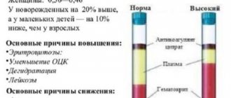
As a result of some changes in the body, the composition of the blood “adjusts” to them, as a result of which an increase in hematocrit in the blood may occur. These factors include:
- Age
Newborns have a large volume of red blood cells in their blood, so at first glance we can conclude that there is an increased hematocrit in the baby's blood. However, the figure of 44-62% is actually within the norm. After three months, the hematocrit drops to 32-44%, and after another 9 months – to 31-40%.
- Dehydration
The body seeks to compensate for water deficiency by reducing plasma in the blood, therefore the total volume of circulating blood (TBV) decreases, and the proportion of red blood cells in it increases, and therefore an increased hematocrit in the blood is observed. Dehydration can occur with low water intake, excessive sweating, frequent diarrhea and vomiting. Hematocrit is increased during pregnancy at the stage of toxicosis.
- Hypoxia
Smokers often experience a lack of oxygen in the body - hypoxia. Hemoglobin, which is part of red blood cells, is responsible for transporting oxygen to all organs and removing carbon dioxide from them. If less oxygen is supplied than necessary, the body tries to compensate for this by producing additional hemoglobin. As a result, the number of red blood cells increases and the hematocrit HCT in the blood test is increased.
Hypoxia can also occur in people who have spent several days in the mountains. Their lungs have not yet had time to adapt to the rarefied mountain air, which contains less oxygen, so the hematocrit increases.
Conducting a hematological study
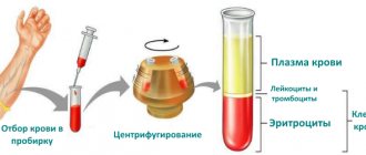
In order to find out the value of hematocrit, doctors can remove a small portion of blood from both the finger capillary and the cubital vein. In laboratory conditions, a substance that prevents premature clotting - an anticoagulant - is added to the sample. The test tube is then placed in one of the centrifuge cells and the apparatus is activated.
The drum begins to accelerate around its own axis, and as a result of the centrifugal force, heavier blood cells (red blood cells) settle at the bottom, and a light liquid appears on the surface - translucent plasma. Other components (leukocyte cells, platelets) form a thin intermediate film.
After completing this stage, the specialist assesses the level of red blood cells in the blood using special markings and writes down information about the hematocrit on a form. Some clinics use modern devices to detect blood counts that perform the counting function automatically. The results of the study are most often given to the attending physician who referred the patient for the procedure the very next day.
The principle of establishing the level of hematocrit in the blood
Level down
A condition in which the HCT level is below normal also has its own characteristic symptoms:
- shortness of breath and difficulty breathing;
- excessive fatigue and a persistent feeling of tiredness;
- hair loss;
- cardiopalmus;
- pallor of the skin.
These symptoms manifest themselves especially strongly during pregnancy and with acute anemia. By the way, during pregnancy the hematocrit value always becomes smaller. The reason is an increase in the volume of circulating blood. The normal blood viscosity level for pregnant women is 33%.
The cause of a decrease in hematocrit level can be any disease that leads to the destruction or shortening of the lifespan of red blood cells. One of them is anemia.
There are other reasons for the development of this condition:
- Large blood loss.
- As mentioned above, pregnancy.
- Lying down for too long.
- Slow production of red blood cells.
- Increased amount of blood.
- Fasting or unbalanced nutrition, for example, during a diet.
- The breakdown of red blood cells due to certain diseases.
- Blood thinning.
- Tumors and other similar diseases of the bone marrow, due to which fewer red blood cells are produced than normal.
- Diseases of the excretory system, in particular renal failure or polycystic disease.
- Inflammatory processes that have entered the chronic stage.
- Anemia, which is characterized by a decrease in the number of red blood cells.
- Diseases of blood vessels and liver.
A reduced level of blood viscosity is a reason to consult a doctor and undergo additional examination. Don't panic.
Sometimes you can normalize the condition in simple ways:
- Add to your daily menu foods that increase hemoglobin levels and activate the process of red blood cell production. These include chicken meat, dried fruits, legumes, eggs, red meat, liver, greens, etc.
- You can also improve your well-being with the help of vitamin C, which helps iron to be absorbed.
- Sometimes dietary supplements that include iron may be prescribed.
There are also traditional medicines that have a beneficial effect on blood composition.
One of them is prepared from 300 grams of garlic and 500 ml of alcohol. The ingredients must be mixed and placed in a dark place for 21 days. Take 20 drops of the product, mixing it with 100 ml of milk.
Like medications, such products must be used under the guidance of a physician.
What is hematocrit? This is the percentage of red blood cells in the entire blood volume. The hematocrit norm depends on the age and gender of a person, as well as on his lifestyle. In some cases, there is nothing wrong with a decrease or increase, while in others you should consult a doctor.
Hematocrit
Hematocrit (packed cell volume, PCV) is the volume of formed elements expressed as a percentage of whole blood in a given sample. Usually, with a small error, it is customary to express the volume of red blood cells through the hematocrit value. Venous hematocrit is practically no different from capillary hematocrit.
The microcentrifuge technique, as well as the methods used in hematology analyzers, have been approved as unified methods.
Unified microcentrifuge technique
Principle. Centrifugation of a capillary with blood for a certain time, followed by determination of the ratio of the volume of formed elements (the height of a column of cells) to the volume of blood (the total height of the entire contents) on a special scale.
Reagents: 1) heparin—500 U/ml, diluted with distilled water in a ratio of 1:5 or 2) EDTA (Trilon B)—40 g/l.
Equipment: 1) special hematocrit microcentrifuges; 2) capillary tubes (complete with centrifuge).
Progress of determination. The capillary, pre-treated with an anticoagulant (reagents 1 or 2) and dried, is filled with blood obtained from a finger prick or venipuncture to 7/8 of its length. Close the capillary at one end with a special paste or plasticine and place it in a centrifuge rotor so that the sealed end rests against the rubber gasket, centrifuge for 5 minutes at 8000 rpm. The hematocrit value is determined using the reading scale attached to the centrifuge.
Determination on hematology analyzers
Hematocrit testing is included in the programs of all hematological analyzers produced by various companies.
Principle. Usually the conductometric or microcentrifuge method is used. In some analyzers, hematocrit is determined by calculation, based on the number of red blood cells in a certain volume of blood (for example, 1 μl) and the average volume of one red blood cell.
The determination process is carried out in accordance with the instructions supplied with the device.
Normal values. In healthy people, the hematocrit of venous and capillary blood is 40–48% for men and 36–42% for women.
Clinical significance. A decrease in hematocrit is characteristic of anemia (up to 20–25%), and its value can be used to judge the degree of anemia. A significant increase (up to 65%) is observed with polycythemia, a less significant increase (50–55%) is observed with symptomatic erythrocytosis accompanying heart defects, pulmonary insufficiency, and some hemoglobinopathies. The hematocrit value characterizes the degree of hemoconcentration and hemodilution. Hematocrit values are used for calculated indicators characterizing red blood cells: average volume, average hemoglobin concentration, etc.
Platelets
Platelets are anucleate cells with a diameter of 2-4 microns, which are “fragments” of the cytoplasm of bone marrow megakaryocytes. Physiological fluctuations in the number of platelets in the blood during the day are approximately 10%. In women during menstruation, the platelet count can decrease by 25-50%. Platelets perform angiotrophic, adhesive-aggregation functions, participate in the processes of coagulation and fibrinolysis, and ensure retraction of a blood clot. They are able to carry circulating immune complexes on their membrane and maintain vasospasm.
Level increased
The main symptoms of increased hematocrit in the blood are dizziness, nausea, numbness in the arms and legs, and difficulty breathing. This is explained by the fact that much more oxygen enters the body than is needed. So-called poisoning occurs, and hyperoxia develops.
It is worth noting that the hematocrit or HCT value can increase for physiological or internal reasons.
The first include several factors:
- Smoking. The body receives little oxygen, resulting in tissue oxygen starvation. To normalize the nutrition process, the body provokes increased production of red blood cells.
- Work or prolonged stay at height. And again the person receives less oxygen than needed.
- Use of anabolic steroids. These substances help athletes build muscle and, as a result, increase hematocrit levels.
- In infants, blood viscosity is always higher than in people of other age categories.
Internal causes of deviation from the norm of hematocrit are diseases or pathological conditions developing in the body:
- Dehydration. Blood viscosity is normal if the body receives enough fluid. But it also happens that it is not enough. This happens with heavy sweating, vomiting, diarrhea, prolonged exposure to heat, etc. The amount of plasma decreases, followed by the amount of blood in the circulatory system. Because of this, the percentage of red blood cells increases and the blood becomes more viscous.
- Diseases characterized by a decrease in plasma levels. These include severe burns, peritonitis, thrombosis and diabetes. As in the previous case, HCT increases due to a decrease in the amount of plasma.
- Hypoxia or oxygen starvation. Most often, this condition is observed in heavy smokers or those who suffer from diabetes. Due to lack of oxygen, the body activates the process of producing red blood cells. “Thanks” to this, the blood becomes thicker.
- Long-term treatment with glucocorticoids and any other hormone-based drugs. This also includes hormonal drugs.
- Kidney diseases, such as hydronephrosis or polycystic disease. In the first case, the body becomes unable to absorb liquid. And in the second, it produces too much erythropoietin. This is the hormone responsible for the production of red blood cells.
- Anemia.
- Leukemia and other blood diseases.
- Acute or chronic form of polycystic disease.
- Erythremia. What it is? This is the same polycythemia or leukemia in the chronic stage with a characteristic disturbance in the process of cell division.
A high hematocrit level increases the risk of thrombosis and therefore requires immediate medical attention.
What to do if the hematocrit norm is exceeded?
- First of all, it is necessary to find the cause of this condition.
- Sometimes no special treatment is required. Frequent walks in the fresh air, quitting smoking and a healthy lifestyle can improve the condition.
- It is also necessary to establish a drinking regime. The body should not suffer from fluid deficiency.
- If improvement does not occur, you can resort to drugs from the group of anticoagulants. They thin the blood quite effectively. You can take such medications only under the supervision of a doctor.
Treatment must be comprehensive and timely, otherwise serious complications may develop.
Red blood counts
Hemoglobin (Hb or HGB)
- in healthy people, the hemoglobin content fluctuates in the range: for women - 120-140 g/l, for men - 130-160 g/l. In healthy people, hemoglobin is found in red blood cells in the form of oxyhemoglobin, reduced hemoglobin and in small quantities in the form of metabolically inactive metabolites: methemoglobin, carboxyhemoglobin, sulfhemoglobin and others.
Clinical significance.
A decrease in hemoglobin content in the blood (hypochromemia) is observed with anemia of various origins, an increase (hyperchromemia) is observed in newborns; in adults this occurs with polycythemia vera and symptomatic erythrocytosis. A relative increase in hemoglobin concentration is observed with a decrease in plasma volume (hemoconcentration). To determine hemoglobin manually, the most widely used method is the hemoglobin cyanide method (the method is based on the conversion of all forms of hemoglobin into the form of hemoglobin cyanide), and when using hematological analyzers (hemichrome method) in which a stable compound of hemoglobin and higher fatty acid is formed.
Red blood cells in peripheral blood smear
R.B.C.
— the normal number of red blood cells in men is 4.0–5.1x10 12 /l; in women - 3.7–4.7x10 12 /l.
Clinical significance.
A decrease in the number of red blood cells (erythrocytopenia) in the blood is one of the main criteria for anemic syndrome.
Erythrocytosis
- a combined increase in the number of red blood cells, hemoglobin levels and hematocrit in the blood. Erythrocytoses represent a heterogeneous group of diseases and physiological conditions, the common feature of which is an increase in the number of red blood cells per unit volume of blood.
Erythrocytosis may be relative
(dehydration of the body due to loss of fluid (vomiting, fever, hyperventilation, diarrhea), loss of fluid through non-physiological means (burns, removal of ascites, inadequate diuretic therapy) and
absolute:
a) reactive increase in erythropoietin during hypoxemia (in residents of high mountain areas, with lung diseases, congenital heart defects) b) pathological production of erythropoietin (kidney disease, endocrine system, tumor), c) tumors of the blood system with increased production of red blood cells (erythremia).
Advice for pregnant women
Experts emphasize that a decrease in hematocrit often causes a decrease in iron levels in the body. This causes the development of first degree anemia
It is very important that during pregnancy a woman receives iron in sufficient quantities, otherwise after childbirth she may face a severe shortage of iron.
Increasing iron levels can be achieved with the help of medicinal-type drugs that contain the necessary element in the required concentration. The course of treatment is selected individually by a specialist. It’s a good idea to add a complex of juices and multivitamins to your medications.
If a woman suffers from poor glandular absorption, then parenteral administration is used. The course of treatment usually does not exceed three weeks. Moreover, despite the presence of anemia in the mother, the child will not have problems with the hematocrit number. However, due to the lack of large reserves of this element in the body, its level can drop sharply, which is normalized with the help of a balanced diet.
In women with this condition, a lack of iron and folic acid can be observed. This is a fairly common occurrence. Such insufficient indicators indicate a low number of red blood cells in the blood.
An increase in hematocrit during pregnancy occurs in some women. This suggests that the density of the blood has increased, and accordingly, a risk is created for the functioning of the cardiovascular and nervous system of the expectant mother
Equally important, increased HCT can cause fetal hypoxia. This entails developmental problems and can also cause death.
An increased hematocrit in pregnant women is most often observed due to dehydration, which is caused by early toxicosis. Vomiting, especially in combination with diarrhea, quickly dehydrates the body. All this may be accompanied by excessive sweating, which is caused by hormonal imbalance. Also, do not forget about maintaining a drinking regime, especially in hot weather conditions.
Late toxicosis is observed much less frequently in women, but it cannot be excluded from the list of possible causes. Late toxicosis is a consequence of improper functioning of the kidneys, liver or heart. It is also worth checking the nervous system and placenta. An unhealthy diet, in particular, excessive consumption of salty foods, can increase HCT levels.
When the hematocrit is reduced
A low level indicates an insufficient number of blood cells (mostly red blood cells). A similar condition occurs with anemia (anemia).
Fluid retention in the body and disruption of its removal causes a state of hyperhydration (high tissue saturation with water). This happens when:
- kidney diseases;
- renal failure;
- acute poisoning;
- viral infections;
- heart failure.
In such cases, the percentage of formed elements in the blood falls, the hematocrit is reduced.
Liver diseases are accompanied by an increase in the total amount of protein; it “pulls” water with it and reduces blood viscosity.
A decrease in hematocrit in a patient undergoing intravenous infusion therapy requires the doctor to adjust prescriptions, use hypertonic solutions, and red blood cells.
The danger is associated with the development of anemia and degenerative changes in organs.
Performing a hematocrit test is much simpler than counting red blood cells. It can be used in diagnostics in the absence of a laboratory. Any deviations require the attention of a doctor and further examination.
Normal hematocrit number
Although the norm is considered a relative concept, depending on the circumstances, a blood test is taken under conditions where pathological abnormalities are easy to notice. It is administered on an empty stomach and abstains from drinking alcohol the day before. If an hct test is taken, there are suspicions that something is wrong with this indicator, it is advisable not to smoke, reduce the dosage or completely give up this bad habit - it can affect the indicator. A gradual deterioration in the condition of the mucous membranes of the lungs, which inevitably occurs if a person smokes and does not take breaks for several months to recover, entails a gradual deterioration in the condition of the blood.
In terms of hematocrit, the normal ratio is approximately 55-60% fluid and 40-45% cells.
Nutrients are dissolved in the liquid. Cells perform their specific functions. Red blood cells transport oxygen, white blood cells protect the body from harmful substances and foreign microorganisms, platelets promote the healing of damage. A healthy balance of cells and fluids ensures that the blood is doing its job.
For a newborn, a hematocrit number of 35% - 65% is considered normal. For children under 1 year of age, 32%-40% is considered normal. The norm for children aged 1 to 11 years is 32-41. In adolescence, the hematocrit number should vary from 34 to 45. In women from 18 to 45 years old, the number should be diagnosed as 39-50, after 45 – 35-46. For men aged 18 to 45, this figure is 34-45, after 45 years – 40-50.
The normal hematocrit number depends on the load, needs, and activity, which change over the years. Body weight and blood volume increase. And yet, a newborn’s hematocrit number is much higher than an adult’s, because the lungs have not strengthened.
How to calculate hematocrit formula
Hematocrit
reflects the volume of red blood cells in a certain volume of blood, in common practice it is expressed as the volume of red blood cells per 100 ml of blood.
Principle of determining hematocrit
. The blood is decoagulated, then centrifuged to separate red blood cells from the plasma and determine the percentage of red blood cells to the volume of whole blood. The anticoagulant must be isotonic and dry. In order to obtain optimal erythrocyte sedimentation, centrifugation should be carried out within a standard time and at a certain number of revolutions (for example, 30 minutes at 3000 rpm).
Methods for determining hematocrit
: 1) Wintrobe macromethod (macrohematocrit) and 2) micromethod described by Guest (microhematocrit). The results obtained by the micromethod are slightly less than those obtained by the macromethod.
After centrifugation the blood
is divided into the following layers: a) red blood cell mass with a red translucent aspect, in the lower part; b) a narrow strip of dark color (Braunberger layer) consisting of red blood cells, the hemoglobin of which is restored by the metabolic activity of nearby leukocytes; c) a gray-whitish layer consisting of leukocytes and platelets; d) plasma. The hematocrit is read along the upper edge of the black stripe.
The normal hematocrit for adults is as follows:
: for men 45%+/-7, for women 42%+/-5.
The newborn is normal
equals 54%+/-10; then the indicator decreases until one year of age, reaching 35% ± 5, and then again increases slowly to the values of an adult. It is not customary to make corrections for somatic hematocrit and interstitial plasma.
Method error
due to tube vibrations (±0.79%) and due to hematocrit reading (±0.34%) is ±1.13%.
Clinical significance of hematocrit
: a) hematocrit is the most accurate method for determining the state of anemia or polyglobulia since it is an erythrocytometric method with the smallest error (±1.13%; b) a necessary indicator in the process of calculating erythrocyte constants; c) the thickness of the leukocyte platelet layer can attract attention to the presence of leukemia and makes it possible to carry out a leukocyte concentrate; d) the plasma aspect gives valuable indications (yellow with jaundice, pale with iron deficiency anemia, cloudy with hyperlipemia, etc.).
It should be noted that the hematocrit
does not always reflect the actual number of red blood cells in the body. There are cases of underestimated hematocrit, not accompanied by a reduction in the total volume of red blood cells - due to an increase in plasma volume (during pregnancy), or normal or elevated hematocrit - in post-hemorrhagic shock, due to hemoconcentration.
Hematocrit
(
hematocrit value
,
hematocrit number
) - the volume of red blood cells in the blood. Sometimes hematocrit is defined as the ratio of the total volume of all formed elements (erythrocytes, leukocytes, platelets) to the total volume of blood [1]; the difference, however, is small, since 99% of the total volume of formed elements is erythrocytes. Hematocrit (Ht) is expressed as a percentage of the total blood volume (then it is indicated in %), or in liters per liter (l/l) - then it is indicated as a decimal fraction (accurate to hundredths), corresponding to the proportion of formed elements in 1 liter of blood (450 ml of cells in 1 liter of blood = 0.45 l/l = 45%).
Human blood consists of 36-48% of formed elements, 52-64% of plasma - a liquid intercellular substance containing 90-93% water and 7-10% dry matter (proteins, carbohydrates, salts) [2]. Hematocrit is the ratio of the volumes of formed elements and blood plasma. Normally, a man’s hematocrit is 0.40–0.48, and a woman’s is 0.36–0.46. In newborns, the hematocrit is approximately 20% higher, and in young children, approximately 10% lower than in adults.
Determination of hematocrit is carried out using a special glass graduated tube - hematocrit
, which is filled with blood and centrifuged, after which it is noted which part of the tube is occupied by blood cells. The use of automatic analyzers is also becoming more widespread.
Increased level
Blood thickening is a dangerous condition. The risk of blood clots increases.
Of course, there is nothing good about a high level of hematocrit in the blood, although this is part of the mechanism of adaptation to environmental conditions - too stuffy rooms, air with a low oxygen content. It is known that animals, some species, can safely spend the night in stuffy burrows. The composition of their blood changes faster than that of humans. But this is also characteristic of humans.
When the number of cells increases in comparison with the volume of liquid, at consistently high levels, the concentration of nutrients decreases, blood circulation becomes difficult, and the walls of blood vessels can become deformed and damaged. There is most likely no oxygen starvation, but hypovitaminosis may be observed.
An increase in hematocrit level may indicate that a person smokes or has hypoxia. It may also indicate that he returned from a vacation in the mountains. Now he has a lot of red blood cells in his blood, although there is no need for them, and soon the body will stop producing them in large quantities. An increased hematocrit in tests sometimes indicates dehydration.
In an obese person, this figure should be higher, because the blood volume is larger and proportional to the body. On the other hand, deviations cannot be significant. In fatty tissues, blood circulation is slow. Blood volumes are slightly higher than normal.
Important! Obesity requires an individual approach. It is advisable to seek help from a specialist before drawing conclusions and interpreting the results yourself
This also applies to very thin people - there is less blood in the body than an average person with well-developed muscles.
Symptoms of blood thickening and a decrease in its volume are hypertension, swelling of the veins on the right or left side of the body, rapid and severe numbness of some part of the body while resting or sleeping on the side. For example, it can be difficult to squat down and sit like that for several minutes.
Thus, a change in the hematocrit number up or down always affects your well-being. But the symptoms are mild. A person most often cannot say that his blood is thickening; the tissues do not receive vitamins in full. The same applies to liquefaction - oxygen starvation is difficult to feel. Against the background of these changes, diseases of internal organs may develop and chronic problems may worsen. By regularly undergoing medical examinations, you can receive timely information and control negative processes.
A healthy lifestyle reduces the risk of deterioration in health, but is not a guarantee of health. Good habits, sports and other activities speed up the healing process. Proper nutrition, quitting smoking, regular rest, love of indoor plants is the key to success, normal hematopoiesis, hematocrit number.
A high result as a sign of the development of pathologies
What diseases can increase the density of the physiological composition? There are not many of them, so it is possible to diagnose the exact pathological process with the help of several additional examinations.
First of all, if the rise in hematocrit is caused by a disease, patients experience unpleasant symptoms. In mild forms, dizziness is felt because red blood cells saturate the body with too much oxygen. This is how hyperoxia occurs. When the disorder provokes serious changes, the symptoms include nausea, difficulty breathing, and limbs may go numb. The person becomes restless and anxious.
The main diseases that contribute to an increase in the indicator include:
- Peritonitis, burns, diabetes, thrombosis - all conditions cause a decrease in plasma volume;
- Oncological neoplasms (erythremia, leukemia), which change the functionality of the hematopoietic system;
- Hydronephrosis, polycystic kidney disease, accompanied by accelerated urination and the production of erythropoietin, which promotes the production of red cells;
- Chronic hypoxia, forcing the hematopoietic system to produce more formed elements to saturate tissues with oxygen (possible with long-term smoking, diabetes);
- Taking medications, especially hormonal and diuretic medications;
- Iron-deficiency anemia.
After receiving the test results, the doctor will not immediately tell you why exactly the patient’s Ht has increased, since the cause may be either a physiological factor or a serious pathology. To clarify the diagnosis, additional examinations are necessary.
Hematocrit: normal, above normal, decreased
Hematocrit is the ratio of the volume of blood cellular elements to plasma. For the study, either venous blood is taken or capillary blood is collected in a special glass capillary treated with heparin.
• male hematocrit – 41–53%; • woman’s hematocrit – 36–46%; • hematocrit of newborns – 54–68%.
Reasons for increased hematocrit
- loss of fluid and thickening of the blood with repeated vomiting or severe diarrhea (diarrhea),
- erythremia,
- dehydration,
- burn disease,
- peritonitis,
- kidney neoplasms accompanied by increased formation of erythropoietin,
- polycystic kidney disease and hydronephrosis;
Hematocrit norm
Since hematocrit reflects the proportion of red blood cells in the blood volume, it is logical to express this indicator as a percentage.
For example, a hematocrit of 25% means that in 100 ml. There is 25 ml of blood present. red blood cells. It should be noted that the norm of hematocrit in men and the norm of hematocrit in the blood of women are two different values that are determined by physiological differences between the sexes. If for men the hematocrit number for the norm will be in the range from 40% to 48%, then for women this value is in the range from 36% to 42%, and even then, during pregnancy this figure drops to values from 31% to 35%.
Separately, we note that the hematocrit norm in children depends only on age and does not take into account gender differences. The division is approximately like this:
- newborn child – the norm is from 44% to 62%;
- child under 3 months – the norm is from 32% to 44%;
- age from 3 months to a year – the norm is from 36% to 44%;
- from one to 5 years – the norm is from 36% to 44%;
- from 6 to 11 years – the norm is from 37% to 44%.
Attention! The determination of the hematocrit number as a percentage in some cases can be replaced by the ratio of liters to liters. If your test results show 0.43 l/l, then do not be alarmed, because this is equivalent to 43%.
Red blood counts
Hemoglobin (Hb or HGB)
- in healthy people, the hemoglobin content fluctuates in the range: for women - 120-140 g/l, for men - 130-160 g/l. In healthy people, hemoglobin is found in red blood cells in the form of oxyhemoglobin, reduced hemoglobin and in small quantities in the form of metabolically inactive metabolites: methemoglobin, carboxyhemoglobin, sulfhemoglobin and others.
Clinical significance.
A decrease in hemoglobin content in the blood (hypochromemia) is observed with anemia of various origins, an increase (hyperchromemia) is observed in newborns; in adults this occurs with polycythemia vera and symptomatic erythrocytosis. A relative increase in hemoglobin concentration is observed with a decrease in plasma volume (hemoconcentration). To determine hemoglobin manually, the most widely used method is the hemoglobin cyanide method (the method is based on the conversion of all forms of hemoglobin into the form of hemoglobin cyanide), and when using hematological analyzers (hemichrome method) in which a stable compound of hemoglobin and higher fatty acid is formed.
Red blood cells in peripheral blood smear
R.B.C.
— the normal number of red blood cells in men is 4.0–5.1x10 12 /l; in women - 3.7–4.7x10 12 /l.
Clinical significance.
A decrease in the number of red blood cells (erythrocytopenia) in the blood is one of the main criteria for anemic syndrome.
Erythrocytosis
- a combined increase in the number of red blood cells, hemoglobin levels and hematocrit in the blood. Erythrocytoses represent a heterogeneous group of diseases and physiological conditions, the common feature of which is an increase in the number of red blood cells per unit volume of blood.
Erythrocytosis may be relative
(dehydration of the body due to loss of fluid (vomiting, fever, hyperventilation, diarrhea), loss of fluid through non-physiological means (burns, removal of ascites, inadequate diuretic therapy) and
absolute:
a) reactive increase in erythropoietin during hypoxemia (in residents of high mountain areas, with lung diseases, congenital heart defects) b) pathological production of erythropoietin (kidney disease, endocrine system, tumor), c) tumors of the blood system with increased production of red blood cells (erythremia).
NST hematocrit %
N - female. = 35–47% N - male.
= 39–50% Reflects the proportion (%) of red blood cells in the total blood volume and directly depends on their number and volume.
Hematocrit is determined to assess the intensity of blood loss in surgical, trauma and gynecological patients.
Hematocrit decreases in anemia with a low content of red blood cells: posthemorrhagic, B12 and folate deficiency, aplastic, anemia in myelodysplastic syndrome and conditions with an increase in plasma volume (pregnancy, overhydration).
An increase in hematocrit level is observed in polycythemia vera, secondary erythrocytosis and conditions (chronic myeloid leukemia) accompanied by a decrease in plasma volume (burn disease, dehydration).
Both traditional methods and the use of hematological analyzers determine the number of red blood cells and hemoglobin concentration.
However, when using traditional methods, hematocrit is determined by centrifuging whole stabilized blood on a hematocrit centrifuge, and when using a hematology analyzer, hematocrit is a calculated parameter, which is calculated using the formula based on the number of cells with a volume of more than 20 μm (RBC) and their average volume (MCV) ).
Of great importance in the characteristics of erythrocytes and the differential diagnosis of anemia are erythrocyte indices (Wintrobe indices) - calculated indicators characterizing the indicators of erythrocytes.
The most commonly used indicators are:
- CPU
- color index - MCH
- average hemoglobin content in an erythrocyte - MCHC
—mean erythrocyte volume - MCV
- average hemoglobin concentration in 100 ml of red blood cells - RDW
—erythrocyte heterogeneity index by volume (indicator of erythrocyte heterogeneity by volume)
1. Color index
The color indicator reflects the relative content of hemoglobin in one red blood cell in arbitrary units. It is calculated using the formula:
For example, hemoglobin - 120 g/l, erythrocytes - 4.12x10 12 / l. Therefore, the color index is: (120 x 3):412 = 0.87.
Normally, the color index is in the range of 0.86–1.05.
Depending on the value of the color index, anemia is classified into: - hypochromic (below 0.8), - normochromic (0.8-1.1), - hyperchromic (above 1.1) anemia.
Other erythrocyte indices are calculated automatically by hematology analyzers.
2. Average hemoglobin content in one red blood cell
(MCI - Mean Cell Hemoglobin content).
This indicator reflects the absolute content of hemoglobin in one red blood cell in picograms (pg). It is determined by the formula:
For example, hemoglobin - 120 g/l, erythrocytes - 4.12x10 12 / l. Average hemoglobin content in an erythrocyte: 120:4.12 = 29.1 pg.
Normal MSI is 27–31 pg. The value of the index is similar to the color index. Depending on the value of MCH, anemia is divided into: - normochromic (MCH = 27–31 pg), - hypochromic (MCH 31 pg).
The MCH value in picograms successfully replaces the color index (CI) value.
3. Average hemoglobin concentration in a red blood cell
The average hemoglobin concentration in an erythrocyte (MCHC - Mean Cell Hemoglobin Concentration) reflects the degree of saturation of the erythrocyte with hemoglobin as a percentage. The average concentration of hemoglobin in a red blood cell is calculated using the formula:
For example, hemoglobin concentration is 146 g/l, hematocrit is 40%. Therefore, MCHC = (146 g/l x 100%):40% = 340 g/l.
However, given that hematology analyzers do not determine hematocrit, but calculate it, the formula for calculating MCHC is more complex:
The average hemoglobin concentration ranges from 320–380 g/l. This value is quite stable. Saturation of more than 380 g/l practically does not occur, since MSHC more than 370 g/l is the upper limit of saturation of an erythrocyte with hemoglobin, and a further increase in concentration can lead to hemolysis of the erythrocyte. Saturation less than 320 g/l occurs with severe hypochromic iron deficiency anemia.
An increase in the MCHC index more often indicates problems at the preanalytical and analytical stages (hemolysis of some red blood cells, cold or immune agglutination of red blood cells) - single samples or incorrect calibration of the device - multiple samples.
4. Average erythrocyte volume
Mean erythrocyte volume (MCV – Mean Cell Volume) is an important indicator in the diagnosis of various forms of anemia. It is calculated using the formula:
1 fl = 10-15 l In healthy people, the MCV norm differs between men and women: N men = 80–90 fl; N women = 75–85 fl.
In the programs of modern hematological analyzers, MCV is often determined directly and amounts to 80–95 fl (femtoliters - volume unit micron 3).
5. Index of erythrocyte heterogeneity by volume
RDW - the degree of difference in size of red blood cells (the Red cell Distribution Width) in modern hematology analyzers is determined automatically. In healthy people, the majority of red blood cells (68%) are normocytes; microcytes make up about 15.5%, macrocytes - 16.5%.
With an increase in the number of red blood cells that differ in volume from the norm, the RDW indicator increases.
RDW Index and Price-Jones Curve
The characteristics of differences in red blood cell size can be described in various ways.
- When using hematological analyzers based on the RDW index and constructing a cellular histogram of cell distribution by volume.
- Based on the results of assessing the diameter of red blood cells (erythrocytometry) and constructing the Price-Jones curve.
In the absence of an automatic analyzer for determining the size of red blood cells, their average diameter is measured by constructing an erythrocytometric Price-Jones curve (a histogram of the distribution of red blood cells by size). The diameter of normal red blood cells in a smear is 7–8 µm, the average diameter is 7.55 µm.
Red blood cells larger than 8 microns are called macrocytes
(large red blood cells with preserved clearing in the center), more than 12 microns -
megalocytes
or
gigantocytes
(giant red blood cells without clearing in the center).
Why is increased hematocrit dangerous for us?
An increase in hematocrit is directly related to an increase in blood viscosity. Thick blood is prone to the formation of clots, which, having collected in a certain amount, already form a thrombus. Small blood clots begin to block the flow of blood in thin capillary vessels, thereby cutting off the supply of initially small areas of tissue. A person can feel such an overlap in the form of numbness, for example, in the fingers and toes, as well as the death of small areas of tissue.
But large blood clots can cause, first of all, major heart problems, including death.
The way out of this situation is to thin the blood. This is a well-known recommendation.
Normal indicators and increase
Speaking about the HCT norm, we can distinguish the so-called conditionally normal level. In particular, for women it ranges from 35 to 42 percent, for men from 40 to 47 percent. The children's norm should not exceed 44 percent and be below 32. However, you need to understand that such an average figure means that the change in HCT level depends on gender and age.
For men, the norm from adulthood to 45 years is no less than 39 and no more than 49 percent. For the older generation, only the upper limit changes; it should not exceed 50 percent.
However, HCT does not always remain within the normal range; sometimes the interpretation of the analysis indicates an increase in the indicator. There may be several reasons for this:
- prolonged hypoxia;
- dehydration;
- oncological diseases of the blood and kidneys.
When we talk about prolonged hypoxia, we mean a lack of air. We are not talking about suffocation, but about the fact that insufficient oxygen enters the blood. As a result, hemoglobin increases, and the body also begins to increase the number of red blood cells. A general analysis will reveal such an increase in HCT in people suffering from respiratory problems, smokers, mountaineers or tourists who often stay in mountainous areas.
Dehydration is another common cause of increased HCT. Dehydration can often be encountered with peritonitis, infectious diseases, problems with housing and communal services and burns.
Unfortunately, deciphering an analysis with a high hematocrit may be the first step in determining cancer of the blood and kidneys. Increased blood density is a mandatory attribute of leukemia or a tumor in the kidneys. If the transcript leads the doctor to exactly this conclusion, the patient will be prescribed additional studies that will reveal the exact cause of the increase.