0
Author of the article: Marina Dmitrievna
2017.09.29
3 319
Blood analysis
Anisochromia in a general blood test can occur in all patients, regardless of gender and age. The very concept of anisochromy determines the heterogeneity of the color of the resulting biomaterial.
When examining blood serum, this term refers to uneven, insufficiently pigmented or, conversely, excessively bright blood color.
One of the reasons for this deviation is a change in the concentration of hemoglobin in red blood cells. Most often, this is provoked by some kind of pathological process or is regarded as an acceptable response of the body to a stimulus.
Let's find out what anisochromia is
A blood test is prescribed to the patient to assess his health . Anisochromia in blood tests is quite common. This is not an independent disease, but a condition that may indicate various pathologies. In a healthy person, anisochromia is also present, but in a very small amount.
Treatment of hyperchromia
There is no isolated treatment for hyperchromic anemia, since treatment of the condition that led to hyperchromatosis is necessary.
Since the most common causes of the pathological syndrome are hypovitaminosis B12 and B9, treatment of these conditions is provided:
- if there is a lack of folic acid, the administration of medications based on B9 is indicated;
- the lack of cyanocobalamin is eliminated by its additional administration;
- good nutrition, refusal of diets, vegetarianism.
Dosages of medications are calculated individually, depending on the severity of the patient’s condition. This applies to the duration of treatment and dosage forms of medications.
In severe conditions, patients are advised to receive red blood cell transfusions.
In patients with myelodysplastic syndrome, the treatment process is long. In addition to vitamin therapy, it is necessary to use not only red blood cells, but also platelets. Immunoglobulins are widely used. One of the most effective treatment methods in such cases is the use of drugs to activate the processes of maturation and division of blood cells. The goal of treatment for myelodysplastic syndrome is to prevent the condition from developing into acute forms of leukemia.
What it is?
Anisochromia is a blood condition in which red blood cells have different colors . The degree of coloring depends on the hemoglobin content. If the hemoglobin content is increased or decreased, then the red blood cells (erythrocytes) are colored unevenly, that is, they may be paler or brighter than normal.
Red blood cells are red blood cells that are filled with hemoglobin.
It is red blood cells, due to their round shape and large size, that deliver oxygen to the tissues. The more hemoglobin contained in red blood cells, the faster the tissues are saturated with oxygen. The amount of hemoglobin in red blood cells is called the color index.
Normally, the color index (CI) in a healthy person is in the range of 0.86-1.04, this is defined by the term “normochromia”. In this case, the red blood cells have a uniform bright color with a slight clearing in the middle of the cell.
If the CP does not reach 0.79 , then this is called “hypochromia”, the red blood cells have a pale pink color, and at the same time their size decreases. Exceeding the indicators is also a deviation from the norm. If the CP is more than 1.5, then they talk about hyperchromia. In this case, the central light part of the red blood cell will be smaller in size or completely absent.
At the same time, an increase in cell thickness is observed.
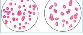
Anisochromia of red blood cells
Anisochromia of erythrocytes is a different degree of staining of erythrocyte cells.
The coloring of red blood cells depends on the concentration of hemoglobin in them, the shape of the cell and the presence of a basophilic substance.
Red blood cells, normally saturated with hemoglobin, in a blood smear have a uniform, medium-intensity pink color with a slight clearing in the center - normochromic red blood cells.
Hypochromic red blood cells
– red blood cells with a pale pink color and pronounced (to a greater or lesser extent) clearing in the center. Hypochromia is caused by low saturation of the erythrocyte with hemoglobin and is often combined with microcytosis.
Hypochromia is characteristic of iron deficiency anemia, and also occurs with lead intoxication, thalassemia and other hereditary anemias associated with impaired synthesis of the globin part of hemoglobin.
The analysis form notes not only the presence of hypochromia, but also its degree:
- hypochromia 1
– clearing in the center of the red blood cell is clearly marked and larger than normal, - hypochromia 2
– only the peripheral part of the erythrocyte is colored in the form of a ribbon, - hypochromia 3
– only the erythrocyte membrane is stained. The red blood cell has the shape of a ring (annulocyte).
Photos of hypochromic red blood cells
Hypochromic RBC
Hyperchromic red blood cells
– red blood cells with a more intense color than normal; their central lumen is reduced or absent. Hyperchromia is associated with an increase in the thickness of red blood cells and is often combined with macrocytosis. Megalocytes and microspherocytes are usually hyperchromic.
Different colors of individual red blood cells in a blood smear are called anisochromia.
Normally, in a smear of blood or bone marrow, you can find single red blood cells colored light purple or lilac.
These are polychromatophiles
– red blood cells with a basophilic substance (with special supravital staining these are
reticulocytes
). An increase in their number is called polychromasia or polychromatophilia.
Its presence must be recorded in the analysis form, and its degree is also indicated:
- polychromasia 1
– single polychromatophiles every 2 – 3 fields of vision; - polychromasia 2
– there are from 1 to 10 polychromatophiles in each field of view; - polychromasia 3
– more than 10 polychromatophiles in each field of view.
Polychromatophilia and reticulocytosis are usually detected in parallel and have the same clinical significance.
- L. V. Kozlovskaya, A. Yu. Nikolaev. A textbook on clinical laboratory research methods. Moscow, Medicine, 1985
- Guide to practical exercises in clinical laboratory diagnostics. Ed. prof. M. A. Bazarnova, prof. V. T. Morozova. Kyiv, “Vishcha school”, 1988
- Handbook of Clinical Laboratory Methods. Ed. E. A. Kost. Moscow “Medicine” 1975
Pathological forms of red blood cells are detected in the form of changes in the size, color, shape of red blood cells, as well as the appearance of inclusions in them.
Section: Hemocytology
Read
Morphologically identifiable cells of the erythrocyte lineage include erythroblast, pronormocyte, normoblasts (basophilic, polychromatophilic and oxyphilic), reticulocytes and erythrocytes.
Section: Hemocytology
Read
Jolly bodies (Howell-Jolly bodies) are small round violet-red inclusions 1 - 2 microns in size, found 1 (less often 2 - 3) in one erythrocyte. They represent the remainder of the nucleus after removal of its RES. They are detected with intense hemolysis and “overload” of the RES, after splenectomy, with megaloblastic anemia..
Section: Hemocytology
Read
Monoblast is the parent cell of the monocytic series. Size 12 – 20 microns. The nucleus is large, often round, delicately reticulate, light purple in color, containing 2–3 nucleoli. The cytoplasm of the monoblast is relatively small, without granularity, and colored in bluish tones.
Section: Hemocytology
Read
Poikilocytosis is a change in the shape of red blood cells. Normal red blood cells are round or slightly oval in shape. A change in the shape of red blood cells is called poikilocytosis. In a healthy person, a small proportion of red blood cells may have a shape that differs from the usual one. Poikilocytosis, in contrast to anisocytosis, is observed with severe anemia and is a more unfavorable sign.
Section: Hemocytology
Read
Source: https://www.clinlab.info/Hemocytology/Anisochromasia-of-erythrocytes-3
General definition
Anisochromia in a general blood test is a condition in which there is uneven coloration of red blood cells. This is due to the hemoglobin content in them. The more of it there is in red blood cells, the brighter their color. Those red blood cells that contain insufficient hemoglobin look paler. In a blood test, this indicator is determined as color.
The main function of red blood cells is to transport oxygen from the lungs to the body tissues. The more hemoglobin these blood cells contain, the faster the body is saturated with oxygen. But there must be moderation in everything. Therefore, experts have identified the optimal hemoglobin content in red blood cells, which allows for the most efficient functioning of the entire body. Deviation from normal values may indicate the presence of pathological processes.
Functions and structure of the erythrocyte
Human erythrocytes are biconcave elastic postcellular (nuclear-free) blood structures 7–10 µm wide, containing hemoglobin in their cytoplasm.
Due to biconcaveness, the working surface area of the erythrocyte increases, and its structure becomes more mobile and elastic, which allows it to better cope with its functions, penetrating into the smallest vessels and capillaries (up to 2-3 microns).
In men, the red blood cell cell is slightly smaller in volume than in women. Also, the size of these cells is affected by the saturation of the body with water - when dehydrated, they decrease, and the amount of hemoglobin in their composition decreases.
Photo:
Hemoglobin, as a pigment, gives the red blood cell body its red color. But the main function of this element is to help transport oxygen to tissues and organs.
Hemoglobin contains iron elements, which helps it bind and retain oxygen. Each hemoglobin molecule consists of 4 iron atoms and 103 atoms of S, N, O, H and C.
Red blood cells are saturated with oxygen in the lungs, after which they carry it throughout the body through the bloodstream.
READ Main causes of increased mean platelet volume
The more hemoglobin they contain, the more saturated the color of the body of red blood cells becomes and the better they can perform their transport function.
But everything is good in moderation. There are standards for the content of its elements in the blood, which ensure the most efficient functioning of the body and interaction between organ systems.
The amount of hemoglobin contained in a red blood cell is reflected by the so-called blood color index. Normally it is 1.
This indicator implies that a red blood cell should contain 33.34 pg of hemoglobin. It has been revealed that 1 red cell is filled with approximately 340 million hemoglobin molecules.
Red blood cells make up approximately 25% of the cells in the entire body. They are produced by the bone marrow of the skull, ribs and spine, after which the working cycle of red blood cells is 100 – 120 days.
After this period, they are utilized by macrophages. Men, women and children have different levels of red cells and hemoglobin in the blood.
Photo:
An adult man normally needs (4 - 5.1) x 1012/l red blood cells per liter of blood, while a woman needs a little less - (3.7 - 4.7) x 1012/l.
In children, these indicators change with age, and in adolescents they gradually approach adult values. The average levels of red bodies in the blood of children will be (3.8 - 4.9) x 1012 per liter.
At the same time, changes in hemoglobin content may not depend on the number of red blood cells (with iron deficiency, anemia or thalassemia).
READ Blood test standards for the detection of Helicobacter pylori
Norm
Normal hemoglobin levels in red blood cells depend on a person’s gender and age. Below is a table of normal values.
Anisochromia in a general blood test can manifest itself in the form of normochromia, hypochromia and hyperchromia. Let's consider these phenomena in more detail:
1. Normochromia is a normal state in which red blood cells are uniformly pink in color with a small spot of light color in the middle.
2. Hypochromia - a decrease in the level of hemoglobin in red blood cells. In this case, there is a disruption in the delivery of oxygen to the tissues, which leads to hypoxia of the organs. As a rule, anisochromia in a general blood test of this type indicates anemia. Currently, experts distinguish three degrees of hypochromia:
- The middle part of the blood cell is significantly lighter than normal.
- Only the periphery of the erythrocyte is colored red.
- The red blood cell remains light, only redness of the cell membrane is observed.
3. Hyperchromia. This type of blood anisochromia indicates oversaturation of red blood cells with hemoglobin. The blood cell has a bright red color without clearing in the middle. The red blood cell itself is increased in size. This condition is quite dangerous, as it can lead to the development of dangerous conditions due to the fact that cells become unable to perform their transport function.
Symptoms
Before anisochromia is detected in a general blood test, a person may notice symptoms that indicate the development of this pathological condition. These include:
- Fast fatiguability.
- Decreased concentration.
- Prostration.
- Mood swings.
- Dizziness.
- Rapid heartbeat for no apparent reason.
- Dyspnea.
- Headache.
- Noise in ears.
- Paleness of the skin.
- Sleep disorders.
- Increased skin sensitivity.
- Hair loss.
- Numbness of the limbs.
- Loss of smell and taste.
If the above symptoms appear, it is recommended to consult a doctor as soon as possible and undergo the necessary tests.
Anisochromia in children
Diagnosing anisochromia in a general blood test in children in most cases indicates the development of anemia. This is a fairly common pathology in childhood, which occurs due to intensive growth of the body against the background of an underdeveloped hematopoietic system. This can also be contributed to by poor nutrition and various pathological processes occurring in the body.
Manifestations of pathology include pale skin, some developmental delay, lethargy, apathy, the appearance of cracks in the corners of the lips, prolonged and frequent colds.
Parents should monitor their children more closely and consult a doctor at the first suspicious signs.
Diagnostics
Anisochromia is detected using a general blood test, which pays special attention to the level of red blood cells and hemoglobin. To identify the cause of deviations in indicators, the following laboratory and instrumental examinations may be prescribed:
- Analysis of urine.
- Fecal occult blood test.
- Ultrasound examination of the kidneys.
- Fluorography.
- Blood serum examination for iron content.
- Gynecological examination.
- Bone marrow samples.
Possible treatment
If anisochromia is detected in a blood test, treatment is aimed at eliminating the root cause and relieving unpleasant symptoms. In most cases, complex therapy is prescribed, which includes medication, adherence to a certain diet and the use of traditional medicine. It is worth noting that if anisochromia is detected in a general blood test, therapy is prescribed only by the attending physician. Self-medication can lead to aggravation of the situation and provoke the development of more serious diseases. Let's look at the most common treatment methods.
In most cases, the following is prescribed:
- Iron preparations (for example, Ferrum-Lek, Hemofer, Ferrofolgama and others). When a drug is prescribed in the form of a dropper or injection, treatment is carried out in a hospital setting.
- Vitamin B12. As a rule, this drug is prescribed by injection (for example, Cyanocobalamin).
- Folic acid preparations.
There are combination preparations that include both vitamin B12 and folic acid. For example, "Maltofer".
In most cases, the above drugs are prescribed in the form of capsules or tablets. In case of complicated anemia, a decision is made to undergo treatment in a hospital setting.
When blood anichochromia is detected, adherence to a special diet is of great importance for normalizing the condition. Let's look at the general rules:
- Animal protein must be present in your daily diet.
- It is necessary to limit your fat intake.
- The diet should contain the required amount of foods rich in B vitamins.
- It is recommended to use fish, meat and mushroom broths.
The following products are allowed:
- Eggs.
- Cottage cheese.
- Liver (every other day or every day in small quantities).
- Brewer's yeast.
- Mushrooms.
- Red meat.
- Fish.
- Beet.
- Apples.
- Pomegranate juice (can be mixed with beet juice or diluted a little with water).
- Legumes.
- Rose hip.
- Currant.
- Pumpkin.
The following products are not recommended:
- Tea.
- Some greenery.
- Fatty foods.
- Dairy products.
- Coffee.
- Oatmeal or millet porridge.
- Alcohol.
Folk remedies are used as an additional treatment to the main therapy or as a preventive measure. The following recipes have worked well:
- Brew 10 grams of nettle leaves in a glass of boiling water. Let it brew, then apply 1 tablespoon three times a day.
- Mix dried fruits with honey and take 1 tablespoon 3 times a day.
Why is hyperchromic anemia dangerous and how to treat it?
This group of anemias, such as hyperchromic ones, is evidence of a violation of hematopoietic processes. The main causes of such conditions are a lack of vitamins B12 and B9. Such hypovitaminosis is extremely dangerous, especially for children and pregnant women. In this category of patients, in the absence of proper control of the condition, the risk of death increases.
Complications
If treatment for anisochromia is not started in a timely manner or is completely absent, more serious pathological conditions may develop. These include:
- Decreased immunity.
- Enlarged liver.
- Decreased quality of life due to unpleasant manifestations of the pathological condition.
- Growth retardation in children.
- Mental and mental retardation in a child.
- Chronic anemia.
Possible consequences and complications
If left untreated, hyperchromic anemia in children can lead to delayed mental and physical development. A decrease in immunity will cause frequent, long-term colds with a considerable number of complications.
Hyperchromia is no less dangerous for pregnant women and the fetus. Lack of proper treatment can lead to spontaneous abortion, early birth, fetal development abnormalities, and death of mother and child due to bleeding.
Also, complications of hyperchromic anemia may include cardiomyopathies, severe cardiac arrhythmias, and heart failure.
What is hypochromia in a blood test, diagnosis and treatment
Hypochromia in a general blood test indicates problems with hemoglobin.
This pathology has other names - hypochromia of erythrocytes or hypochromic anemia. All these names refer to several types of anemia, each of which requires special diagnosis and treatment. Let us remember that anemia in any form, especially with prolonged exposure to the body, leads to oxygen starvation of cells, which cannot but affect their condition. This is especially true for brain cells. In advanced cases, it is possible to develop a stroke with a transition to the “vegetable” state followed by the terminal stage. Most often, this scenario concerns the female body.
What is hypochromia
This condition is called so because it is only due to the normal content of hemoglobin, and therefore iron, that healthy red blood cells have a red color. And with a syndrome such as hypochromia of erythrocytes, their color is lost.
In addition to the fact that hypochromic anemia is characterized by a deficiency of hemoglobin in red blood cells, the following symptoms are also inherent:
- Change in the shape of red blood cells.
- Loss of color in red blood cells - the red blood cell becomes two-colored, with a dark red outer ring and a discolored middle.
- The acquisition of an abnormal shape by red blood cells.
When examining a general blood test for the level of hypochromia, it is always necessary to pay attention to the color indicator, which directly characterizes the level of hemoglobin in red blood cells. These two indicators should always be tested together for an accurate diagnosis. An important diagnostic sign is a decrease in color index below 0.8.
Causes
A common symptom for all hypochromic anemias is a decrease in the level of hemoglobin in red blood cells.
The leading causes of this pathology are considered to be:
- Low iron levels in the body.
- Chronic lead intoxication.
- Vitamin B6 deficiency.
- Inflammatory processes in the body (more typical for chronic diseases, for example, hepatitis, intestinal lesions).
- Heredity.
- Menses.
- Extensive blood loss after trauma and surgery.
- Chronic internal bleeding.
- Unbalanced diet, low in protein.
- Pregnancy.
- Blood diseases.
- Helminthic infestations.
- Autoimmune diseases.
- Long-term donation.
There is such a thing as “pseudo-blood loss”. This phenomenon occurs with cystic transformation of the ovaries and benign neoplasms in the uterus. In this case, the resulting cavities are filled with blood, which stagnates. In it, hemoglobin breaks down into various compounds and disappears over time.
Why does it occur
Hypochromic anemia can occur for many reasons:
- Loss of blood volume, leading to bleeding, during long and heavy menstruation, during surgical interventions as a result of injury.
- With poor nutrition, this is due to a deficiency of vitamin and mineral complexes in the body, often observed in vegetarians and those on a strict diet.
- Internal bleeding of a hidden nature, often occurring or constant, the patient simply may not see this situation, the cause may be inflamed gums, hemorrhoids and other diseases of the intestines, gastrointestinal tract, and female organs of the reproductive system.
- Chronic infectious diseases, such as pulmonary tuberculosis, various types of hepatitis, in this case, iron elements are poorly distributed and enter organs and tissues.
- When the body is intoxicated with substances of chemical origin.
- The risk of anemia is associated with pregnancy, since during this time the female body needs several times more iron.
- Helminthic infestations.
- Autoimmune lesions of the body.
Hypochromia of erythrocytes in children often occurs in premature infants, due to a conflict of the Rh factor, intrauterine transmission of infections to the mother, unbalanced nutrition during gestation, or birth injury. During adolescence, most adolescents are susceptible to anemia; this is due to hormonal changes and an increased period of growth.
Important information: What are red blood cells in the blood (what are red blood cells responsible for)
Classification of hypochromia (hypochromic anemia)
- Iron deficiency is also known as microcytic. Is the most common. Typical for children and young women. Occurs during pregnancy, breastfeeding, bleeding, poor absorption and lack of iron, prolonged diarrhea, high need for iron, limited nutrition (absence or low content of meat products).
- Iron-saturated – sideroachrestic (sideroblastic). In this condition, there is no lack of iron in the body. The condition develops because iron is not recycled. Such changes occur when there is a large deposition of iron due to the massive death of red blood cells. The most common causes of this process are chemical intoxications and long-term use of certain groups of medications.
- Iron distribution - in this situation, there is insufficient supply of iron in the blood. The reason for this is its retention by cells. Factors leading to this pathology are tuberculosis, infectious diseases of various etiologies and other inflammatory reactions
- Mixed - includes any combinations. A feature of this condition is the difficulty in diagnosis, since when one cause is identified, treatment is often immediately prescribed, which, unfortunately, turns out to be not completely effective, since the therapy affects only one link in the development of the disease. Most often, this condition is observed with a lack of cyanocobalamin (vitamin B12) and iron.
Causes of the condition
Anisochromia can normally be present in an absolutely healthy person, but the percentage of unevenly colored cells is so small that they are practically not detected by a blood test.
The causes of anisochromia in a general blood test will be discussed below.
Hypochromia
The reasons are the following:
- Anemia. This is the main reason for the development of this condition. They can be of several types: iron deficiency, iron-saturated (in the body the concentration of iron is within normal limits, but under the influence of certain factors it is poorly absorbed by cells) and iron redistribution (develops when red blood cells are destroyed under the influence of various pathological processes).
- Bleeding.
- Pregnancy and adolescence.
- Diseases of the gastrointestinal tract.
- Chronic diseases (for example, bronchitis or heart disease).
- Chronic purulent inflammatory processes occurring in the body.
- Poor nutrition with lack of protein.
- Taking certain medications.
- Poisoning.
Hyperchromia
The reasons for detecting anisochromia in a general blood test, which is defined as hyperchromic, are the following conditions:
- Deficiency of vitamin B12 and folic acid.
- Intestinal diseases.
- Congenital diseases.
- Hereditary factor.
- Malignant tumors of the stomach or lungs.
- Hepatitis.
- Presence of worms.
- Blood diseases.
- Pathological conditions of the kidneys.
- Consequences of radiation or chemotherapy used in the treatment of leukemia.
- Disturbances in the normal functioning of the bone marrow.
The phenomenon of anisochromia in a general blood test
The phenomenon of anisochromia in a general blood test can be found in both adults and children.
At its core, the very concept of anisochromy implies heterogeneity of color.
In blood tests, this term refers to uneven, insufficiently pigmented or, conversely, too brightly colored red blood cells.
The reasons for this phenomenon are associated with changes in the saturation of the cell body with hemoglobin, which can be caused by pathological processes or be a normal response of the body to the environment.
Varieties
The condition of hypochromia is divided into several different types of anemia that can be experienced by patients.
Iron-deficiency anemia
Anemia with iron deficiency is the most common type of all; its causes are bleeding, low intake of iron elements (especially insufficient consumption of meat products) with difficulty absorbing this element. Often observed during pregnancy and breastfeeding, young women are also susceptible.
Sideroblastic anemia
This species is characterized by a normal level of iron in the blood, but its absorption into the blood is impaired, and this reduces the number of hemoglobin cells included in the general blood test. Elderly people suffering from alcohol addiction, poisoning with substances of chemical origin, or long-term drug therapy are prone to this type.
Important information: What does low platelet count mean in an adult?
Ferro redistribution anemia
With Ferro redistribution anemia, an increase in the number of iron-containing elements is observed, with the death of the red blood cells themselves. Occurs against the background of tuberculosis of the lungs, diseases with a purulent course.
Mixed anemia develops with a lack of B vitamins 12 and iron compounds; its signs are:
- increased fatigue;
- decrease in the body's immune forces;
- frequent swelling in the upper extremities.