Why are blood tests needed during a heart attack?
During an attack, a number of characteristic changes occur in the composition of the blood fluid - certain proteins are thrown into it, CPK enzymes are increased, etc. Depending on the stage of development of the heart attack, blood tests may show:
- During the acute period, the amount of myoglobin (a type of protein) increases by almost 20%. Based on the degree of increase in protein, the exact time of onset of the attack can be determined. Neutrophilic leukocytosis is also detected in the blood, erythrocyte sedimentation accelerates, and the leukocyte formula shifts to the left. At the end of this stage, phagocytosis begins to be activated, as a result of which the number of eosinophils, troponin I and T increases. Total creatine kinase is noted.
- In the subacute period, the number of CPK and myoglobin decreases, but the level of troponin remains the same.
- In the post-infarction state, a minimal number of cells are found that function.
Determination of the level of cardiac-specific troponins T and I
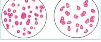
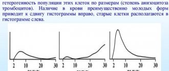
The contractile apparatus of myocytes includes a troponin complex formed by three proteins: troponin T (TrT), which forms a bond with tropomyosin, troponin I, which inhibits ATP activity, and troponin C, which has a significant affinity for Ca2+. The latter is also found in skeletal muscles. The content of TpT in cardiomyocytes is approximately two times higher than the level of troponin I. In the blood of healthy people, even after excessive physical activity, the level of troponin T ns exceeds 0.2 - 0.5 ng/ml, so its increase above this limit indicates damage to the heart muscle . The specificity of this marker is 90–100%, which allows it to be considered the “gold” standard for diagnosing AMI. The development of a heart attack is accompanied by extensive destruction of cardiomyocytes and a significant release of TpT into the blood, the level of which increases 20-40 times. An increase in TrT is detected within 3-4 hours after the onset of a painful attack. The maximum increase in TrT levels is determined on days 3-4, remains high for a week, and then gradually decreases, remaining elevated until days 10-18. Determination of troponin I can be used to diagnose myocardial infarction in patients with concomitant skeletal muscle damage. It has been shown that acute and chronic injuries to skeletal muscles, excessive physical activity, surgical operations, excluding cardiac surgery, and muscle injuries do not cause an increase in troponin 1 levels.
General blood test indicators
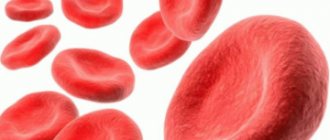
This study makes it possible to recreate an accurate picture of the patient's condition. With myocardial infarction, anemia most often occurs, so an important point is to establish the level of hemoglobin and red blood cells. Leukocyte counts are also important.
The following indicators indicate myocardial infarction:
- White blood cells - leukocytes - increase. In an adult healthy person, the indicators correspond to the figures 4-9/109 (for 1 liter of blood). During a heart attack, the first indicators change, they increase to 10-15.
- The leukocyte formula shifts to the left.
- Eosinophils, that is, phagocytic cells, increase. A heart attack is diagnosed if there are more than 5% of eosinophils in the blood, more than 12% of neutrophils (band form), and more than 80% of neutrophils.
- The erythrocyte sedimentation rate changes, but on the second day after the attack. Normal ESR values for women: from 3 to 15 mm/h; for men: from 2 to 10.
- The patient does not have a heart attack if the red blood cells correspond to the following norm: in women – 3.7-4.7/1012; in men – 4.5-4.7/1012.
- Hemoglobin should normally have the following indicators: female gender - from 120 to 140 g/l; male gender - from 129 to 160.
- If the cause of myocardial infarction is thrombosis, then the platelet level will be increased, starting at 320/109.
Pre-infarction period
The concept of “pre-infarction state” includes a number of different pathological conditions in which the risk of developing myocardial infarction is significantly increased.
Clinical forms of this period: 1) first-time angina; 2) progressive exertional angina; 3) variant angina.
Differential diagnosis with myocardial infarction in the pre-infarction period should consist of dynamic monitoring of the electrocardiogram, blood enzymes (CPK, troponin, LDH, AST, ALT, etc., temperature changes, blood leukocytes, ESR).
Blood chemistry
This study reveals changes in blood plasma proteins. Proteins are released rapidly, but this speed largely depends on the location of the substances in the cells. Molecular weight, local blood supply and lymph outflow also play a significant role in this. It is extremely important to carry out biochemical analysis in the early stages of the examination.
During myocardial infarction, there is an increase in the following substances:
- fibrinogen;
- haptoglobin;
- seromucoid;
- sialic acid;
- gamma globulin;
- C-reactive protein.
Ultrasound of the heart
Ultrasound examination of the heart or echocardiogram (ECHOCG) is a mandatory diagnostic method for myocardial infarction and any other heart diseases. Its importance is explained by the high accuracy of the research carried out.
Using echocardiography you can establish:
Modern ultrasound machines allow not only to see the current state of the heart, but also to assess the likelihood of complications and make a forecast of the patient’s quality of life in the future.
Methods for differential diagnosis of acute myocardial infarction
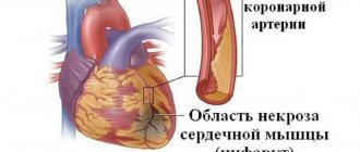
This disease is characterized by impaired patency of the coronary vessels, resulting in necrosis of the heart muscle. The larger the area of myocardial damage, the more severe the pathological process, and the less chance the patient has for recovery.
Myocardial infarction is diagnosed based on three main types of diagnostics. First of all, this is the assessment and correct interpretation of the patient’s clinical condition, symptoms of the disease and main complaints. Instrumental examination methods are no less important. ECG, ultrasound of the heart, coronary angiography and other techniques allow you to quickly and with a high degree of probability establish the correct diagnosis.
Laboratory methods for studying myocardial infarction also remain effective. In addition to the clinical blood and urine tests that are familiar to most patients, special markers of damage to heart muscle tissue are widely used. This includes determining the level of enzymes or troponins CPK, AST, LDH and others.
Symptoms of myocardial infarction
Description
Infarction of the thick muscle layer of the heart (myocardium) occurs due to blockage of the coronary artery
Myocardial infarction occurs due to a blockage of the coronary artery, the blood vessel that supplies the heart muscle. The main provoking factor in the development of an acute crisis is atherosclerosis - deposits on the walls of blood vessels in the form of cholesterol plaques.
How clinical blood and urine tests react to the development of myocardial infarction
Considering that the main pathomorphological manifestation of this disease is acute necrosis of areas of the heart muscle, a general blood test during myocardial infarction will show a picture of the presence of an inflammatory process in the body. Already 4 - 6 hours after the onset of the acute phase of heart disease, the number of leukocytes in this study will increase by 2 - 3 times. The growth of white blood cells occurs mainly due to young forms of neutrophils. A similar symptom in clinical laboratory diagnostics is called a neutrophilic shift of the leukocyte formula to the left.
Read also: Acute inferior myocardial infarction
The following picture is observed with the percentage of eosinophils in the blood: 24 hours after the onset of the disease, their number drops sharply and is practically undetectable in a blood test. As soon as the patient’s body begins to receive appropriate treatment, and the regeneration processes in the myocardium intensify, these blood elements are restored to normal values. The process may take up to 2 - 4 weeks.
As soon as the inflammatory process in the heart muscle begins to subside under the influence of specific treatment, the ESR returns to its normal numbers.
A general urine test does not carry any special meaning for this disease. Specific changes in this study may occur if myocardial infarction is accompanied by the development of acute renal failure.
In this rather rare case, the general analysis of urine may contain a large number of white blood cells, mucus, and a rapid increase in specific gravity will also be characteristic.
Electrocardiography
Electrocardiography may reveal:
- arrhythmias;
- extrasystoles;
- atrial fibrillation.
ECG is the most informative method, which should be done in the first 10 minutes from the moment the patient enters the clinic.
ECG signs indicating the development of an ischemic process in the heart muscle:
- elevation (rise from the isoline) of the ST segment by more than 1 mm in two or more leads;
- the presence of pathological Q waves (not a mandatory criterion - in the clinic, myocardial infarction without a pathological Q wave is distinguished).
Electrocardiography should be performed on the patient over time: every 8 hours on the first day of the disease (in 15 leads), and then daily. This allows us to further confirm the patient’s diagnosis.
Biochemical blood test for myocardial infarction
Most of the indicators of this analysis do not have specific values; experts estimate the level of their fluctuations between the maximum and minimum figures.
Acute myocardial infarction is characterized by a violation of the following parameters:
- Total blood protein, namely albumin and globulin, is characterized by an increase during the active phase of myocardial ischemia. This is due to metabolic disorders in the patient’s body.
- An increase in indicators such as urea and creatinine is possible. If in the normal state these substances characterize the functioning of the kidneys and urinary system, then during myocardial infarction an increase in the levels will indicate the development of heart failure.
- A fairly important symptom in acute pathology of the heart muscle will be an excess of cholesterol concentration in the blood. Normally, its quantitative indicator is 3.5 - 6.5 mmol, and with the development of coronary artery disease and atherosclerosis, the numbers increase by 2 times.
- Absolutely necessary for the timely diagnosis of this acute process are enzyme data for ischemia of the heart wall. A sharp increase in ALT and AST always accompanies necrosis of the heart muscle, since the concentration of these enzymes in the myocardium exceeds their content in the blood plasma by 2,000 - 3,000 times.
- Another enzymes during myocardial infarction, amylase and phosphatase, can increase only with a pronounced necrotic process in the heart and indicate the inadequacy of the therapy.
- Just a few years ago, it was believed that if myoglobin was detected in the patient’s blood during myocardial infarction, this indicated the pronounced severity of the pathological process. However, recent studies reject this claim and do not recommend that clinicians respond particularly to this indicator. Myoglobin is excreted from the body in the urine within 1 to 2 hours and cannot be a 100% criterion for the presence of myocardial ischemia in a patient.
A biochemical blood test for myocardial infarction is an important component in the diagnosis of this terrible disease.
Specific laboratory tests for suspected myocardial infarction
Analyzes for myocardial infarction can be divided into general and specialized studies. The latter allow doctors with a high degree of probability to establish a diagnosis and prescribe timely treatment. These include:
- Analysis for CRP or C-reactive protein. The level of this substance in the human body usually increases sharply during various inflammations. Myocardial infarction is no exception in this case. The norm of SRP is 2.5 - 5 mg/l.
- The enzyme creatine phosphokinase or CPK is a specific test for cardiac muscle necrosis; it can be an indicator not only of acute myocardial infarction, but also of major trauma. For a clearer diagnosis, the CPK-MB fraction is determined; this is a direct cardiac market for myocardial infarction.
- The most common test for diagnosing acute ischemia of the heart muscle is the determination of the tissue enzyme “Tropanin”. The level of this substance in the patient’s blood increases within a few hours after the development of myocardial necrosis. Normally, it reaches 0.4 μg/l; any excess of this indicator indicates the development of myocardial infarction.
However, good old blood and urine tests cannot be completely ignored. Thanks to the introduction of new methods of laboratory examination, they remain in demand in the differential diagnosis of many diseases of the human body.
Troponin test for myocardial infarction: indications for use. Which test will be more reliable and how it is carried out. Norm and deviations of indicators.
Not 100%, but enzymes will be quite effective in case of a heart attack. Cardiac-specific enzymes help determine the extent of myocardial necrosis and distinguish it from angina pectoris and other problems.
Read also: Myocardial infarction vitamins
Under the influence of external factors, a pre-infarction state may occur. The signs are similar in women and men; recognizing them can be difficult due to the location of the pain. How to relieve an attack, how long does it last? At the appointment, the doctor will examine the ECG readings, prescribe treatment, and also tell you about the consequences.
If coronary angiography of the heart vessels is performed, the study will show structural features for further treatment. How is it made? How long does it last, likely consequences? What preparation is needed?
The causes of small-focal myocardial infarction are similar to all other types. It is quite difficult to diagnose; acute on ECG has an atypical picture. The consequences of timely treatment and rehabilitation are much easier than with a regular heart attack.
Post-infarction cardiosclerosis occurs quite often. He may have an aneurysm or ischemic heart disease. Recognizing symptoms and timely diagnosis will help save lives, and ECG signs will help establish the correct diagnosis. Treatment is lengthy, rehabilitation is required, and there may be complications, including disability.
It is quite difficult to diagnose, since subendocardial myocardial infarction quite often has an abnormal course. It is usually detected using ECG and laboratory examination methods. An acute heart attack threatens the patient's death.
If there is a suspicion that a patient has heart failure, diagnostics will help confirm the suspicion. It is prescribed for acute and chronic forms. Laboratory differential diagnosis includes urine analysis, bnp, and additional cardiac examination methods are used.
Normally, coagulogram indicators show the characteristics of the blood, which allows timely treatment of many dangerous diseases. Their interpretation is different for children and adults, as well as pregnant women. What will the extended coagulogram tell you about, mno, achtv, d dimer, fibrinogen?
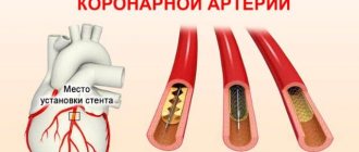
Coronary angiography
Coronary angiography is usually performed in conjunction with percutaneous coronary intervention (PCI). It is desirable that these studies be carried out as soon as possible from the onset of acute myocardial infarction. They allow the doctor not only to verify the diagnosis, but are also used as the most effective method of treatment. Coronary angiography has low morbidity and mortality rates. Differs in long-term results.
Percutaneous coronary intervention is a method that involves angioplasty with the installation of stents.
Indications for coronary angiography (after drug treatment has been started):
- persistence of myocardial ischemia, which is confirmed by electrocardiography data and the clinical picture;
- unstable hemodynamics;
- ventricular rhythm disturbances.
Are blood tests important in diagnosing a heart attack?
If a blood clot enters the coronary artery, blood flow to the heart muscle is hampered or completely disrupted. This condition is called a heart attack. This is the name for disturbances in blood flow to the heart muscle. Lack of oxygen and nutrients lead to tissue death. Depending on the degree of hemodynamic disturbances, location and size of the lesion, the pathology has different consequences.
The timeliness of the diagnosis plays an important role, since the risk of complications and death increases with every minute. To confirm the problem, laboratory and instrumental studies are prescribed.
If an attack is suspected, then first of all there is a need for a general and biochemical blood test.
In a general analysis, an increase in the content of white blood cells is observed during a heart attack. A biochemical study shows that cytolysis enzymes have entered the general bloodstream, and their number is increasing. Usually they do not extend beyond the lung, cardiac tissue and tissues of other organs. But if muscle damage is observed, it enters the blood serum.
Cytolysis enzymes contained in the myocardium are called:
- lactate dehydrogenase;
- aspartate aminotransferase;
- creatine phosphokinase.
The degree of increase in indicators can be different, and the standards also differ depending on the equipment and reagents used in laboratories.
Blood test indicators during myocardial infarction make it possible to detect the pathological process in the first hours of its development. Although it is usually accompanied by bright manifestations, sometimes they are absent.
If we take into account the fact that the disease manifests itself as acute necrosis of areas of the myocardium, using a general blood test it is possible to determine the development of the inflammatory process.
After 4 hours from the onset of development of necrotic disorders, the content of leukocytes in the blood increases. Their level increases several times.
Young neutrophils act in such a way that the number of white blood cells steadily increases.
The concentration of eosinophils in the blood also changes:
- During the first day, their level decreases sharply, and it is almost impossible to determine them in a blood test.
- With the start of treatment, the processes of regeneration of heart muscle tissue intensify, and indicators are restored to normal values. The process continues for a month.
As a result of a heart attack, an inflammatory process begins in the body. This is accompanied by a violation of the erythrocyte sedimentation rate. With the onset of an acute attack of ischemia, a sharp increase in ESR occurs. The indicator remains at a level 2-3 times higher than normal for a month. Thanks to proper treatment, the course of the inflammatory process subsides, which helps to reduce the erythrocyte sedimentation rate. Therefore, the patient will undergo blood tests several times during the treatment process.
Read also: Rehabilitation of patients who have suffered myocardial infarction
A general blood test for myocardial infarction is mandatory, as for any other disease. This study is included in the list of those prescribed during the examination of patients.
Conducting research
A blood test for CPK is carried out in the morning before the first meal. Biological material is taken from the vein of the elbow. In the laboratory where the blood is sent, decoding is carried out. To do this, the liquid is divided into two parts: plasma and a fraction containing cells. Enzyme CPK indicators are recorded in units of enzyme activity (abbreviated as Units) per 1 liter of serum.
In some cases, it is indicated to repeat the analysis to confirm the diagnosis and monitor treatment. Additional analysis for CPK is usually carried out after two days.
Biochemical
A biochemical blood test for myocardial infarction does not allow one to determine specific indicator values. During the study, the maximum and minimum values are determined and the difference between them is determined. If an acute attack of ischemia occurs, then the following is determined:
- The level of total protein albumin and globulin, the concentration of which increases during the active development of ischemic disorders. This occurs due to a violation of metabolic processes in the body.
- Urea and creatinine. These indicators increase during an attack, which indicates serious disruptions in the functioning of the heart.
- Cholesterol. An increase in cholesterol concentration also indicates acute pathology. Normally it should be from 3.5 to 6.5 mmol/l. If coronary heart disease and atherosclerosis develop, the values increase several times.
- Enzymes aspartate aminotransferase and alanine aminotransferase. Their sharp increase always indicates a necrotic process in the heart muscle.
- Amylase and phosphatase enzymes. They increase if tissue dies in the heart muscle. Deviation from the norm may also indicate improper treatment.
- Myoglobin. Previously, it was believed that its presence in the blood indicates a complicated course of a heart attack. But this theory was refuted through numerous tests. Doctors have found that the period of myoglobin elimination is several hours. Therefore, it cannot provide accurate information about the development of the pathological process.
It is impossible to diagnose acute ischemic disorders without a biochemical blood test. Using these indicators, you can confirm the presence of a problem and choose the appropriate treatment.
With cardiovascular diseases, the level of heavy metals in the blood often increases. If there is more cadmium or aluminum in the body, then these substances have a toxic effect. At the same time, the amount of microelements important for health decreases. This is noticeable by a drop in the level of chromium, copper, and manganese.
High content has a negative effect on the body:
- Lead. This substance causes serious disruption to the endocrine system. In addition to damage to the heart and blood vessels, there is a deterioration in the condition of the liver. Large amounts of lead cause arterial hypertension and atherosclerotic changes in blood vessels.
- Cadmium. Excessive content of the element is accompanied by liver poisoning and the development of cardiomyopathy.
- Arsenic. Because of it, there is a decrease in vascular permeability. If their walls thicken, the likelihood of cardiogenic shock increases.
Changes in the concentration of heavy metals occur in connection with an attack of myocardial infarction. This significantly increases the risk of complications.