967
0
Leukemia or leukemia, which is also popularly called blood cancer, refers to malignant diseases of the human hematopoietic system. During its development, the blood and bone marrow cells are the first to be affected.
According to statistics, the disease affects every 25 adults per 100 thousand people. Moreover, men are susceptible to this malignant pathology one and a half times more often than women. And among children, leukemia is considered the most common type of oncology.
The success of treatment and further prognosis directly depend on how early the disease was detected. Therefore, it is important to pay attention to the first symptoms of leukemia in adults, not to delay a visit to an oncologist, and to receive adequate therapeutic prescriptions as soon as possible.
What is leukemia?
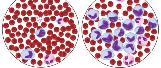
This is a cancer of the blood and bone marrow, a malignant disease that causes the abnormal growth of certain blood cells. As unhealthy cells crowd out healthy ones, blood functions begin to fail and you may notice physical symptoms. The disease can progress quickly if you have a type such as acute leukemia. Or it will develop slowly and get worse over time if you have chronic leukemia. But no matter what type is present, if you notice some of these subtle signs of leukemia, see your doctor for an evaluation.
Symptoms of blood cancer
Hyperleukocytosis, known as leukemia, affects 9,200 people a year. It is a disease of the lymphatic and hematopoietic system with type-dependent but often identical symptoms to leukemia.
Symptoms of leukemia are often nonspecific
The first signs and symptoms of hyperleukocytosis (leukemia) are, for example, anemia or an increased number of infections. Firstly, there are no signs of chronic leukemia. This is why doctors usually discover them later.
"White blood" due to hematopoietic disorders
Many symptoms of leukemia are caused by the patient having too few healthy blood cells or too many diseased leukemia cells. Specific symptoms depend on whether it is acute or chronic leukemia. Acute symptoms of leukemia do not appear gradually, but occur suddenly. The patient's condition may worsen in just a few days.
Fatigue and weakness
These are the most common symptoms for all types of leukemia, and are often caused by anemia (a deficiency of red blood cells), which only worsens physical exhaustion. In both the chronic and acute stages of the disease, symptoms may range from mild to extreme fatigue and physical weakness, but in all cases these symptoms worsen over time. The problem is that leukemia creeps up unnoticed. But fatigue and weakness are often ignored, since this is a common sign of many diseases, and can also appear due to lack of normal rest.
A rare 1932 Ford is being sold in Illinois for $4,300 in disassembled condition.
Brakes upgraded: new tuning package introduced for NC Mazda Miata
Baking will be fluffy if you extinguish the soda correctly: life hack from grandma
Acute leukemia and its symptoms
The accompanying symptoms of acute leukemia do not appear; they arise suddenly. The patient's condition may worsen in just a few days or weeks and result in death without treatment.
Typical but nonspecific symptoms of acute hyperleukocytosis include:
- Fatigue,
- Decrease in efficiency,
- Pallor,
- Increased susceptibility to infection,
- Constant fever
- Frequent bleeding of the nose and gums,
- Petechiae (point hemorrhages of the skin),
- Gingivitis,
- Hematomas (bruises),
- Delayed hemostasis (hemostasis),
- Not self-directed weight loss,
- Anorexia (loss of appetite),
- Bone pain (Pain caused by leukemia occurs in the bone tissue, and the patient perceives deep somatic pain as dull).
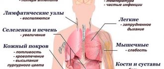
- Swollen lymph nodes in the neck, armpits and groin,
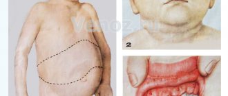
- Pain or pressure in the upper abdomen due to an enlarged spleen (splenomegaly),
- Enlargement of the liver (hepatomegaly)
- The closures of small blood vessels, rarely itchy, brownish-red,
- purple spots, blisters or nodules in the skin due to leukemia cells,
- Rarely, severe headache due to involvement of the meninges (leukemic meningitis),
- Photosensitivity, facial paralysis, blurred vision, nausea and vomiting,
- Dizziness or emotional disturbances due to involvement of the central nervous system,
- Dyspnea.
Unusual bleeding
Grandparents built a “park” for their granddaughter on an old playground
Leps has a new pet - puppy Joni (cute photo of a new family member)
Survived captivity and became a hero: the son prepared a surprise for his father for his 100th anniversary
Like bruising, bleeding from the nose, gums, intestines, lungs or head can be signs of platelet deficiency and blood clotting problems, which may indicate acute forms of leukemia.
Disease syndromes
Syndromes of a pronounced nature in leukemia can occur in a very serious form. The most common symptoms of blood cancer are:
- Hemorrhagic syndrome as a sign of blood cancer - exhibits severe symptoms in leukemia that affects the blood. This is a tendency to such a phenomenon as skin hemorrhage. It manifests itself in leukemia in adults in the form of bleeding of the mucous membranes, bruising appears too much, it can rash, etc.
- Anemic syndrome - this occurrence of a symptom of leukemia affects the body in the form of pallor of the skin and mucous membranes, loss of strength, fatigue, shortness of breath, irritability, increased heart rate even under conditions of minor exertion, a burning sensation on the tongue, loss of sensitivity, and taste disorders are possible.
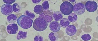
The leading role in the primary diagnosis of the pediatrician. Subsequently, the examination will be undertaken by a pediatric oncohematologist. Diagnostic methods are based on examination of bone marrow and peripheral blood. In acute leukemia, changes in the general blood test will be characteristic, such as anemia, increased ESR, reticulocytopenia; a typical indicator can be when the so-called “leukemic failure” phenomenon appears.
“Leukemic failure” represents a condition in which the analysis reveals mature cells in small numbers, and the youngest ones are in advantage, while transitional forms of cells are absent. The decisive argument that allows us to make a decision that a child is developing leukemia is blast cells in a content of 30% or more.
However, it is important to remember that this type of disease can be cured and not show up in a blood test in about 10% of cases. Therefore, if there are any other clinical manifestations that indicate a problem, bone marrow puncture should be done, as well as cytochemical tests.
Petechiae
These are small red spots under the skin caused by bruising. You may not even notice these spots due to their size, painlessness, and location on the lower extremities, but they also indicate a low platelet count. Petechiae are commonly found around the ankles because gravity causes fluid to accumulate in the lower legs throughout the day.
Signs of leukemia in women
The general condition of the woman becomes very apathetic and lethargic. The patient feels overwhelmed and emotionally depressed. It becomes difficult for a woman to concentrate. All this is explained by a lack of oxygen in the body. Due to disruption of the hematopoietic system, women experience problems with menstruation. Menstruation becomes more abundant and longer. Spontaneous uterine bleeding is also possible. They are typical for older women. Younger girls suffer from heavy menstruation. The main symptoms of the disease in women include:
- sudden causeless loss of body weight;
- lack of appetite;
- fever for no obvious reason;
- shortness of breath without exercise;
- enlarged lymph nodes;
- swelling of the groin area;
- decreased visual acuity;
- joint pain;
- seizures;
- blurred vision;
- causeless appearance of bruises;
- violation of taste reflexes, aversion to food, gag reflex.
Swelling and enlargement of gums
Although increased gum size, also known as hyperplasia, typically appears in only a small number of patients with acute leukemia, it is one of the most obvious signs of the disease. If a patient has leukemia, doctors always check their mouth to see if their gums have increased in size. They may appear swollen, and patients almost always feel a strange heaviness in the mouth.
Listen to yourself and play sports: how to clear your head of many thoughts
I apply a beautiful gold foil print to clothes myself: step-by-step instructions
The first thing you see in the picture will show your secret fear in love
Diagnostics
Bruises due to leukemia are often only harbingers of serious health problems. This disease is characterized, for example, by liver enlargement, hemorrhages in the abdominal cavity and many other complications. This is why it is especially important to start treatment as early as possible. If a rash is detected, you must visit a doctor and get diagnosed.
- External examination, including various organs. For example, an enlarged spleen sometimes causes pallor, nausea, and other symptoms. It often results from leukemia. It is detected by palpation and other studies.
- A general blood test is required. Allows you to get a first idea of the composition of blood cells, for example, there are indicators of the number of red blood cells.
- Biochemical blood test - reveals damage to the kidneys and liver.
- Spinal puncture and its analysis (myelogram). May confirm acute leukemia.
- Trephine biopsy allows us to judge the extent of the spread of mutated cells.
- Cytochemical study of punctate. With its help, a type of acute leukemia is determined.
- Ultrasound, MRI - determine the condition of the organs, the degree of development of the disease.
- X-ray is a means of recognizing concomitant infections in the lungs in some forms of leukemia. It also reveals other signs of the disease, such as the growth of lymph nodes.
Fever or chills
Fever or chills are not the most common symptoms. As a rule, they are absent in a quarter of cases of acute leukemia, and almost never in chronic cases. However, fever and chills indicate infection and a weakened immune system, which can also be associated with leukemia.
In Crimea, two friends built and launched a copy of a Greek galley
Dad's view: Vitas' daughter is becoming more and more like him (new photos)
Greens are perfectly stored with honey and lemon: my grandmother taught me this method
Leukemia rash
Manifestations of leukemia on the skin, such as a rash, may not always occur until other psychosomatic signs are present. Most often, this problem already occurs with a full clinical picture of the symptoms of blood cancer.
The largest group of people with leukemic rashes belongs specifically to the hemorrhagic syndrome. Psychosomatic changes in the skin can be in the following form:
- Urtico-pruriginous elements;
- Pemphigoid rash;
- Trophic skin changes;
- Eczematous changes.
This rash can appear mainly on the torso; it usually does not appear on the skin of the face or extremities. Depending on the type of leukemia, skin-related disorders can be observed both in the early stages of the disease and in the later stages.
Excessive pallor
As with headaches, fatigue, and shortness of breath, unusual pallor can be a sign of anemia in acute and chronic forms of leukemia. If the patient looks very pale, then the disease is progressing. Patients in such cases almost always feel extremely tired. If the red blood cell level is very low, the patient will have difficulty breathing even when moving from one room to another.
Impact of the Chernobyl accident
After all that has been said above, we should not forget about the Chernobyl nuclear power plant. The Chernobyl accident has made or will continue to make a significant contribution to the increase in the prevalence of leukemia. This is especially true for Europe, the CIS countries and even Asia. As is known, the accident at the Chernobyl nuclear power plant occurred in 1986. On the night of April 25-26, the fourth power unit of the power plant exploded. The Chernobyl nuclear power plant used a reactor of the RMBK-1000 type. In reactors of this type, the main fuel is uranium-235. And now we are gradually approaching the essence of the issue. During the explosion at the station, 5.7% of all radioactive substances released into the atmosphere was strontium-90.
Together with precipitation and wind, these radioactive isotopes spread and settled over a vast area of Europe, Russia and Asia. Strontium-90 belongs to a group of radionuclides nicknamed “Bone Finders.” They received this name due to the fact that, like calcium, they accumulate in the bones of people and animals. Moreover, at the cellular level they cause irreversible damage to the bone marrow of the body. The consequence is chronic and acute forms of leukemia and radiation sickness. The half-life of strontium-90 is about 28 years, and the complete decay and purification of this radioactive isotope takes several hundred years. This information is rarely mentioned, probably so as not to once again remind us of the consequences of the Chernobyl disaster.
Skin rash
About 20% of people with leukemia may develop a skin rash that falls into one of two categories. This is a skin leukemia or rash caused by Sweet's syndrome, which is commonly associated with leukemia. Leukemia skin almost always appears as if lumps have started to form inside the skin. Over time they begin to grow. Sweet's rash syndrome, on the other hand, is similar to an allergic reaction. But while skin rashes come in all shapes and sizes, when they are associated with leukemia they have one thing in common: they will continue to grow and spread.
Leukemides
Leukemids
is the general name for focal and diffuse lesions of the skin, subcutaneous tissue, genital mucosa and oral cavity, occurring in places of malignant proliferation of cells of the hematopoietic organs and reticular tissue.
They are extramarrow foci of hematopoiesis that develop in the dermis during the transition of leukemia to the terminal stage. The lesions have the appearance of erythema, erythroderma, raised papules, vesicles or bullae of light pink or dark red color, covered with smooth shiny skin, less often with crusts.
In the presence of a detailed clinical picture of leukemia, the diagnosis of leukemias does not cause difficulties. Radiation and PUVA therapy are used for treatment.
In the scientific literature, several synonyms are used to designate leukemias: specific hemoderma, hematoderma, hematodermatosis, reticulosis, reticulohemoblastosis. Depending on the type of leukemia, skin manifestations are found in 3-50% of patients.
In monocytic leukemia, damage to the dermis and hypodermis is observed in every second patient, in chronic lymphocytic leukemia - in every fourth. More often, leukemia is diagnosed in men. In patients of both sexes over 50 years of age, the likelihood of the formation of foci of extramedullary hematopoiesis is higher than in young people.
In children, and less often in adults, skin infiltrates can appear at the very beginning of the disease, forming one of the so-called “debut masks” of acute leukemia.
Leukemides
The development of blood cancer is based on malignant degeneration of hematopoietic cells of the bone marrow. Their active division leads to the formation of a clone of cancer cells, which within a short time displace normal elements from the bone marrow and begin to metastasize. Metastases of blasts and granulocytes into the dermis give rise to leukemias.
The appearance of metastatic foci in the lungs, heart, intestines, and brain leads to infiltration and disruption of the functioning of these organs. For some forms of leukemia, for example, for primary ulcers, the causative factors of appearance have been established: previous trauma, skin irritation with chemicals, rubbing parts of clothing.
But most forms of rash appear on unchanged skin.
Cancer cells enter the dermis and subcutaneous tissue through the blood and lymph. Having established themselves in the tissues, the cells of the metastatic focus begin to actively divide, forming large infiltrates, single or multiple elements of the rash.
Each of the foci of proliferation of the leukemic cell clone forms one element. The growth rate of elements is largely determined by the type of cancer cells and the rate of progression of the underlying disease.
As a rule, skin infiltrates are formed by blasts that are characteristic of this type of myeloid leukemia and have lost tissue specificity. Less common are formations consisting of mature granulocytes: promyelocytes and myelocytes, up to segmented ones.
Mature cell leukemias grow relatively slowly until they transform into blasts.
Accumulations of blasts in the dermis actively disseminate with the formation of new foci on the surface of the body and in the internal organs. In parallel with the dissemination of blasts, there is a process of disruption of blood circulation in the tissues. This is facilitated by thrombocytopenia and coagulopathy characteristic of blood diseases.
Thrombocytopenia leads to the appearance of subcutaneous hemorrhages, petechiae and ecchymoses. Coagulopathy is the cause of blood clots that block blood flow in vessels of different sizes.
As a result of a local circulatory disorder, leaf-shaped crusts form on the surface of the papules, and foci of leukemic infiltration become ulcerated.
The rash appears suddenly against the background of a previous illness. In one patient, the rash may be formed by elements of different sizes and types. New elements appear within a few days.
All this time, as a rule, there is a disturbance in the general condition and signs of intoxication.
In cases where leukemides appear at the onset of the disease, health can remain satisfactory for a long time.
Skin tumors do not cause any discomfort. Infiltrates on the mucous membranes are sharply painful; with their necrotic disintegration, the pain can become excruciating.
The shade of the skin over the neoplasms can vary from light green and pale pink to bluish-burgundy in blastic leukemias.
Mature cell formations are covered with unchanged skin of normal shade. The surface of leukemias is smooth and hairless.
Most often, small papules and plaques occur with leukemia. The elements are slightly raised above the surface of the skin, which is clearly visible in side lighting, have a rounded shape, and are located symmetrically on different parts of the body.
Consistency – from soft, doughy to dense. The attachment of the hemorrhagic component is characterized by the formation of blisters with a flaccid cap. For a long time, the elements remain unchanged or gradually increase in size.
Their spontaneous regression is possible.
Nodes can be single or multiple, isolated or drain. They often penetrate deeply into the hypodermis. Their size varies from millet grain to walnut. The shape is hemispherical or truncated.
The consistency is dense, in chronic myeloid leukemia and acute leukemia it is woody. The color is bright, rich red, less often burgundy. The clogged ducts of the sebaceous glands on the surface of the formations look like whitish grains or inclusions.
Individual nodes erode and undergo purulent-necrotic decay. Ulcers formed at the site of the nodes are resistant to local treatment.
Local leukemic infiltration, primary ulcers, and specific erythroderma are rare variants of hemodermatosis. Infiltrates are formed mainly on the torso and scalp, covered with longitudinal folds and deep grooves.
The usual localization of primary ulcers in myeloid leukemia is the lower extremities, groin area, and gums. Their diameter is 6-7 cm. The edges are uneven and undermined in places. The bottom is smooth, shiny, bright red, covered with blood crusts, purulent-necrotic plaque, and less often with granulations.
Specific erythroderma in leukemia is indistinguishable from nonspecific erythroderma in its clinical picture.
Damage to the skin in patients with leukemia occurs against the background of reduced immunity.
In this regard, the likelihood of a bacterial, fungal or viral infection increases many times over, which aggravates the patient’s general condition, complicates treatment, and worsens the prognosis regarding remission of the underlying disease and survival.
Severely painful leukemic infiltration of the oral cavity disrupts the process of chewing and eating. Weight loss can reach 10 kg per month, which quickly leads the patient to exhaustion. The spread of the purulent-necrotic process to the jaw bones increases the mobility of the teeth, provokes their loosening and loss.
Algorithms for carrying out diagnostic procedures in patients who initially consult a doctor about a skin disease and in patients with an already established diagnosis of myeloid leukemia are significantly different. In the first case, the examination is carried out by a dermatologist, in the second by a hematologist or oncologist. With damage to the mucous membranes, the patient can consult a dentist.
Primary diagnosis of leukemias is difficult. This is due to the numerous forms of skin rashes characteristic of hemoderma, the similarity of almost each of the forms with other dermatological diseases, and in some cases also to the insufficient sensitivity and specificity of diagnostic tests.
An accurate diagnosis can be made by:
- Careful history taking.
It involves identifying symptoms characteristic of the onset of leukemia, factors that increase the likelihood of developing oncology, conditions of infection with syphilis, HIV infection, leprosy, and other significant data. A detailed questioning of the patient allows us to reasonably prescribe diagnostic procedures and tests and interpret them correctly. - Blood tests
. A general blood test is prescribed, which can reveal abnormalities that allow one to suspect or completely exclude the diagnosis of myeloid leukemia, a serological test for syphilis, and HIV. If necessary, a blood test may be performed for Mycobacterium leprosy and other infections. - Histological examination
. Tissue samples are taken from the largest leukemias with the least pronounced inflammatory changes in the elements themselves and the surrounding skin. In punctates from leukemias, accumulations of blasts are detected, less often granulocytes at different stages of maturation. The method makes it possible to make a diagnosis in 50% of cases, since changes detected by microscopy are not always specific. - Molecular genetic research.
The polymerase chain reaction (PCR) method makes it possible to register clonal rearrangements of the T-cell receptor of lymphocytes and to establish the fact of monoclonal proliferation in lesions in the early stages of the disease. The probability of false positive results is 5%: monoclonal rearrangement is detected in a number of benign dermatoses, inflammatory processes, and systemic connective tissue diseases. - Immunohistochemical study.
The method is based on the use of fluorescent dyes, enzymes, or electron-dense particles labeled to identify tumor cells of a specific type. Antibodies selectively bind to blasts and make them visible under microscopy.
In dermatology, in patients without a history of cancer, leukemides are differentiated primarily from benign processes, such as diffuse neurodermatitis, lymphoplasia or primary reticulosis, mycosis fungoides, sarcoidosis.
In second place are infectious diseases with characteristic skin manifestations: lenticular papular syphilide, lepromatous type of leprosy. In patients with myeloid leukemia, leukemides must be differentiated from cutaneous manifestations of infections and complications of chemotherapy.
These are viral and bacterial rashes, side effects of medications, and other dermatological pathology that developed simultaneously with leukemia.
Therapeutic effects for hemoderma are aimed at treating the underlying disease. Putting myeloid leukemia into stable remission leads to a gradual regression of skin manifestations. If, as a result of chemotherapy, the manifestations of hematodermatosis persist, a local effect on the lesions is prescribed. For this purpose:
- Radiation therapy.
Cancer cells, unlike other cells in the body, are more sensitive to any damaging influences. The radiation therapy method is preferable for eliminating large single lesions. The power of X-ray radiation is selected in such a way as to affect the entire depth of leukemia. The number of procedures is determined individually, taking into account previously performed treatment, the patient’s condition, and the rate of reverse development of formations. - PUVA therapy.
The method involves exposing skin tumors to long-wave ultraviolet radiation, which is preceded by the administration or local application of drugs that increase the photosensitivity of tissues. PUVA therapy is used to treat multiple elements or large infiltrative lesions.
The prognosis is determined by the underlying disease. The use of modern treatment methods helps to increase the life expectancy of patients with leukemia. However, this increases the likelihood of developing serious complications of the disease, for example, damage to the central nervous system.
Leukemids, which appear in the early stages of leukemia development, with timely consultation with a doctor and proper diagnosis, allow you to quickly establish a diagnosis and begin treatment as early as possible.
The development of hemodermatosis with diagnosed blood cancer indicates the transition of myeloid leukemia to the final stage, which is an unfavorable prognostic sign.
Source: //www.KrasotaiMedicina.ru/diseases/zabolevanija_dermatologia/leukemid
Frequent and recurring infections
If you have an infection that doesn't go away no matter how many antibiotics your doctor prescribes, you may need to have a complete blood count done to check for abnormalities in your white blood cell, hemoglobin, and platelet levels. Abnormal levels of white blood cells impair the immune system, which may explain frequent infections and feeling like you have the flu or a cold. Combined with other signs such as fatigue and bruising, this may be enough to warrant a trip to the doctor. Leukemia always appears unexpectedly and is an unpleasant surprise for patients.
Because signs of leukemia may be asymptomatic or common to many diseases, it is important to get tested, have blood tests and bone marrow biopsies to help diagnose the exact type of leukemia and find the best treatment.
Found a violation? Report content
Possible causes of leukemia development
If 60-40 years ago the diagnosis of leukemia was tantamount to a death sentence, then today the situation has changed radically - the problem with the disease has been solved, albeit not perfectly, but the results speak for themselves: 90% of patients return to normal activities.
Regarding the reasons that provoke the appearance of leukemia (leukemia), there is no exact data in medicine; there are a lot of assumptions and factors that provoke the appearance of the disease, but in essence, these are in most cases just guesses. Scientists suggest that environmental and genetic factors combine to trigger the mechanics of change, causing DNA mutations that cause cells to grow abnormally, losing the functional characteristics of the white blood particles in the cell. The causes of changes in the DNA of cells are attributed to chromosome translocation; these changes lead to the rupture of one of the chromosomes and attachment to another.
Another possible cause, experts say, is exposure to radiation, which increases the risk of developing the disease. To the same list, doctors add the abuse of bad habits, for example, smoking, or the consequences of taking chemotherapy drugs.
IMPORTANT! 16 signs of leukemia you need to know about
Leukemia is a very dangerous and insidious disease! At the initial stage, the disease is asymptomatic. What you should pay attention to in order to seek help in time.
Leukemia, or in other words leukemia, is an oncological disease in which a mutation occurs in bone marrow cells, and therefore they do not develop into normal leukocytes
. The distinctive features of this disease are the absence of tumors, but the presence of cancer cells in the bone marrow and blood, sometimes their presence is observed in the spleen, lymph nodes and other organs.
Prevention of leukemia in adults
In order to avoid leukemia, adults need to follow a number of preventive measures:
- Get rid of bad habits.
- Spend more time in the fresh air, walk and exercise.
- When working in particularly hazardous chemical areas, personal protective equipment should be used. You should always adhere to the instructions given before starting work.
- Eat properly. Include more fruits and vegetables in your diet. Do not eat highly fatty, spicy and salty foods. You should also avoid drinking carbonated drinks.
- It is necessary to be examined in a clinic at least once a year for those people who have a high hereditary factor for the development of the disease.
Leukemia in adults manifests itself with clear and characteristic symptoms, so it is not difficult to detect this disease in yourself.
It is imperative to consult a doctor to undergo all diagnostic measures. Treatment of leukemia takes quite a long time, and the recovery period can also last more than one year. To avoid this disease, you need to carefully monitor your health and treat diseases of any nature in a timely manner.
Treatment of leukemia in adults
Treatment depends on many factors, such as age, immune status, and the results of the diagnosis.
There are several types of treatment for leukemia in adults:
- Radiation therapy or radiotherapy. A malignant tumor is removed by destroying pathogenic cells. The method has many side effects: nausea, hair loss, brittle nails.
- Chemotherapy. It is used more often than other methods of treating the disease. Certain drugs are introduced into the body over several courses to destroy the source of infection. It involves the introduction of drugs into the body. It is carried out through a special needle in the area of the spinal canal, or through a special catheter, also located in part of the spinal canal.
- Bone marrow transplantation. A very serious operation during which the removed part of the affected bone marrow is replaced with healthy donor cells.