1. 2013 Clinical recommendations “Diagnostics and tactics for stroke in the context of general medical tactics, including primary and secondary prevention” (Association of General Practitioners (Family Doctors) of the Russian Federation)
A stroke is an acute cerebrovascular accident (CVA), which is characterized by the sudden (within minutes, less often - hours) appearance of focal neurological symptoms (motor, speech, sensory, coordination, visual and other disorders) and/or general cerebral disorders (changes in consciousness, headache, vomiting, etc.), which persist for more than 24 hours or lead to the death of the patient in a short period of time due to a cause of cerebrovascular origin.
There are two clinical and pathogenetic forms of stroke:
- ischemic stroke (cerebral infarction), caused by acute focal cerebral ischemia leading to infarction (area of ischemic necrosis) of the brain;
- hemorrhagic stroke (non-traumatic intracerebral hemorrhage), caused by rupture of an intracerebral vessel and penetration of blood into the brain parenchyma or rupture of an arterial aneurysm with subarachnoid hemorrhage.
ACVA also includes transient cerebrovascular accidents, which are characterized by the sudden onset of focal neurological symptoms that develop in a patient with cardiovascular disease (arterial hypertension, atherosclerosis, atrial fibrillation, vasculitis, etc.), lasting several minutes, less often hours, but not more than 24 hours and ends with complete restoration of impaired functions.
Transient cerebrovascular accidents include:
- transient ischemic attack (TIA), which develops as a result of short-term local ischemia of the brain and is characterized by sudden transient neurological disorders with focal symptoms;
- hypertensive cerebral crisis, which is a condition associated with an acute, usually significant rise in blood pressure (BP) and accompanied by the appearance of cerebral (less often focal) neurological symptoms secondary to hypertension. The most severe form of hypertensive crisis is acute hypertensive encephalopathy, the basis of the pathogenesis of which is cerebral edema.
Cerebral infarction is, as a rule, the result of the interaction of many diverse etiopathogenetic factors, which can be divided into local and systemic:
- local: morphological changes in the brachiocephalic or intracerebral arteries, atherosclerotic lesions of the vessels of the aortic arch and cerebral arteries, cardiac lesions as a source of thromboembolic cerebral infarctions, fibromuscular dysplasia of the walls of the brachiocephalic and cerebral arteries, arteritis, changes in the cervical spine, structural anomalies of the vessels of the neck and brain, etc. .;
- systemic factors: disturbances of central and cerebral hemodynamics, coagulopathies, polycythemia, certain forms of leukemia, hypovolemia, etc.
In every second case, the cause of intracerebral non-traumatic hemorrhage is arterial hypertension, about 10-12% are due to cerebral amyloid angiopathy, about 10% are due to the use of anticoagulants, 8% are due to tumors, and all other causes account for about 20%. Intracerebral hemorrhages can develop either as a result of vessel rupture or by diapedesis, usually against the background of preexisting arterial hypertension.
Spontaneous subarachnoid hemorrhage in most cases (70-85%) is caused by the rupture of a saccular aneurysm, the size of which can range from 2 mm to several centimeters in diameter, more often 2-10 mm. Saccular aneurysms are most often localized in the arteries of the circle of Willis, and their formation appears to be caused by a congenital defect of the vascular wall, usually occurring at the site of bifurcation or branching of the artery. Over time, there is a gradual increase in the size of the aneurysm. Approximately 30% of all aneurysms are localized on the posterior communicating artery (at the point of its origin from the internal carotid artery), 20-25% on the middle cerebral artery, 10-15% on the arteries of the vertebrobasilar system (mainly the basilar and inferior cerebellar arteries).
Read also: Stroke China
The main risk factor (RF) for rupture of a saccular aneurysm is arterial hypertension, additional factors include smoking and alcohol abuse.
The clinical course of a stroke includes the following periods:
- 1-3 days - the most acute period;
- up to 28 days – acute period;
- up to 6 months - early recovery period;
- up to 2 years - late recovery period;
- after 2 years - the period of residual effects.
How to recognize a stroke?
A characteristic sign of a stroke is a complaint of weakness in the limbs. You need to ask the person to raise both hands up. If he really has a stroke, then one arm can rise well, but the other may either not rise, or the movement will be difficult.
With a stroke, facial asymmetry is observed. Ask a person to smile, and you will immediately notice an asymmetrical smile: one corner of the mouth will be lower than the other, and the smoothness of the nasolabial fold on one side will be noticeable.
Stroke is characterized by speech impairment. Sometimes it is obvious enough that there is no doubt about the presence of a stroke. To recognize less obvious speech disorders, ask the person to say, “Thirty-Thirty-third Artillery Brigade.” If he has a stroke, impaired articulation will become noticeable.
Even if all these signs appear in a mild form, do not expect that they will go away on their own. You need to call an ambulance using the universal number (both from a landline and a mobile phone) – 103.
What are the standards and tactics for treating hemorrhagic stroke? Recommendations for recovery
Hemorrhagic stroke (HS) is characterized by the formation of a blood clot followed by rupture of a cerebral artery. To relieve the pathology, emergency therapy is carried out, aimed at restoring cerebral circulation and stopping further hemorrhage into the cranial cavity. This is necessary to prevent swelling of the brain with subsequent death.
Features of female stroke
Women are more susceptible to stroke, take longer to recover and are more likely to die from its consequences.
Increase the risk of stroke in women:
- smoking;
- use of hormonal contraceptives (especially over the age of 30);
— hormone replacement therapy for menopausal disorders.
Atypical signs of female stroke:
- an attack of severe pain in one of the limbs;
- sudden attack of hiccups;
- an attack of severe nausea or abdominal pain;
- sudden fatigue;
- short-term loss of consciousness;
- sharp chest pain;
- attack of suffocation;
- suddenly increased heart rate;
- insomnia (insomnia).
Causes and characteristics of the disease
Hemorrhagic stroke is a non-traumatic (not associated with trauma) form of cerebral hemorrhage.
There are several types of GI depending on the location of the hemorrhage:
- parenchymal (intracerebral);
- subarachnoid (under the arachnoid membrane);
- ventricular (ventricular);
- mixed.
But most often the term “hemorrhagic stroke” refers to intracerebral hemorrhage.
The causes of hemorrhagic stroke are pathological conditions that lead to thinning and weakening of the walls of blood vessels, which ultimately leads to their rupture.
These reasons are:
- arterial hypertension;
- aneurysm;
- vasculitis;
- arteriovenous malformation;
- systemic connective tissue diseases;
- oncological diseases (tumor decay);
- taking certain medications (anticoagulants, fibrinolytics);
- drug use (amphetamine, cocaine).
The most common of them is arterial hypertension, in which hypertension is one of the most dangerous and frequent complications.
Principles of treatment
Future prospects depend on the early start of stroke treatment. With regard to stroke (as with most diseases), there is a so-called “therapeutic window” when the treatment measures are most effective. It lasts 2-4 hours, then the part of the brain dies, unfortunately, completely.
The treatment system for patients with cerebral stroke includes three stages: prehospital, inpatient and recovery.
At the prehospital stage, a stroke is diagnosed and the patient is urgently transported by an ambulance to a specialized facility for inpatient treatment. At the stage of inpatient treatment, stroke therapy can begin in the intensive care unit, where emergency measures are carried out aimed at maintaining the vital functions of the body (cardiac and respiratory activity) and at preventing possible complications.
Consideration of the recovery period deserves special attention, because its provision and implementation often falls on the shoulders of the patient’s relatives. Since strokes occupy the first place in the structure of disability among neurological patients, and there is a tendency towards “rejuvenation” of this disease, each person should be familiar with the rehabilitation program after a cerebral stroke in order to help his relative adapt to his new life and restore self-care.
What to do to prevent stroke
Stroke prevention comes down primarily to lifestyle modifications. Here's what, according to experts from the authoritative research organization Mayo Clinic Stroke, should be done first:
Watch your weight
Excess weight brings with it several factors that increase the risk of stroke. This includes an increase in blood pressure, cardiovascular diseases, and the possible development of diabetes... Losing even 4-5 extra pounds will significantly improve your chances of avoiding a stroke.
- How to lose weight in a month: working instructions →
Eat more vegetables and fruits
At least 4-5 servings (apple, coleslaw, grilled vegetables, etc.) per day. Plant foods lower blood pressure and improve vascular elasticity. And this, in turn, is Fruits and Vegetables Can Lower Stroke Risk - Infographic an excellent stroke prevention.
- 11 original vegetable dishes that can be prepared without any hassle →
Quit smoking
And with visiting smoking rooms for company too. Passive smoking, like active smoking, is destructive. Smoking and stroke: the more you smoke the more you stroke affects blood vessels.
- How to quit smoking: 11 best ways, according to scientists →
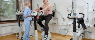
Exercise regularly
Exercise as Stroke Prophylaxis reduces the risk of developing all types of stroke. Aerobic workouts are especially good: walking, running, swimming, cycling, low-impact fitness...
The exercises work comprehensively. They help reduce weight, improve the overall condition of blood vessels and the heart, and reduce stress. Try to increase the duration of your daily workouts to at least 30 minutes.
- How to train so as not to turn into a wreck at 40 →
Drink less
There is an interesting point here: doctors do not urge you to stop drinking altogether. No, drinking is definitely dangerous, if only because it increases blood pressure. But one serving of alcohol a day can even be beneficial: according to some data, Alcohol consumption and stroke: benefits and risks. , moderate alcohol consumption reduces the risk of blood clots and may prevent ischemic stroke.
Yes, one serving, according to Fact Sheets - Alcohol Use and Your Health doctors, is 17 ml of pure alcohol or:
- 350 ml beer;
- 147 ml wine;
- 44 ml of something strong - vodka, cognac, whiskey and so on.
But keep in mind: this does not yet give carte blanche to moderate alcohol consumption. Whether you can drink or not, it’s best to discuss it with your therapist.
- How to drink less →
Eat less trans fats
Trans fats reduce the lumen of blood vessels. This means that the formation of a blood clot will become more likely. Therefore, down with fast food, store-bought baked goods, chips, crackers and margarine.
- Are saturated fats really killing us →
Monitor your blood pressure levels
Don't let it be more than 130/80. If such situations occur, be sure to consult a therapist for advice.
- How to lower blood pressure: 6 quick ways that are sure to work →
Rehabilitation of patients who have suffered a cerebral stroke
The World Health Organization (WHO) defines medical rehabilitation as follows.
Medical rehabilitation is an active process, the goal of which is to achieve complete restoration of functions impaired due to illness or injury, or, if this is not possible, the optimal realization of the physical, mental and social potential of a disabled person, his most adequate integration into society.
There are some patients who, after a stroke, experience partial (and sometimes complete) independent restoration of damaged functions. The speed and extent of this recovery depend on a number of factors: the period of the disease (duration of the stroke), the size and location of the lesion. Restoration of impaired functions occurs in the first 3-5 months from the onset of the disease. It is at this time that restoration measures should be carried out to the maximum extent - then they will have maximum benefit. By the way, it is also very important how actively the patient himself participates in the rehabilitation process, how much he understands the importance and necessity of rehabilitation measures and makes efforts to achieve the maximum effect.
Conventionally, there are five periods of stroke:
- acute (up to 3-5 days);
- acute (up to 3 weeks);
- early recovery (up to 6 months);
- late recovery (up to two years);
- period of persistent residual effects.
Basic principles of rehabilitation measures:
- earlier start;
- systematicity and duration;
- complexity;
- phasing.
Rehabilitation treatment begins already in the acute period of a stroke, during the patient’s treatment in a specialized neurological hospital. After 3-6 weeks, the patient is transferred to the rehabilitation department. If after discharge a person needs further rehabilitation, then it is carried out on an outpatient basis in the rehabilitation department of the clinic (if there is one) or in a rehabilitation center. But most often such care is shifted onto the shoulders of relatives.
The objectives and means of rehabilitation vary depending on the period of the disease.
How to treat the disease?
For hemorrhagic stroke, therapy is focused on restoring normal cerebral circulation and preventing recurrent thrombus formation in the arteries of the brain. For this purpose, complex treatment with drugs and physical procedures is carried out. If conservative therapy is ineffective, surgery is prescribed.
Medications
The basic principles of drug therapy are the restoration of normal blood pressure and the elimination of hemorrhage in brain tissue.
Lowering blood pressure helps reduce blood pressure on the vascular walls of the cerebral arteries, which reduces the risk of relapse and stops further hemorrhage. To stabilize blood pressure in hemorrhagic stroke, antihypertensive drugs are prescribed:
- Beta blockers. Usually prescribed are Atenolol, Obzidan or Vasocardin. Depending on the dosage and quantity of the drug, the price of beta-blockers varies from 60 to 1000 rubles.
- ACE inhibitors. Among them are Enap, Kapoten, Renitek. The latter sells for 120 rubles. The cost of Enap is 80 rubles, Kapoten - from 160 to 306 rubles.
- Calcium channel blockers: Adalat, Isoptin, Cardizem, Corinfar. The price of drugs reaches up to 300 rubles.
- Diuretics. Hypothiazide and Triampur are prescribed. The cost of medicine is 400 rubles.
To stop bleeding, medications that increase blood clotting are used:
The latter is the cheapest - its cost reaches 80 rubles. The price for Kontrikal and Ditsinon varies from 400 to 800 rubles.
Therapeutic procedures
There are several types of therapeutic procedures used to treat hemorrhagic stroke and its complications:
Rehabilitation in acute and early recovery periods of stroke
It is carried out in a hospital setting. At this time, all activities are aimed at saving lives. When the threat to life passes, measures to restore functions begin. Positional treatment, massage, passive exercises and breathing exercises begin from the first days of a stroke, and the time for the start of active recovery measures (active exercises, transition to a vertical position, standing up, static loads) is individual and depends on the nature and degree of circulatory disorders in the brain, from the presence of concomitant diseases. Exercises are performed only on patients who are clearly conscious and in satisfactory condition. For small hemorrhages, small and medium-sized heart attacks - on average from 5-7 days of stroke, for extensive hemorrhages and heart attacks - on 7-14 days.
In the acute and early recovery periods, the main rehabilitation measures are the prescription of medications, kinesitherapy, and massage.
Medications
In its pure form, the use of drugs cannot be classified as rehabilitation, because it is more of a treatment. However, drug therapy creates the background that ensures the most effective recovery and stimulates the disinhibition of temporarily inactivated brain cells. Medicines are prescribed strictly by a doctor.
Kinesiotherapy
In the acute period, it is carried out in the form of therapeutic exercises. Kinesitherapy is based on positional treatment, passive and active movements, and breathing exercises. On the basis of active movements, carried out relatively later, learning to walk and self-care is built. When performing gymnastics, the patient must not be overtired; efforts must be strictly measured and the load gradually increased. Treatment with position and passive exercises for uncomplicated ischemic stroke begin on the 2-4th day of illness, for hemorrhagic stroke - on the 6-8th day.
Treatment by position. Purpose: to give paralyzed (paretic) limbs the correct position while the patient lies in bed. Make sure that your arms and legs do not remain in one position for a long time.
Laying in a supine position. The paralyzed arm is placed under a pillow so that the entire arm, including the shoulder joint, is at the same level in a horizontal plane. Then the arm is abducted to the side to an angle of 900 (if the patient has pain, then they start with a smaller angle of abduction, gradually increasing it to 900), straightened and turned outward. The hand with the fingers extended and spread apart is fixed with a splint, and the forearm with a bag of sand. The leg on the side of paralysis (paresis) is bent in a log at an angle of 15-200 (place a bolster under the knee), the foot is in dorsiflexion at an angle of 900 and is held in this position by resting on the headboard or using a special case in which it is placed foot and lower leg.
Laying in the position on the healthy side is carried out by giving the paralyzed limbs a flexion position. The arm is bent at the shoulder joint and elbow, placed on a pillow, the leg is bent at the hip, knee and ankle joints, placed on another pillow. If muscle tone has not yet increased, the position on the back and healthy side is changed every 1.5-2 hours. In cases of early and pronounced increase in tone, treatment in the supine position lasts 1.5-2 hours, and on the healthy side - 30-50 minutes.
There are other styling options. J.Vantieghem et al recommend alternating the patient's position on the back, on the healthy side and on the paralyzed side.
Laying on the back: the patient's head lies on a pillow, there is no need to bend the neck, the shoulders are supported by the pillow. The paralyzed arm lies on a pillow at a short distance from the body, straightened at the elbow and wrist joints, fingers straightened. The thigh of the paralyzed leg is extended and placed on a pillow.
Positioning on the paralyzed side: the head should be in a comfortable position, the torso should be slightly turned and supported by pillows behind and in front. The position of the paralyzed arm: it rests completely on the bedside table, at the shoulder joint it is bent by 900 and rotated outward, at the elbow and wrist joints it is extended as much as possible, the fingers are also extended and spread apart. Position of the paralyzed leg: the hip is extended, the knee is slightly flexed. The healthy arm rests on the body or on a pillow. The healthy leg lies on a pillow, slightly bent at the knee and hip joints (step position).
Laying on the healthy side: the head should lie in a position comfortable for the patient in line with the torso, slightly turned forward. The paralyzed arm lies on a pillow, bent at the shoulder joint at an angle of 900 and extended forward. Position of the paralyzed leg: slightly bent at the hip and knee, lower leg and foot placed on a pillow. The healthy arm is placed in a position comfortable for the patient. The healthy leg is extended at the knee and hip joints.
When treating with position, it is important that on the side of paralysis the entire arm and its shoulder joint are located at the same level in a horizontal plane - this is necessary to prevent stretching of the shoulder joint bursa under the influence of gravity of the arm.
Passive movements improve blood flow in paralyzed limbs, can help reduce muscle tone, and also stimulate active movements. Passive movements begin with the large joints of the arms and legs, gradually moving to the small ones. Passive movements are performed slowly (a fast pace can increase muscle tone), smoothly, without sudden movements, on both the sick and healthy side. To do this, the methodologist (the person who performs rehabilitation measures) clasps the limb above the joint with one hand, and with the other below the joint, then making movements in this joint to the fullest extent possible. The number of repetitions of each exercise is 5-10 times. Passive movements are combined with breathing exercises and teaching the patient to actively relax muscles. When performing passive movements in the shoulder joint, there is a high risk of injury to the periarticular tissues, so there is no need to perform sharp abduction and flexion of the paralyzed arm at the shoulder joint, or sharp movement of the arm behind the head. To prevent stretching of the shoulder joint bursa, the technique of “screwing” the head of the humerus into the glenoid cavity is used: the methodologist fixes the shoulder joint with one hand, with the other hand clasps the patient’s arm bent at the elbow joint and makes circular movements, pressing towards the shoulder joint.
Among the passive exercises, it is necessary to highlight a passive imitation of walking, which serves to prepare the patient for real walking: the methodologist, clasping the lower third of the shins of both legs, bent at the knee joints, alternately flexes and extends them at the knee and hip joints with simultaneous sliding of the feet along the bed.
When performing passive movements, it is important to suppress synkinesis (cooperative movements) in paralyzed limbs. When performing exercises on the leg in order to prevent synkinesis in a paretic arm, the patient is told to clasp his fingers in the “lock” position and clasp his elbows with his palms. To prevent concomitant movements in the leg when performing movements with the arms, the leg on the side of the paresis can be fixed with a splint.
Following the passive movements with which therapeutic gymnastics begins, they move on to performing active ones.
Active gymnastics, in the absence of contraindications, begins in case of ischemic stroke after 7-10 days, in case of hemorrhagic stroke - after 15-20 days from the onset of the disease. The main requirement is strict dosing of the load and gradually increasing it. The load is dosed by amplitude, tempo and number of repetitions of exercises, and the degree of physical stress. There are static exercises, accompanied by tonic muscle tension, and dynamic exercises: during them the movements themselves are performed. With severe paresis, active exercises begin with those of a static nature, since they are easier. These exercises involve holding your arms and legs in their given position. The table shows exercises of a static nature.
Exercises of a dynamic nature are performed primarily for muscles whose tone usually does not increase: for the abductor muscles of the shoulder, supinators, extensors of the forearm, hand and fingers, abductors of the thigh, flexors of the leg and foot. With severe paresis, they begin with ideomotor exercises (the patient first mentally imagines the movement, then tries to perform it, while pronouncing the actions performed) and with movements in facilitated conditions. Facilitated conditions imply the elimination in various ways of gravity and friction, which make it difficult to perform movements. To do this, active movements are performed in a horizontal plane on a smooth slippery surface, systems of blocks and hammocks are used, as well as the help of a methodologist who supports the limb segments below and above the working joint.
Towards the end of the acute period, the nature of active movements becomes more complex, the pace and number of repetitions gradually but noticeably increase, and exercises for the torso begin (light turns, bends to the sides, flexion and extension).
Starting from 8-10 days (ischemic stroke) and from 3-4 weeks (hemorrhagic stroke), if the patient feels well and is in satisfactory condition, they begin to teach sitting. Initially, he is helped 1-2 times a day for 3-5 minutes to take a semi-sitting position with a seating angle of about 300. Over the course of several days, while monitoring the pulse, both the angle and the sitting time are increased. When changing body position, the pulse should not increase by more than 20 beats per minute; if pronounced palpitations occur, then reduce the landing angle and duration of the exercise. Usually, after 3-6 days, the elevation angle is increased to 900, and the procedure time is up to 15 minutes, then they begin learning to sit with their legs down (the paretic arm is fixed with a scarf to prevent stretching of the joint capsule of the shoulder joint). When sitting, the healthy leg is placed on the paretic leg from time to time - this is how the patient is taught to distribute body weight on the paretic side.
Next, they begin to learn to stand next to the bed on both legs and alternately on the paretic and healthy leg (the knee joint on the affected side is fixed using the hands of a methodologist or a splint), walking in place, then walking around the room and corridor with the help of a methodologist, and with improving gait - using a three-legged crutch or stick. It is important to develop in the patient the correct walking stereotype, which consists of friendly flexion of the leg at the hip, knee and ankle joints. For this purpose, footprints are used, and to train “triple bending of the leg” on the side of the paresis, wooden planks 5-15 cm high are installed between the footprints. The last stage of learning to walk is training to walk on stairs. When walking, the patient's paretic arm must be secured with a scarf.
The rehabilitation measures carried out should bring the maximum possible restorative effect. The most gentle care techniques are shown in the table below.
Along with teaching the patient to walk, exercises are carried out to restore everyday skills: dressing, eating, and performing personal hygiene procedures. Exercises and techniques for restoring self-service are reflected in the table below.
Massage
Massage begins for uncomplicated ischemic stroke on days 2-4 of illness, for hemorrhagic stroke - on days 6-8. The massage is carried out when the patient lies on his back and on his healthy side, daily, starting from 10 minutes and gradually increasing the duration of the massage to 20 minutes. Remember: vigorous tissue irritation, as well as a fast pace of massage movements, can increase muscle spasticity! When selectively increasing muscle tone, massage should be selective.
On muscles with increased tone, only continuous planar and grasping stroking is used. When massaging opposing muscles (antagonist muscles), stroking is used (deep planar, tong-like and intermittent grasping), gentle transverse, longitudinal and spiral rubbing, light shallow longitudinal, transverse and tong-like kneading.
Direction of massage: shoulder-scapular girdle → shoulder → forearm → hand; pelvic girdle → thigh → lower leg → foot. Particular attention is paid to massage the pectoralis major muscle, in which the tone is usually increased (slow stroking is used), and the deltoid muscle, in which the tone is usually reduced (stimulating methods in the form of kneading, rubbing and tapping at a faster pace). Massage course 30-40 sessions.
In a hospital setting, rehabilitation measures are carried out no longer than 1.5-2 months. If it is necessary to continue rehabilitation treatment, the patient is transferred to an outpatient rehabilitation facility.
Rules for conducting exercises for recovery after ischemic stroke
The greatest difficulty is motivating a seriously ill patient to perform exercises. This requires persistence and support. You need to focus on the successes achieved and explain why the classes are needed.
Basic principles of conducting exercise therapy classes:
- in the initial stages, a healthy limb is trained;
- all classes are conducted constantly and on schedule so as not to lose the progress achieved;
- constant increase in intensity and complexity of exercises;
- psychological support, especially important in case of low success, when the patient quickly loses faith in the usefulness of exercise therapy.
Rehabilitation and full recovery after an ischemic stroke are unthinkable without gradually more complex exercises and exercise therapy complexes.
Exercise therapy technique for stroke
If mobility is limited, stroke survivors need to engage in physical therapy daily. With paralysis, exercises are performed in a passive form, that is, with the help of another person. You need to start with flexion-extension manipulations on the limbs. It is important to work out the joints: arms, legs, fingers. Strengthen muscles and restore blood flow in affected limbs. In addition, passive exercise prevents the occurrence of bedsores and skin wounds characteristic of bedridden patients.
As soon as the patient begins to assume a sitting position, it is time to begin active physical therapy. The stroke patient must bend and turn from side to side, swing his arms, flexion and extension of the upper and lower extremities, and turn his head to the right and left.
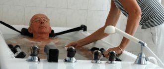
Then they begin to develop the mobility of the body. The patient stands with support on his legs, it is important to feel them. Afterwards you need to alternately tense and relax your fingers. Then they move on to learning to stand without support and walk. To enhance the effect, exercises are combined with massage and therapeutic baths. This way muscles and joints become more flexible, pain and tension go away.
The main thing is to follow all the advice of a neurologist and take medications strictly according to the prescribed regimen. Other rehabilitation methods should also be carried out under the supervision of a specialist.
Forecast
In contrast to ischemic brain damage, the prognosis for hemorrhagic cerebral stroke is much more serious. In approximately 60-80% of cases, the prognosis is unfavorable for life, and the outcome of a vascular accident is the death of the patient. Fatal outcomes are especially common in hemorrhagic stroke localized in the brain stem, blood breakthrough into the ventricular system of the brain. When aggravated by decompensated somatic pathology and extensive hemorrhage, death occurs in almost one hundred percent of cases. The prognosis for work ability is also unfavorable. Although, in general, the prognosis for functional restoration is better than for ischemic stroke. With speech disorders, severe paresis of the limbs due to hemorrhagic stroke, patients become disabled in most cases. Only in small areas of hemorrhage that do not affect important speech and motor areas does the patient return to work after long-term rehabilitation. I would especially like to touch upon the issue of patients in a coma. The prognosis for a hemorrhagic stroke in a comatose patient is very difficult to predict. Coma is not at all an indicator that a person will die. Attention should be paid to the state of hemodynamics, electrolyte metabolism, renal and pulmonary functions. If blood saturation reaches 95-96%, creatinine clearance is normal, and the patient’s blood pressure and heart rate are adequate without hardware support, then the prognosis is generally satisfactory. The prognosis worsens when artificial ventilation is required, air oxygenation with humidified oxygen is required, and the acid-base balance is unstable.
Treatment in the intensive care unit of the Yusupov Hospital
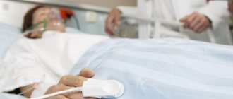
The most severe patients with ischemic stroke are hospitalized in the intensive care wards of the Yusupov Hospital. The department is equipped with the most modern medical equipment in accordance with the latest European standards: the wards are equipped with mainline oxygen, there is a 24-hour nursing station. Using modern cardiac monitors, resuscitators monitor the functional activity of the cardiovascular system and the level of oxygen saturation in the blood. If necessary, doctors use stationary or portable ventilators.
Thrombolytic therapy in patients with ischemic stroke in the first hours of the disease can prevent the development or reduce the amount of irreversible damage to the brain. This makes it possible to reduce the degree of neurological deficit. To prevent further thrombus formation and re-embolism, direct anticoagulants are used: sodium heparin or low molecular weight heparin. In order to prevent thrombus formation and embolism of cerebral arteries, antiplatelet agents are widely used, which are prescribed in combination with anticoagulants or in isolation.
Another direction in the treatment of ischemic stroke is neuroprotective therapy with the aim of increasing the survival of neurons under conditions of ischemia and hypoxia. To prevent the death of viable neurons near the source of the infarction (in the area of the “ischemic penumbra”), drugs with vasoactive and neurometabolic effects are prescribed.
At the Yusupov Hospital, doctors use the most effective drugs to treat ischemic stroke; treatment regimens are selected individually.