Cardiac catheterization is a cardiac examination by inserting a catheter into the arteries of the heart. Another name is probing. This procedure is of utmost diagnostic importance for identifying various heart diseases.
A catheter is a hollow flexible tube inserted into the channels and cavities of the human body for emptying, washing, administering medications, or for diagnostic purposes. Catheterization is the insertion of a catheter into a natural canal or cavity of the human body for diagnostic or therapeutic purposes (for example, cardiac, arterial, urethral catheterization, etc.).
In 1929, at the Eberswald Surgical Clinic near Berlin, the German physician Werner Forsmann (1904-1979) first used a catheter to study the anatomical and functional features of the human heart. In 1956, he was awarded the Nobel Prize in Physiology or Medicine in the field of cardiac catheterization together with the American D. Richards and the Frenchman A. Cournan. Currently, many different cardiac catheters have been created. With their help, and after performing other tests, the doctor can conduct a thorough examination of the heart and its functions.
Main types of procedure
Experts distinguish the following sensing methods:
- extensive catheterization (or catheterization of the left chambers of the heart) is carried out most often; specialists advance the catheter for cardiac sounding along the aorta to the left ventricle until it reaches the coronary vessels;
- small catheterization (or catheterization of the right heart) - the catheter into the right heart and pulmonary arteries passes through special veins located in the groin area or in the elbow, but in some cases floating catheters are used, which enter the heart together with venous blood.
In addition, the doctor may prescribe synchronous (or simultaneous) catheterization, in which several catheters are inserted into the heart through an artery and vein. During the examination, the catheters can be placed opposite each other so that they are separated only by the cardiac channel (for example, mitral or aortic). This diagnostic method helps determine the pressure gradient that occurs in the openings of the heart valves.
Specific tests during cardiac catheterization
Angiography
Coronary angiography by left heart catheterization is used to evaluate coronary artery anatomy in a variety of clinical situations, such as patients with suspected atherosclerotic or congenital coronary artery disease, valvular disorders prior to valve replacement, or unexplained heart failure.
X-ray contrast agent is injected into the coronary or pulmonary arteries, aorta and heart chambers. Digital subtraction angiography is used for fixed arteries and for chamber cine angiography.
Pulmonary angiography using right heart catheterization can be used to diagnose pulmonary embolism. A radiopaque contrast agent is usually selectively injected into one or both pulmonary arteries and their segments. However, computed tomographic pulmonary angiography (CTPA) has largely replaced right heart catheterization for diagnosing pulmonary embolism.
Aortic angiography by left heart catheterization is used to evaluate aortic regurgitation, coarctation, patent ductus arteriosus, and dissection.
Ventriculography (LV Gram)
Ventriculography is used to visualize the movement of the ventricular wall and ventricular tract, including the subvalvular, valvular, and supravalvular regions. It is also used to assess the severity of mitral valve regurgitation and determine its pathophysiology. Once left ventricular mass and volume are determined from a single planar or biplanar ventricular angiogram, end-systolic and end-diastolic volumes and ejection fraction can be calculated.
Coronary arterial flow measurements
Coronary angiography shows the presence and extent of stenosis, but not the functional significance of the lesion (that is, how much blood flows through the stenosis) or whether a particular lesion may be causing symptoms. , measurement of coronary arterial blood flow is used .
The measurement is carried out with pressure sensors or Doppler flow sensors. Data from these sensors can be used to estimate coronary artery blood flow, which is expressed as fractional flow margin (FFR). FFR is the ratio of the maximum flow through the stenotic area to the normal maximum flow. FFR <0.75 to 0.8 is considered abnormal.
These flow scores correlate well with the need for intervention and long-term outcome. Thus, patients with FFR >0.8 do not seem to benefit from stent placement. These flow measurements are most useful in intermediate lesions (40 to 70% stenosis) and in multiple lesions (to identify those that are most clinically significant).
Intravascular ultrasound (IVUS)
Miniature ultrasound transducers at the tip of coronary artery catheters can create images of the lumen and walls of coronary vessels and determine blood flow. Intravascular ultrasonography is increasingly being used concomitantly with coronary angiography.
Optical coherence tomography (OCT)
Optical coherence tomography is an optical analogue of intracoronary ultrasound imaging that measures the amplitude of scattered light to determine the temperature of coronary plaques. The method helps determine whether lesions have a high risk of future rupture (leading to acute coronary syndromes).
Tests for cardiac shunts
Measuring blood oxygen at successive levels in the heart and great vessels can help determine the presence, direction, and extent of central shunts. The maximum normal difference in oxygen content between structures is as follows:
- pulmonary artery and right ventricle: 0.5 ml/dl;
- right ventricle and right atrium: 0.9 ml/dl;
- right atrium and superior vena cava: 1.9 ml/dl;
If the blood oxygen level in the distal chamber exceeds its content in the proximal chamber by more than these values, a left-to-right shunt is likely at this level.
Right-to-left shunts are suspected when LA, LV, or arterial oxygen saturation is low (≤92%) and does not improve when pure oxygen is given (fractional inhalation O2 = 1.0).
Desaturation of the left heart or arteries plus elevated oxygen levels in blood samples taken outside the shunt node on the right side of the circulation indicate a bidirectional shunt.
Measurement of cardiac output and blood flow
Cardiac output (CO) is the volume of blood ejected by the heart per minute (normal at rest: 4 to 8 L/minute). Methods used to calculate CO include:
- method of direct determination of cardiac output (Fick method);
- indicator dilution technique;
- thermodilution technique.
When using the Fick technique, CO is calculated proportionally to oxygen consumption divided by the arteriovenous oxygen difference.
Dilution methods are based on the assumption that once an indicator is introduced, it appears and disappears in proportion to CO.
Endomyocardial biopsy
Endomyocardial biopsy helps evaluate graft failure and myocardial impairment due to infection or infiltrative disease. A biopsy catheter (bioptome) can be inserted into any ventricle, usually the right one. Three to five samples of myocardial tissue are removed from the septal endocardium. The main complication of endomyocardial biopsy, cardiac perforation, occurs in 0.3–0.5% of patients. This can cause hemopericardium, leading to cardiac tamponade. Damage to the tricuspid valve and supporting chordae may also occur, which can lead to tricuspid regurgitation
Who should conduct it?
Probing of the heart of a child and an adult is carried out for diagnostic purposes if a specialist needs to obtain detailed information about the coronary vessels and heart, and other research methods cannot provide complete information about the degree of development of the disease, its causes and distinctive features.
After receiving the results of the study, the doctor will be able to make an accurate diagnosis and prescribe correct and effective treatment (for example, an operation to probe the cavities of the heart).
Indications
Catheterization can be performed in a variety of situations where it is critical to obtain more information about the patient's heart and blood vessels.
The peculiarity of this procedure is that catheterization is carried out not only for diagnostic, but also for therapeutic purposes.
The main indications when this procedure is necessary for diagnostic purposes:
- heart failure;
- cardiomyopathy;
- cardiac amyloidosis;
- congenital heart defects;
- pulmonary hypertension;
- pathology of heart valves.
In these cases, catheterization is necessary for therapeutic purposes. It can be used to:
- opening of canals that are narrowed (that is, stenotic);
- supervision of a number of heart defects;
- stenting or angioplasty of arteries that are unhealthy.
Also, sometimes catheterization can be performed in conjunction with other diagnostic and therapeutic procedures as an additional diagnostic or therapeutic tool. It is permissible to carry out the procedure even at a young age.
When is it appointed?
Diagnostic cardiac catheterization may be prescribed in the presence of the following diseases:
- congenital heart defects;
- diseases of the cardiac system (valve damage);
- ischemic diseases;
- cardiomyopathy;
- heart failure;
- mild form of hypertension;
- cardiac amyloidosis.
The diagnostic measure helps to accurately identify the type of damage to the coronary vessels, myocardial tissue and heart valves, which cannot be determined using simpler examinations (or when they show an inaccurate result). In addition, the procedure for probing cardiac vessels helps to fully assess the severity of damage and examine the pathophysiological mechanisms of changes in myocardial function. Most often, this diagnostic method is prescribed to patients undergoing cardiac surgery.
Cardiac catheterization
What is the purpose of cardiac catheterization?
The cardiac catheterization procedure is performed for both diagnostic and therapeutic purposes for diseases of the cardiovascular system. This procedure is widely used in modern cardiology, since, carried out in a minimally invasive manner, it provides detailed information about the condition of the chambers, valves and vessels of the heart and allows, if necessary, to carry out effective intervention and optimally solve the detected problem.
Cardiac catheterization allows for the diagnosis and treatment of the following cardiovascular diseases:
- And myocardial infarction
- With tenocardia
- With carotid tenosis
- Mitral valve regurgitation
- Heart rhythm disturbances
- Damage to venous shunts
- Congenital heart defects (PDA – patent ductus arteriosus;
- P FO - patent foramen ovale)
Diagnostic and hemodynamic cardiac catheterization
The purpose of diagnostic catheterization is to study the function of the heart and its structural units: heart chambers, coronary vessels, valves, etc. The procedure is undertaken with the aim of accurately identifying existing defects, congenital or acquired, or in cases where there is a suspicion of narrowing of the coronary vessels (coronary angiography). During cardiac catheterization, Doppler can be used to measure blood current and pressure inside the chambers and vessels of the heart, as well as perform a biopsy of the heart muscle.
During the test, a thin, long and flexible catheter tube made of elastic material is inserted and advanced from the groin area to the mouth of the coronary vessels. Through this catheter, a contrast agent is injected into the bloodstream and, using a miniature video camera, an image of the heart is obtained in various projections to obtain the most accurate picture and localization of the vessel stenosis, its length and diameter. Through the introduction of a contrast agent, the size of the heart is determined, the contraction of the heart muscle and the functioning of the valves are assessed.
The entire inspection process is recorded on video and displayed on the monitor screen in real time.
Therapeutic cardiac catheterization with the introduction of supporting devices (stents)
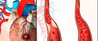
This therapeutic cardiac catheterization, also called coronary angioplasty, is performed to restore blood flow in one or more coronary vessels that supply the heart muscle.
The procedure is performed when a blockage in a coronary artery has not been cleared with thrombolytic drugs or when there is severe disease in a heart vessel or vessels discovered during diagnostic cardiac catheterization. During coronary angioplasty, a balloon is inserted to expand the blocked artery and a special coil is inserted to act as a support device (stent).
A stent is installed at the site of narrowing of the vessel in order to preserve the lumen of the vessel and ensure unimpeded blood flow. The high efficiency and clear benefits of stents have been studied and proven by scientific research. The procedure to widen the narrowed vessels of the heart, with or without the installation of a stent, is performed on both coronary arteries and arteriovenous shunts and veins, as well as on shunts created during previous heart operations.
Modern stents of the latest generation can significantly reduce the likelihood of re-narrowing of blood vessels compared to previous installations and, as a result, reduce the need for repeated catheterizations. Such innovative stents (“Cypher”, “Taxus”, “Endeavour” and others) are coated with a special substance that releases a drug that prevents re-stenosis of the vessel. This drug was developed only in recent years and is already actively used in the invasive cardiology department of the medical center.
The success rate of the procedure using a balloon and stent is very high. We are talking about immediate opening of narrowed vessels in 95 - 97% of cases.
What is a stent
A stent is a structure made of plastic or metal used to restore the lumen of a vessel. Currently, mesh structures made from the most inert materials have become widespread to minimize the reaction of surrounding tissues. In Israel, a unique development of Israeli scientists is used with great success - a stent called “Cypher”. The clinic's specialists have extensive experience in the field of coronary angiography, angioplasty and stenting.
A coronary stent is a small tube made of stainless mesh. It is installed on the balloon catheter in a “crimped” or rolled form. When the balloon is inflated, the structure opens and is pressed into the inner wall of the coronary artery, thus restoring the lumen of the vessel. The development of coronary stents has become a solution to many problems associated with angioplasty.
Advantages of a drug-eluting stent
The drug coating the stent is dosed into the blood, reducing the proliferation of cells inside the stent, which sharply reduces the possibility of developing a blockage after the procedure.
Drug-eluting stents are more expensive than bare stents. However, a comparative analysis conducted by investigator Dr. David Cohen of the Harvard Clinical Research Institute confirmed the cost-effectiveness of the Cypher Sirolimus stent. Dr. Cohen used actual cost data from the first hospitalization to one year after stent implantation. He reported that a twelve-month follow-up of Cypher Sirolimus stents in patients showed a significant reduction in the need for repeat treatments and hospitalizations.
Drug eluting stents (DES) reduce the likelihood of restenosis and the risk of revascularization, which has been proven in studies.
Bare metal stents are more resistant to thrombus formation, so patients are allowed to stop taking Plavix two months after stenting.
After the installation of drug-eluting stents, it is possible to stop taking Plavix 6-12 months after the operation with the doctor’s permission, otherwise a blood clot may form and myocardial infarction may develop.
The third generation of stents are biostents made of bioabsorbable polymers. The innovation is that a year after implantation, the frame of the structure gradually dissolves, while maintaining lumen in the vessel.
Israeli cardiologists remind that even the most advanced methods of treating cardiovascular diseases do not cancel preventive measures and a healthy lifestyle. Physical activity, proper nutrition, weight control, alternating mental and physical work will allow you to live a long and fulfilling life.
Angioplasty is a method of restoring the lumen of an artery using a catheter and an inflatable balloon. It has been used for more than 20 years, and is still in demand for angiography using a contrast agent to determine the location of the narrowing. It is possible to combine angioplasty with other procedures, in particular stenting of the coronary arteries.
It should be borne in mind that coronary angioplasty has a number of disadvantages, for example, blockage of the vessel below the site of narrowing due to the rupture of an atherosclerotic plaque. It is possible that the artery may re-narrow 6 months after surgery and restenosis may develop.
Coronary stenting is a minimally invasive operation that is characterized by low trauma, high efficiency, and minimal risk of complications. During the procedure, a mesh stent is placed on a special balloon, and when it is inflated, the stent is expanded and pressed into the narrowed lumen of the artery. The use of stents has achieved positive results in restoring vascular patency. Their use in practice has reduced the risk of artery blockage and reduced the likelihood of developing restenosis by almost 50%.
Angioplasty and coronary stenting restore vascular patency, relieve pain in the thoracic region, improve quality of life, and reduce the development of other complications. Stenting is performed on arteries in the groin or arm.
How is coronary stenting performed?
Before coronary stenting, the location and cause of the narrowing, as well as the shape and size of the vessel, are determined. This helps the surgeon make an appropriate decision regarding treatment tactics, choosing the most appropriate procedures in each particular case (stetting, angioplasty, atherectomy or drug therapy).
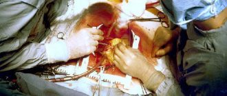
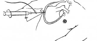
Cardiac catheterization is a specific examination of the heart using a catheter that is inserted into arteries located in the groin area or arm. The operation is performed under X-ray guidance to ensure proper advancement of the catheter. The catheter is a long, flexible, soft plastic tube with a diameter of 2 - 3 mm. To visualize the narrowed area of the vessel, a contrast agent is injected. The cardiologist, using X-ray images, assesses the size of the coronary artery and selects the type of balloon catheter and guidewire that will provide access to the stent, threaded on a deflated balloon, to the narrowed part of the vessel.
Heparin , a drug that prevents blood clots, is given before surgery. In most cases, stenting is preceded by angioplasty, which provides preliminary dilatation of the artery, which facilitates delivery of the stent.
First, a guide catheter with a flexible tip is inserted. A balloon catheter with a stent at the end is then inserted, which is inflated with a special manual syringe pump with a mixture of saline and contrast agent. Once at the site of narrowing of the vessel, the balloon is inflated and the stent is installed in the narrowed area of the vessel, expanding it. All actions are carried out under x-ray control.
This minimally invasive operation is performed under local anesthesia and takes from thirty minutes to an hour. The duration depends on the technical complexity of the operation.
In the hands of experienced cardiologists and with modern technology, the risk of death during stenting is believed to be less than 1%. This is a relatively safe procedure. The risk of complications is negligible.
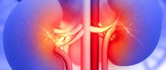
Possible deterioration of kidney function (particularly in patients with diabetes mellitus and patients with kidney disease) due to the larger amount of contrast material. In such cases, additional precautions are taken to prevent this possible complication.
Although coronary artery stenting is superior to angioplasty, it has not lost its importance. According to experts, stents are used in 50-75% of cases.
Rehabilitation after coronary stenting
During the recovery period after the coronary stenting procedure, patients are prescribed aspirin and cholesterol-lowering drugs. Physical therapy, limiting the consumption of cholesterol-containing foods, avoiding smoking and alcohol abuse are recommended.
How to resolve issues with organizing a trip to us for treatment
You will only need medical documents or an extract from them on this disease, which can be prepared in Russian. You send them to our email: [email protected]
We translate them into Hebrew, leading specialists hold a consultation in absentia on them, and after a few days you receive an answer. Where is it reported about the advisability of coming (there are such advanced cases that we are powerless), what length of stay is required, the list of diagnostic and treatment measures that we offer, accommodation conditions, the cost of medical measures, accommodation, transfer and other organizational issues.
We have a special department that deals with organizational issues: meeting at the airport, transfer, ordering accommodation, accompaniment of an interpreter at all stages of treatment and diagnosis, excursion services.
We also have our own housing stock: these are fully equipped 2-3 room apartments. The apartments have everything: TV with satellite TV and Russian channels, washing machine, refrigerator, air conditioning, equipped kitchen, Internet. Usually, living in apartments is cheaper than hotels.
Our patients are provided with the following free of charge:
- accompanied by an interpreter in the medical center.
- transfer place of residence - medical center - place of residence
- mobile phone and local SIM card
- translation into Russian of medical documents received in Israel
Payment methods: no prepayment required, because... Payment for the medical program is made during the implementation of the program to the center’s accounting department.
Payment methods: cash (US dollars, Euros, Israeli shekels), credit card, bank transfer.
Control after treatment (after departure)
After treatment, you leave with documents that are translated into any of the languages you choose. But we are not saying goodbye to you - we continue to monitor your health. To do this, we maintain contact with our specialists, with whom you can contact on any issue regarding your recovery and receive the necessary recommendations.
Main contraindications
There are cases when cardiac probing is prohibited for a patient. These include the following contraindications:
- fever;
- cramps in the limbs;
- dangerous infections;
- systemic lesions;
- swelling in the lungs;
- if the patient has digitalis intoxication or hypokalemia;
- severe form of peripheral atherosclerosis;
- the presence of arrhythmia or hypertension;
- decompensated heart failure;
- severe anemia;
- coagulopathy;
- presence of allergies when taking certain medications;
- bleeding in the gastrointestinal tract;
- acute renal failure;
- carrying a child or lactation period.
The need for cardiac catheterization in the presence of the lesions described above is determined for each patient individually, taking into account the general clinical picture of the disease. As a rule, diagnosis can be carried out after eliminating the contraindication or after preliminary preparation of the human body.
In some cases, doctors are forced not to perform catheterization because the patient independently refuses to perform it.
Expert advice
When prescribing venous cardiac catheterization, the patient must inform the doctor about the following factors:
- bearing a child, especially in the early stages;
- use of medications or dietary supplements;
- taking glucose-lowering medications;
- allergies to iodine, radiopaque agents, seafood, rubber or latex;
- the use of Viagra and other medications that are aimed at restoring the condition of the reproductive system.
Possible complications
Most patients experience normal tolerance to the procedure and no side effects. But you need to understand that the likelihood of complications, although low, remains. The most serious include:
- heart rhythm disturbance;
- blood clot formation;
- stroke;
- heart attack;
- air getting into the catheter and blocking the artery (possibly fatal);
- allergy to contrast, in severe form - anaphylactic shock;
- cardiac tamponade;
- collapsed state or cardiogenic shock;
- infection or bleeding in the area where the catheter was;
- damage to the coronary artery;
- impaired renal function after contrast removal.
Cardiac tamponade is one of the possible complications after catheterization
The importance of preparation
It is especially important to carefully prepare the patient for the examination in the following cases:
- the presence of dangerous pathological diseases (insulin-dependent diabetes, pulmonary and renal failure, serious diseases of the brain and peripheral vessels);
- heart failure;
- severe disturbances in the left ventricle;
- children or old age.
In the presence of the described conditions, it is important to carry out cardiac catheterization with extreme care; in the presence of such lesions, the risk of death greatly increases.
How to prepare for the procedure?
After prescribing cardiac catheterization, the specialist must tell the patient all the examination techniques and warn about possible complications and adverse reactions.
After this, the patient is given documents confirming consent for catheterization and all the basic recommendations on preparing for the diagnostic procedure.
Preparing for the examination
The preparation will include the following rules:
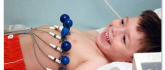
- Two weeks before the procedure, the patient is prescribed blood, urine, ECG, and chest x-ray. In some cases, the doctor prescribes additional tests.
- If necessary, the doctor changes the regimen of certain medications and medications before the procedure.
- The patient can come for diagnostics on the appointed day or be hospitalized several days before catheterization. When registering at the clinic, the patient needs to take with him all the required things (slippers, clean and comfortable clothes, hygiene products). These same items will also be needed if the patient, after the diagnosis, remains in the clinic for medical reasons. It is for this reason that you need to take everything you need with you before visiting the clinic.
- In some cases, the doctor prescribes a test for a local anesthetic, which is used for pain relief during the procedure, or a contrast agent. This is important to prevent an allergic reaction.
- It is important to take the medications prescribed by your doctor on time before the procedure.
- In the evening, before the diagnosis, you should take a shower and remove hair from the catheter insertion area.
- It is important to stop eating and drinking 6-8 hours before the procedure.
- If after catheterization the patient is going to go home, then an accompanying person must be with him.
- Before cardiac probing, the patient must leave dentures, glasses, telephone, hearing aids and other equipment in the room that will not allow the examination to be fully carried out.
Who is not recommended to pass
Cardiac and other somatic pathologies are not an obstacle to inserting a catheter into the left or right side of the heart. Although some body conditions are undesirable for an invasive procedure in the heart. If patients are aware of these conditions, they should warn medical personnel in order to avoid dangerous consequences. In such cases, the problem is solved with an increased degree of caution or rejected until the causes are eliminated.
Problems that allow you to pause preparation for the procedure are:
- external or internal allergic reaction to administered medications;
- individual intolerance to iodine or the administered substance;
- pregnancy status;
- the patient's use of Viagra and its analogues to increase male potency;
- seafood intolerance.
We will talk about complications of electrophysiological and other types of cardiac catheterization below.
Features of the event
The patient must remember that catheterization of cardiac vessels is a painless procedure that does not cause any discomfort. During the diagnosis, the patient will be conscious, can talk with a specialist and perform the actions that the doctor instructs him.
In some cases, during the examination, the patient feels his heartbeat, a slight burning sensation in the area where the catheter was inserted, or feels heat. Such unpleasant sensations should not greatly disturb or make the patient nervous, since they do not indicate any complications during the procedure. After the examination is completed, all discomfort immediately disappears.
The essence of the method
A flexible catheter (hollow tube) is passed through a peripheral artery or vein in the arm (leg) into the vessel. Under the control of an X-ray machine, its movement to the desired cavity of the heart is checked. Further diagnostic studies are carried out in the form of:
- contrasting of the ventricles (ventriculography);
- obtaining images of the coronary bed (coronary angiography);
- taking tissue for analysis (biopsy);
- measuring pressure in cavities;
- blood test for oxygen and carbon dioxide levels.
In addition to diagnosing the condition of the valves, walls and septa of the heart, therapeutic effects can also be carried out during catheterization.
An inflatable balloon is inserted into the coronary arteries and widens the narrowed area, and to secure the result, a metal frame called a stent is installed.
During probing, holes in the septum are eliminated, valves are repaired, or previously implanted prostheses are corrected.
Through an inserted catheter, cauterization of the area of the myocardium that produces abnormal cardiac impulses can be performed. Destruction is carried out by the action of radio waves, laser radiation or freezing with liquid nitrogen. If a blood clot is found in an artery, it can be removed using special hooks placed inside the catheter to the site of the blockage.
We recommend reading the article about heart biopsy. From it you will learn about indications and contraindications, the procedure, and possible complications afterwards.
And here is more information about vein catheterization.
Technique of the procedure
How is cardiac catheterization done? Features of the procedure:
- One hour before the test, the patient is given a sedative.
- Afterwards, the patient is taken to an office equipped with special equipment. He is offered a change of clothes and placed on the doctor's table.
- The nurse punctures the patient's vein to administer the necessary medications.
- If necessary, an additional catheter is inserted into the bladder.
- The treating specialist treats the catheter insertion site (elbow or groin) with antiseptics and administers local anesthesia. After obtaining the desired analgesic effect, the doctor makes a small incision to insert a catheter or punctures the vessel with a thick needle.
- Next, a catheter is inserted into the selected blood vessel, and a special fluoroscopic device is used to help advance the catheter to the ventricles of the heart or coronary vessels.
- After the device is inserted into the left or right ventricle, a special pressure gauge is attached to the catheter, which monitors the patient’s pressure. If necessary, additional procedures are performed.
- For antigraphy, a radiopaque agent is injected into the catheter, which helps to identify the ventricles and coronary vessels on the monitor. Doctors carefully examine the organ, study its condition, take pictures and make the necessary conclusions.
- At the end of the procedure, the doctor removes the heart catheter and, if necessary, applies stitches.
After cardiac catheterization, the patient can go home after the condition has completely stabilized (most often this happens after a couple of hours) or remain in the hospital until the next day.
The cost for the probing procedure will reach up to 15,000 rubles, but additional treatment will not be included in this cost.
Story
The history of cardiac catheterization dates back to Stephen Hayles (1677-1761) and Claude Bernard (1813-1878), who both used it in animal models. Clinical applications of cardiac catheterization begin with Werner Forsmann in the 1930s, who inserted a catheter into a vein in his own forearm, guided it fluoroscopically into the right atrium, and took an x-ray picture. Forsmann received the Nobel Prize in Physiology or Medicine for this achievement, although hospital administrators removed him from his position due to his unorthodox methods. During World War II, Cournan, a physician at NewYork-Presbyterian/Columbia, then Columbia-Bellevue, opened the first catheterization laboratory. Dr. Cournand received the Nobel Prize in Medicine for co-inventing cardiac catheterization in 1956.
Dr. Eugene A. Stead, founder of the physician assistant profession, also conducted research in the 1940s that paved the way for cardiac catheterization in medicine today.