What is the jugular vein
They are also called jugular (jugularis), they are vascular trunks designed to drain carbon dioxide-saturated blood from the head and neck to the subclavian vessel. Sometimes they converge to form the median vein of the neck. The internal sinus, which releases blood from the cranial sinus, begins at the jugular foramen of the skull. Here the vessel that accompanies the occipital artery flows into it, as well as the posterior auricular vein. It then descends to the point where the collarbones and sternum meet. Here it connects with other vessels, forming the brachiocephalic venous line.
The external jugular artery is smaller and its purpose is to drain blood from the outside of the neck and head. Catheters are inserted into this vessel to administer medications. The trunk of the transverse veins of the neck flows into the external one, connecting with the suprascapular vein. The anterior jugular vein is one of the smallest among them. Its origin is located in the chin area.
Types of jugular vessels
The jugular veins are a set of vessels that take blood flow from the head, brain, and neck and carry it further to the area under the collarbone. Their main functional load is to prevent blood stagnation in the head, brain and neck. For a person to feel well, it is necessary that these veins function correctly. The presence of diseases leads to serious consequences. The jugular canals contain three main vessels:
- Anterior (the main functional load is the timely outflow of blood from the chin. Located in the middle part of the cervical region, it also passes through the tongue and jaw muscles. Connects on both sides to form an arch);
- External (located in the tissues of the neck. Blood departs from the facial part, the external part. It is easy to detect during coughing, difficult physical work, when turning the head to the side);
- The internal jugular vein (is a fairly large canal in relation to the other two. It narrows and expands very easily when blood is pushed out. This process is carried out due to thin walls and a diameter of no more than 20 mm. Equipped with a valve system).
Anatomy
The internal vein drains most of the blood from the head. It has a diameter from 11 to 21 mm. The diagram of its location and tributaries is as follows. Having its origin at the cranial jugular foramen, it goes down, forming the sigmoid sinus, and further to the clavicle. Near the place where the subclavian vein joins it, which is formed by the fusion of the external vessel with the axillary one. The internal vein has a thickening called the inferior dilatation, above which the valves are located.
In the jugular fossa of the temporal bone there is the superior bulb of the jugular vein, as its small extension is called. The tributaries of the internal vein include both extracranial and intracranial. The first are tributaries of the facial vessels, connecting by transverse anastomoses with the internal vein along its entire length. In the lower part of the neck, the venous trunks converge into a V-shaped depression called the jugular fossa. The anterior jugular vein is located in the mental part, where it is formed through a superficial plexus of venous trunks in a small area.
With connections in the suprasternal interaponeurotic space, the anterior veins form the jugular venous arch. Intracranial tributaries are sinuses of the dura mater into which veins leading to the brain flow. They are venous collectors. The sinus connects with the trunks and with the venous plexuses. An important transverse sinus is located in the groove of the occipital bone, in the area of the plexus of the occipital vascular trunk with other vessels.
Extracranial tributaries drain blood from the pharyngeal plexus. Intracranial and extracranial veins merge through ligaments that extend through the cranial cavities. The location of the jugular vein directly under the skin makes it easy to feel and notice if a person coughs or screams, and sometimes with any other stress. The transverse sinus is located in the groove of the occipital bone and connects with the sigmoid sinus and the occipital cerebral veins.
In the space between the pterygoid muscles and the branch of the lower jaw there is the pterygoid venous plexus. From here, blood flows through a network of large vessels, to which the anastomoses of the facial vein connect. The superior thyroid vein passes near the artery of the same name and reaches the facial and internal jugular venous trunks. The lingual veins are the dorsal and deep veins of the tongue. At the large horn of the hyoid bone they merge into one trunk of the lingual vein. Jugular is characterized by the presence of a developed anastomosis.
Jugular internal and external veins: structure, functions, pathologies
The section of the venous network, which is commonly called the jugular vein (abbreviated as JV), is a collection of veins that provide the outflow of depleted blood from the cranium and facial part of the head.
Not only well-being, but also a person’s life depends on the functioning of this vessel, because the jugular vein is as important an element of the blood supply system to the head as the artery that supplies the brain.
Any violation in one of them can provoke serious consequences for human health, including disability or death.
Anatomical structure
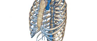
The location of the jugular vein is on the neck along the lateral surfaces, spreading to the front. It consists of three paired branches: the anterior, internal and external jugular vein, each of which is responsible for draining blood from certain areas of the head and skull. The general structure of all branches is standard:
- the inner layer, consisting of endothelium and receptors that respond to humoral and other regulatory mechanisms;
- the middle layer, consisting of collagen and elastic fibers and smooth muscle cells, ensuring constant blood flow from the head to the heart.
- outer layer consisting of connective tissue and collagen fibers that provide strength and stability to blood vessels.
The topography and detailed anatomical structure of the ventricle depends on where the vessel is located and what functions are assigned to it:
Name of the vesselTopography, size and other anatomical characteristicsFunctions and features of the vessel
| Internal jugular vein | The anterior (farthest from the heart) end of the vessel is represented by a bulb, which is located near the jugular foramen of the skull, and its lower end is located under the sternoclavicular joint and connects to the subclavian vein through another bulb. | The internal jugular vein drains blood from the skull, so its diameter is the largest of all the branches of the jugular vein. Since with each push a lot of blood enters it, the walls of the vein are well stretched, and the direction of flow is regulated using two valves. |
| Anterior jugular vein | The location of the anterior jugular vein is limited to a small area on the sides of the midline of the neck. It starts from the jawline and goes down to the sternoclavicular joint. At this point the vessel flows into the external jugular vein. | The function of the vessel is to drain blood from the tongue and lower jaw. A peculiarity is the merging of paired veins into one in the lower part of the neck with the formation of a venous arch. |
| External jugular vein | The diameter of the external jugular vein is smaller than that of the internal one, but larger than that of the anterior one. It is located in the subcutaneous tissue along the anterior surface of the neck and runs from the lower angle of the jaw along the sternoclavicular muscle. | The function of the vessel is to drain blood from the face, surface of the skull and neck. The valves in it are located in the upper and middle parts of the neck. |
The greatest participation in the life support system is played by two pairs of nuclear weapons - external and internal. Their pathologies can lead to irreversible changes that pose a threat to human health and life.
These are one of the few vessels in the body that are equipped with a fairly pronounced muscle layer that can forcefully push blood through.
Internal jugular vein
The internal jugular vein (abbreviated IJV) has many tributaries, as it is responsible for draining blood from the inside and outside of the skull. Intracranial branches flow into it:
- vessels that drain blood from the lining of the brain;
- veins of the cranial bones;
- vessels of the organs of hearing and vision.
It also receives vascular branches coming from the outer surface of the skull and soft tissues of the face. Anatomoses are present between these vessels and the internal jugular vein, resulting in the formation of a multi-tiered vascular network. Such a complex topographical structure determined the anatomical structure of this part of the circulatory system.
Due to the fact that the load on the IJV is quite high, its walls have increased elasticity and stretchability. This is especially noticeable in the terminal sections of the vessel bordering the subclavian arch and the jugular foramen of the skull. At the moment of blood release into its lumen, the walls at both bases stretch, forming “bulbs”. This reduces the load on the vessel as a whole.
The peculiarity of the IJV is a thickened outer shell made of a double layer of connective tissue cells and collagen.
The functional state of the brain, as well as communication with the outside world through vision and hearing, depends on the correct functioning of the internal branch of the neck. Its pathologies can lead to deafness, paralysis, loss of speech or deterioration of cognitive functions.
External jugular vein
The structure and location of the external jugular vein (abbreviated EJV) makes it easy to visualize it, especially if a person talks passionately and loudly for a long time, sings or screams.
It is smaller in diameter than the outer one, which makes its anatomical structure more typical of the venous network: the muscle layer in it is less developed, but there are more valves for directing blood flow, and there are no bulbs at the base.
The external jugular vein has many lateral tributaries that fill it with blood along its entire length. This is what distinguishes it from the IJV, which is filled with venous blood only from one end.
The posterior auricular, occipital, suprascapular and transverse branches, as well as the third anterior UR, flow into the MN. The main function of the external jugular vein is to drain blood from the lower jaw and chin, neck and surface of the skull.
It is the NJV that is used to install a catheter when it is necessary to administer long-term intravenous drug solutions.
: veins of the head and neck
Source: https://bloodvessel.ru/krovenosnaya-sistema-cheloveka/yaremnaya-vena
Pathologies
When screaming, stressing, or crying, all people, from babies to adults, can swell blood vessels, often on the right. This is the norm, although it often worries new parents. Problems with blood vessels often occur in old age, but in the presence of congenital defects they can appear at a young age. Changes include:
- Thrombosis.
- Vessel dilation.
- Consequences of inflammation (phlebitis).
- Congenital defects, dilatation.
Phlebectasia
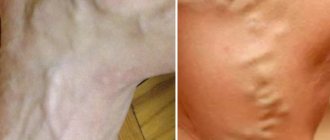
Dilatation of the jugular vein is a common phenomenon. The disease affects people of any gender and age. Jugular vein ectasia occurs due to problems with the valves, leading to stagnation of blood. The disease is often a consequence of diseases. Ectasia often occurs in women and older people. With age, the connective tissue of blood vessels weakens, varicose veins occur, which leads to disruption of the functioning of the valves. In women, similar problems arise with hormonal changes.
Due to the deep location of the vessel inside, it is difficult to distinguish ectasia. Violations of the vascular trunk are visible to the naked eye from the outside. Phlebectasia of the right internal jugular vein is common. It can be almost invisible. There may be unpleasant sensations in the neck, especially strong when screaming. Severe ectasia can change the voice and make breathing difficult.
Among the main causes of the disease:
- Trauma, bruise.
- Passive lifestyle.
- Problems with valves.
- Heart diseases.
- Leukemia.
- Neoplasms.
- Abnormal functioning of the endocrine system.
Phlebitis
The cause of the disease is often an inflammatory process in the middle ear and tissues of the mastoid process. If a blood clot becomes infected, its particles can spread throughout the body along with the infection. With thrombophlebitis, the patient feels pain, swelling, swelling occurs, accompanied by symptoms of intoxication. The spread of infection may be accompanied by tachycardia, rash, fever, and shortness of breath. The cause of phlebitis may be:
- injury or bruise;
- infection;
- distribution of the drug in the tissues around the vessel.
Thrombosis
Blockage of a vessel by a blood clot leads to disruption of blood flow. It is widely believed that blood clots are a pathology of the femoral, inferior vena cava or iliac vein, but blockage can also form in the deep jugular vessels and their branches. It leads to severe headaches and painful sensations in the neck when trying to turn the head, a pronounced venous pattern appears, and swelling of the face appears. In some cases, the pain moves to the arm. Blockage is expressed as compaction. Among the reasons:
- Problems with blood clotting.
- Consequences of operations, installation of catheters.
- Neoplasms.
- Long period of immobility.
- Use of hormones.
- Pathologies of internal organs, inflammation and infections.
Aneurysm
It is a rare pathology that occurs in children aged two to seven years. The probable cause is considered to be an abnormal development of the fetus, leading to improper development of the connective tissue of the vessel. An aneurysm manifests itself as an expansion of the vascular trunk, which intensifies when the child laughs, screams or cries. Symptoms include: sleep problems, increased fatigue, headache, restless behavior.
Jugular vein: internal (vv) and external: anatomy, pathology
The human brain receives nutrients and oxygen through the blood, so its flow to it is extremely important.
No less significant is the outflow of blood. If it stagnates, processes with destructive consequences can begin in the brain. The outflow of blood from the brain is provided by a special vessel.
The internal jugular vein is located on the right side of the neck, weakly covered by the saphenous muscle and is a convenient place for catheterization, along with the cubital fossa.
What is the jugular vein
They are also called jugular (jugularis), they are vascular trunks designed to drain carbon dioxide-saturated blood from the head and neck to the subclavian vessel. Sometimes they converge to form the median vein of the neck.
The internal sinus, which releases blood from the cranial sinus, begins at the jugular foramen of the skull. Here the vessel that accompanies the occipital artery flows into it, as well as the posterior auricular vein. It then descends to the point where the collarbones and sternum meet.
Here it connects with other vessels, forming the brachiocephalic venous line.
The external jugular artery is smaller and its purpose is to drain blood from the outside of the neck and head. Catheters are inserted into this vessel to administer medications. The trunk of the transverse veins of the neck flows into the external one, connecting with the suprascapular vein. The anterior jugular vein is one of the smallest among them. Its origin is located in the chin area.
Anatomy
The internal vein drains most of the blood from the head. It has a diameter from 11 to 21 mm. The diagram of its location and tributaries is as follows.
Having its origin at the cranial jugular foramen, it goes down, forming the sigmoid sinus, and further to the clavicle. Near the place where the subclavian vein joins it, which is formed by the fusion of the external vessel with the axillary one.
The internal vein has a thickening called the inferior dilatation, above which the valves are located.
In the jugular fossa of the temporal bone there is the superior bulb of the jugular vein, as its small extension is called. The tributaries of the internal vein include both extracranial and intracranial.
The first are tributaries of the facial vessels, connecting by transverse anastomoses with the internal vein along its entire length. In the lower part of the neck, the venous trunks converge into a V-shaped depression called the jugular fossa.
The anterior jugular vein is located in the mental part, where it is formed through a superficial plexus of venous trunks in a small area.
With connections in the suprasternal interaponeurotic space, the anterior veins form the jugular venous arch. Intracranial tributaries are sinuses of the dura mater into which veins leading to the brain flow.
They are venous collectors. The sinus connects with the trunks and with the venous plexuses.
An important transverse sinus is located in the groove of the occipital bone, in the area of the plexus of the occipital vascular trunk with other vessels.
Extracranial tributaries drain blood from the pharyngeal plexus. Intracranial and extracranial veins merge through ligaments that extend through the cranial cavities.
The location of the jugular vein directly under the skin makes it easy to feel and notice if a person coughs or screams, and sometimes with any other stress.
The transverse sinus is located in the groove of the occipital bone and connects with the sigmoid sinus and the occipital cerebral veins.
In the space between the pterygoid muscles and the branch of the lower jaw there is the pterygoid venous plexus. From here, blood flows through a network of large vessels, to which the anastomoses of the facial vein connect.
The superior thyroid vein passes near the artery of the same name and reaches the facial and internal jugular venous trunks. The lingual veins are the dorsal and deep veins of the tongue. At the large horn of the hyoid bone they merge into one trunk of the lingual vein.
Jugular is characterized by the presence of a developed anastomosis.
Functions
Vascular trunks are critically necessary for the functioning of the human body. The functions are:
- The removal of blood saturated with carbon dioxide and other waste products from the brain towards the heart.
- Formation of blood circulation in the brain area.
Pathologies
When screaming, stressing, or crying, all people, from babies to adults, can swell blood vessels, often on the right. This is the norm, although it often worries new parents. Problems with blood vessels often occur in old age, but in the presence of congenital defects they can appear at a young age. Changes include:
- Thrombosis.
- Vessel dilation.
- Consequences of inflammation (phlebitis).
- Congenital defects, dilatation.
Phlebectasia
Dilatation of the jugular vein is a common phenomenon. The disease affects people of any gender and age. Jugular vein ectasia occurs due to problems with the valves, leading to stagnation of blood. The disease is often a consequence of diseases.
Ectasia often occurs in women and older people. With age, the connective tissue of blood vessels weakens, varicose veins occur, which leads to disruption of the functioning of the valves.
In women, similar problems arise with hormonal changes.
Due to the deep location of the vessel inside, it is difficult to distinguish ectasia. Violations of the vascular trunk are visible to the naked eye from the outside. Phlebectasia of the right internal jugular vein is common. It can be almost invisible. There may be unpleasant sensations in the neck, especially strong when screaming. Severe ectasia can change the voice and make breathing difficult.
Among the main causes of the disease:
- Trauma, bruise.
- Passive lifestyle.
- Problems with valves.
- Heart diseases.
- Leukemia.
- Neoplasms.
- Abnormal functioning of the endocrine system.
Phlebitis
The cause of the disease is often an inflammatory process in the middle ear and tissues of the mastoid process. If a blood clot becomes infected, its particles can spread throughout the body along with the infection.
With thrombophlebitis, the patient feels pain, swelling, swelling occurs, accompanied by symptoms of intoxication. The spread of infection may be accompanied by tachycardia, rash, fever, and shortness of breath.
The cause of phlebitis may be:
- injury or bruise;
- infection;
- distribution of the drug in the tissues around the vessel.
Thrombosis
Blockage of a vessel by a blood clot leads to disruption of blood flow. It is widely believed that blood clots are a pathology of the femoral, inferior vena cava or iliac vein, but blockage can also form in the deep jugular vessels and their branches.
It leads to severe headaches and painful sensations in the neck when trying to turn the head, a pronounced venous pattern appears, and swelling of the face appears. In some cases, the pain moves to the arm. Blockage is expressed as compaction.
Among the reasons:
- Problems with blood clotting.
- Consequences of operations, installation of catheters.
- Neoplasms.
- Long period of immobility.
- Use of hormones.
- Pathologies of internal organs, inflammation and infections.
Aneurysm
It is a rare pathology that occurs in children aged two to seven years. The probable cause is considered to be an abnormal development of the fetus, leading to improper development of the connective tissue of the vessel.
An aneurysm manifests itself as an expansion of the vascular trunk, which intensifies when the child laughs, screams or cries.
Symptoms include: sleep problems, increased fatigue, headache, restless behavior.
Methods of treating pathologies
Phlebectasia is not life-threatening and is a cosmetic defect. It can be removed through unilateral ligation of the vessel, in which the outflow of venous blood will be taken over by collaterals and vessels located on the other side.
Thrombophlebitis requires a surgical operation to remove the “sick” vessel, thereby eliminating thrombotic formations. Treatment of unilateral thrombosis involves conservative methods.
To eliminate a venous aneurysm, resection of the malformation is used.
The following medications are used for treatment:
- Diclofenac
- Ibuprofen
- Phlebodia
- Detralex
- Trental
- there are no other methods of administering medications into peripheral vessels;
- infusion therapy is coming;
- examinations are required;
- detoxification.
It is an antipyretic, analgesic and anti-inflammatory drug. Used after surgery or injury to relieve pain and swelling. There are contraindications: individual sensitivity to the components of the drug.
Lowers temperature, relieves inflammation, has an analgesic effect. Ibuprofen cannot become addictive; it does not have a depressant effect on the central nervous system.
Used for prevention in the initial stages of vascular diseases, recommended for pregnant women and those who lead a sedentary lifestyle.
The drug is able to eliminate swelling and inflammation, has a beneficial effect on the walls of blood vessels, makes capillaries less distensible, and increases their tone.
By slightly thinning the blood, it promotes its outflow. The drug promotes the saturation of blood vessels with oxygen.
Reduces capillary permeability and is effective if the patient has venous-lymphatic insufficiency or varicose veins. The drug is well tolerated, low toxicity, and is contraindicated only in cases of individual sensitivity to its components and in women who are breastfeeding.
The drug strengthens blood vessels, increases their elasticity, normalizes the supply of tissues with nutrients, and has a beneficial effect on the central nervous system. Trental makes the blood a little more fluid, promotes vasodilation, improves blood flow, and has a beneficial effect on metabolic processes in the cerebral cortex.
Jugular vein catheterization
For injections and punctures, doctors use vessels located on the right.
Catheterization of the internal jugular vein is done in cases where the ulnar or popliteal fossa does not allow the procedure to be carried out or a targeted effect of drugs is necessary.
Performing surgery on the left side can lead to disruption of the thoracic lymphatic duct. The left vein drains the bulk of the blood coming from the brain. The procedure is recommended if:
Photo of the jugular vein in the neck
The information presented in the article is for informational purposes only. The materials in the article do not encourage self-treatment. Only a qualified doctor can make a diagnosis and make recommendations for treatment based on the individual characteristics of a particular patient.
Source: https://varikozz.ru/bez-rubriki/yaremnaya-vena-vnutrennyaya-vyav-i-naruzhnaya-anatomiya-patologiya.html
Methods of treating pathologies
Phlebectasia is not life-threatening and is a cosmetic defect. It can be removed through unilateral ligation of the vessel, in which the outflow of venous blood will be taken over by collaterals and vessels located on the other side. Thrombophlebitis requires a surgical operation to remove the “sick” vessel, thereby eliminating thrombotic formations. Treatment of unilateral thrombosis involves conservative methods. To eliminate a venous aneurysm, resection of the malformation is used.
The following medications are used for treatment:
- Diclofenac
It is an antipyretic, analgesic and anti-inflammatory drug. Used after surgery or injury to relieve pain and swelling. There are contraindications: individual sensitivity to the components of the drug.
- Ibuprofen
Lowers temperature, relieves inflammation, has an analgesic effect. Ibuprofen cannot become addictive; it does not have a depressant effect on the central nervous system.
- Phlebodia
Used for prevention in the initial stages of vascular diseases, recommended for pregnant women and those who lead a sedentary lifestyle. The drug is able to eliminate swelling and inflammation, has a beneficial effect on the walls of blood vessels, makes capillaries less distensible, and increases their tone. By slightly thinning the blood, it promotes its outflow. The drug promotes the saturation of blood vessels with oxygen.
- Detralex
Reduces capillary permeability and is effective if the patient has venous-lymphatic insufficiency or varicose veins. The drug is well tolerated, low toxicity, and is contraindicated only in cases of individual sensitivity to its components and in women who are breastfeeding.
- Trental
The drug strengthens blood vessels, increases their elasticity, normalizes the supply of tissues with nutrients, and has a beneficial effect on the central nervous system. Trental makes the blood a little more fluid, promotes vasodilation, improves blood flow, and has a beneficial effect on metabolic processes in the cerebral cortex.
Diseases of the venous system
It should be clearly understood that any pathological process in the venous system can cause death, so you should not delay the treatment of problems. Among the most common diseases are:
- phlebitis (the disease is characterized by an inflammatory process of the venous walls. The degree of the disease is expressed by various symptoms. Patients note swelling in the neck, pain. In severe situations, acute inflammation is diagnosed, often purulent; with such a disease, the correct outflow of venous blood is disrupted);
- thrombosis (the formation of blood clots causes a narrowing of the lumen or its complete closure. Blood clots can also break off and move towards vital organs. In this case, instant death occurs. Among the accompanying symptoms of thrombosis, there is a sharp pain in the clavicle area, neck radiating to the arm, pain in the cervical area, general malaise, tissue swelling, itching, chills, blue discoloration of the skin);
- ectasia (expansion of the lumen of the vessel. In this case, swelling of the blood vessels becomes clearly noticeable. There is a feeling of compression in the neck area, loss of voice is observed, respiratory function is impaired. Symptoms intensify when turning the head).
The danger of the above diseases lies in their proximity to the brain. What causes the malfunction of many organs and systems. It is necessary to treat diseases in a timely manner and regularly visit a specialist.
Jugular vein catheterization
For injections and punctures, doctors use vessels located on the right. Catheterization of the internal jugular vein is done in cases where the ulnar or popliteal fossa does not allow the procedure to be carried out or a targeted effect of drugs is necessary. Performing surgery on the left side can lead to disruption of the thoracic lymphatic duct. The left vein drains the bulk of the blood coming from the brain. The procedure is recommended if:
- there are no other methods of administering medications into peripheral vessels;
- infusion therapy is coming;
- examinations are required;
- detoxification.
Possible anomalies and pathologies
The jugular veins are subject to the development of inflammatory processes, the formation of blood clots and stretching. The most common pathologies: phlebectasia, aneurysm, thrombosis, phlebitis, thrombophlebitis.
Not only representatives of the older generation, but also young people are at risk. Some of these diseases are also diagnosed in children.
Phlebectasia
Phlebectasia - dilated veins
This pathology is characterized by disruption of the normal functioning of the valves and dilation of the veins. The risk group is men over 50 years old.
Main provoking factors:
- disruption of normal valve operation;
- disruption of normal blood flow (the process is caused by the appearance of tumors or scars).
Initially, the disease is asymptomatic. Then a burning sensation appears in the chest space. This symptom occurs periodically and lasts for several minutes, after which it gradually subsides. From time to time, belching appears with a sour taste.
Phlebectasia is a rather dangerous disease. If not treated promptly, there is a risk of severe bleeding. Without help, the patient dies within a few hours.
Cerebral aneurysms
Types of aneurysms
Arterial aneurysms are a limited or diffuse increase in the lumen of an artery (sometimes we are talking about protrusion of its wall). In 80% of cases, saccular aneurysms are detected. Aneurysms are almost always located in the internal carotid artery system.
The main reason for the development of pathology is the penetration of infected emboli into the brain. In this case, we are talking about the development of mycotic aneurysms. Large aneurysms develop against the background of progressive atherosclerosis.
Saccular aneurysms develop when the arterial system of the brain is incompetent. The main provoking factors: arterial hypertension, atherosclerosis, any injury. Aneurysms can be numerous or single.
At first, the pathology does not manifest itself in any way. Then the following clinical signs appear:
- severe headaches;
- speech disorder;
- loss of coordination;
- visual impairment.
The appearance of these symptoms is due to compression of tissue and brain structures by the aneurysm. The formation takes away some of the blood, which is why a person develops neurological disorders. In the worst case, a stroke develops. The most dangerous consequence is rupture of the vessel. This is fatal.
Thrombosis of cerebral vessels
Thrombosis
Thrombosis is characterized by blockage of blood channels with a blood clot. Against this background, microcirculation is disrupted and ischemia develops.
Thrombosis is diagnosed in older people and young people. There are cases of the disease being detected in newborns.
The diagnosis is made conditionally: the clinical manifestations of the pathology are similar to the symptoms and mechanism of stroke development. Sometimes we are talking about extracranial thrombosis: in this case, minor neurological disorders appear.
With the intracranial form of thrombosis, symptoms characteristic of a stroke appear.
The main reasons for the development of thrombosis:
- infectious processes (encephalitis, sinusitis, otitis media, meningitis);
- diabetes;
- head injuries;
- increased blood clotting;
- thrombophlebitis;
- arrhythmia;
- arterial hypertension.
Symptoms of the pathology depend on where exactly the clot is located and how it moves after separation from the wall.
Patients usually complain of severe headaches. This symptom is combined with dizziness. The person experiences disorientation in space. Sometimes panic fear appears against this background. The pupils narrow, vision deteriorates. The person becomes weak, drowsy, apathetic.
Phlebitis
An inflammatory process that affects the walls of venous vessels. It occurs in acute or chronic form. In the acute form, the affected veins are very painful. The general temperature rises, the person complains of weakness, drowsiness, and apathy.
In the chronic form, the pathology has no signs for a long time. The manifestation of the disease is observed during its exacerbation.
Main types of disease:
- panphlebitis - inflammation affects all membranes of the venous wall;
- periphlebitis – the outer lining of the vein is affected;
- endophlebitis - an inflammatory process affecting the inner lining of the vein wall.
In 85% of cases, cerebral phlebitis is detected. A person complains of dizziness and a severe headache. Arterial and intracranial pressure increases. There is an unpleasant, pressing sensation in the eyes. Sometimes (if the disease develops quickly and aggressively) neurological symptoms appear.
A complication of the pathology is thrombophlebitis (thrombosis).
Photo of the jugular vein in the neck
What is characteristic of phlebitis?
The most common factor in the progression of phlebitis is inflammation in the middle ear, or the tissues of the mastoid process.
When a blood clot becomes inflamed and embolized, infected particles can circulate throughout the bloodstream, settling in unexpected places.
Also, factors may be:
- Infectious lesion;
- Traumatic situations and bruises;
- Distribution of the drug in the tissues around the vessel.
The main features are:
- Painful sensations;
- Swelling;
- Swelling;
- Signs of damage to the body by toxins;
- Acceleration of heart contractions;
- Rash;
- Fever;
- Hard breath.
Anatomy of veins and arteries
Treatment
In most cases, it is not the jugular vein itself that is treated, but the disease that is caused by problems with it.
Both the anterior jugular vein, the external and the internal may be affected.
The drugs used are varied: from anti-inflammatory drugs to medications that improve blood flow.
The system is quite complex; only an experienced doctor can diagnose problems. In addition to the examination, fibroesophagoscopy is usually prescribed, which checks the dilatation of the veins; ultrasound, puncture, phlebography and duplex scanning are also performed.
First of all, you should contact a therapist, then a cardiologist, vascular surgeon, neurologist and other specialized specialists will get involved.
Patients often ask when and why a jugular vein puncture is performed. Puncture is prescribed for small diameter peripheral veins. The procedure has been practiced for a long time and does not pose any health hazard if the doctor is highly qualified. The doctor pierces the vein with his fingers, carries out the treatment and gives an injection; the whole procedure takes only a few minutes and brings little discomfort.
If a puncture is indicated for you, this means that the doctor has no other way to assess the condition of the jugular vein.
Always pay attention to the signals your body sends you - it can save your life.
Vessels
Brachiocephalic Vein (Left): From Which Veins Is Formed?
Feb 06, 2020 Kokh V. A.
7196
Vessels
Subclavian Vein: Topography, Anatomy, Thrombosis, Puncture
Feb 03, 2020 Kokh V. A.
11244
Vessels