What is the apical impulse and where is it located?
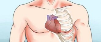
The heart is a muscle whose general function is to constantly pump blood through the body's circulatory system. It is located at the back of the sternum. Its apex is located above the diaphragm. When the heart beats, the muscles touch the chest wall, which can be felt by placing your hand on the left side of the chest.
The human heart is shaped like a pear. The apical or apical pulse is a heartbeat that is heard at the cardiac apex in the region of the left ventricle. This is one of the chambers of the heart that takes blood from the lungs, moves it to the aortic valve, then to the aortic arch, and the blood then flows through the rest of the body. It's a continuous cycle.
Heart rate is determined by the beat heard at the apex of the heart for one full minute. There are several reasons why this pulse is preferable to counting a detectable heartbeat from the wrist or neck. Some people experience irregular heartbeats that are not always transmitted to the arteries of the extremities.
Therefore, the pulse counted at the wrist may differ from the apical pulse.
The heart rate of young children and infants is estimated from the apical pulse because it is difficult to count the beat rate on the arms or legs of infants.
The apical impulse is normally a rhythmically pulsating protrusion of the intercostal space in the area of the cardiac apex onto the chest wall. It is created by tremors generated during systole. During diastole, the heart returns to its normal position. If the beat of the apex of the heart falls on the rib, then the push is invisible to the doctor.
The apical impulse can also be defined as a systolic protrusion of the space between the ribs. The point of the apical, apical pulsation can be found on the left side of the chest, just below the breast nipple. This position roughly corresponds to the lower pointed end of the heart.
Other ripples
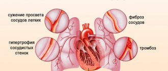
Diagnostically significant pulsations are the pulsation of the aorta, pulmonary artery and epigastric pulsation. The first of them is invisible normally. Pathological pulsation appears in the second intercostal space on the right at the edge of the sternum. The reasons for its occurrence include:
- shrinkage of the right lung;
- enlargement of the aorta (syphilis, aneurysm of the ascending aorta, aortic valve defects).
Pulsation of the pulmonary artery (II intercostal space to the left of the sternum) is the result of pulmonary hypertension with mitral valve defects.
Epigastric pulsation is found in the epigastric fossa. Reasons for its appearance:
- right ventricular prolapse;
- abdominal aortic aneurysm.
Properties normal and abnormal
The apex beat can be heard with your fingers or with a stethoscope. Both options are effective.
When listening to the pulse, you should pay attention to the speed of the heartbeat:
- A slow beat is normal for a person who is in good physical shape. But a slowdown may occur under the influence of certain medications.
- An increased pace in adults indicates that the person has high blood pressure or cardiac problems. It is also typical for people involved in physical exercise and young children.
Before palpation, the pulsation is examined by a doctor under direct and then lateral lighting. The angle of rotation during inspection changes to 90°.
The apical impulse provides a broad picture of cardiac activity to the physician and helps to establish the correct diagnosis.
| Character of the blow | ||
| Fine | Abnormal | |
| Short early (systolic) impulse, felt over an area of 2-3 cm² | Property of momentum | Possible violations |
| Hyperdynamic impulse (very short impulse of large amplitude) | Thyrotoxicosis, anemia, sepsis, vitamin deficiency, ventricular septal defect | |
| Pulsating impulse | Hypertension, aortic stenosis, | |
| Hypodynamic impulse (weak or absent apical impulse) | Myocardial infarction | |
All conclusions about the quality of the apical impulse are based in most cases on subjective analysis and many years of experience of doctors.
Positive and negative
The apical impulse is normally positive, as a rule. In rare cases it may be negative.
Under the influence of the formed pericardial adhesions, a symptom of a negative apical impulse is detected. This occurs when during systole the chest does not protrude at the site of the apical impulse, but is pulled inward.
In the zone of the V intercostal space at the moment of ventricular contraction, a vacuum is formed. As a result, the intercostal space is retracted and a negative apical impulse occurs.
With a positive push, a pulsating protrusion of the space between the ribs occurs.
In what cases is it not determined (not palpated)?
Some physical conditions interfere with the transmission of impulse from the apex to the chest wall:
- obesity;
- pericardial effusion;
- accumulation of fluid in the pleura;
- subcutaneous emphysema.
It is impossible to locate the apical impact site if there is spinal deformity, chest wall deformity, or tracheal deviation.
Why might it be displaced?
The apex beat is normally determined by a limited rhythmic beat. For some people, the impulse may have a diffuse pulsation, be stronger or weaker. The displacement of the apical impulse is influenced by changes in the heart or organs surrounding it.
| Pulse point | Reasons for displacement |
| Shift left | Mitral or aortic valve insufficiency, myocardial damage. Right-sided pleurisy, pneumothorax, hydrothorax, tricuspid valve insufficiency. |
| Offset right | Left-sided pneumothorax, hemothorax, left-sided exudative pleurisy, dextrocardia. |
| Deviation left and down | Aortic valve insufficiency (dilatation of the left ventricle). |
| Mixing towards the center | Displacement of the heart to the right due to left-sided hydrothorax, pneumothorax, pneumosclerosis. |
| Shift up and left | Occurs in conditions when the level of the diaphragm increases (flatulence, obesity, acidosis). |
Do not confuse thrust displacement with displacement. If the patient lies on the left side, then the apical pulsation shifts to the left by 3-4 cm. When lying on the right side, the displacement occurs to the right by 1.5 cm. In pregnant women with increased weight, the pulse moves to the left. For people with nicotine addiction - to the right.
A stable change in the location of the pulse, displacement, occurs due to internal disorders.
Width and area
The area of the apical impulse is determined by the width between II and IV and the fingers located at the outer and inner borders of the visible pulsation. The indicator for a healthy person does not exceed 2 cm. If the width is less than 2 cm, such a push is called limited.
An increase in area was noted among athletes and people engaged in heavy physical labor.
What affects the change in the area of the apex beat:
| Increased area | Reduced area |
| Myocarditis | Well developed muscles |
| Heart defects | Narrow intercostal spaces |
| Hypertension | Enhanced nutrition |
| Tumor of the posterior mediastinum | |
| Left ventricular dilatation |
The boundaries of the push are established at the 1st stage of palpation. The doctor gives a qualitative assessment during the 2nd stage of palpating the patient.
Spilled
A diffuse cardiac impulse can be felt in 2 or more intercostal spaces.
An enhanced apical impulse, the area of which is more than 2 cm² (spread), indicates hypertrophy of the right or both ventricles of the heart.
In case of enlargement of the left ventricle, the apical pulse is shifted downwards, has a local point, and is often strengthened.
Limited
A localized pulse is felt in the 5th intercostal space. The location is influenced by gender, human constitution, and the level of the diaphragm. In some women, the impulse is determined in the area of the IV intercostal space; in hypersthenics, it falls on the midclavicular line.
Height
A high apical pulse is determined by the amplitude of finger lifting and manifests itself in the following cases:
- accelerated heartbeat;
- high diaphragm position;
- deep exhalation, during which the cardiac apex moves closer to the chest wall;
- tumor of the posterior mediastinum;
- left ventricular hypertrophy.
Low shock occurs when:
- obesity;
- left-sided exudative pleurisy;
- pericarditis;
- deep breath;
- emphysema.
The apex beat normally has an average height. It is characteristic of normosthenics, people with a normal width of intercostal distances.
The range of oscillations of the intercostal space does not exceed 2-3 mm and depends on several factors:
- thickness of the chest wall;
- dynamics of myocardial contractility;
- hemodynamic type of state of the cardiovascular system.
Force
The strength of the apical impulse can be determined as follows:
- an increased push raises the finger;
- pulsating, of medium strength, easily identified, but the finger does not lift;
- a weak push is hard to find.
The strength of the pulsating impulse, as well as the area, depends on the location of the heart in the chest, but mainly on the contractile power of the left ventricle.
A pulsation of average strength is determined in a person with a moderate diet, standard physical development, and normal hemodynamics.
Intensified shocks occur with increasing speed and strength of contractile work of the left ventricle. It is observed more often in athletes, in people after emotional stress or workload. Pathological causes are associated with left ventricular hypertrophy, tumor of the posterior mediastinum, thyrotoxicosis, and shrinkage of the lung tissue.
Weak tremors can be detected in completely healthy people. These include patients with enhanced nutrition and powerfully developed muscles.
Pathologies affecting the decrease in thrust force:
- subcutaneous emphysema;
- obesity;
- left-sided pneumothorax;
- adhesive pericarditis;
- cardiosclerosis;
- heart defects.
The most alarming cause of decreased strength is a decrease in myocardial contractility.
If the doctor notices the simultaneity of changes in some parameters, then on this basis the following conclusions are drawn:
| Properties | Volume | Violation | Volume | Violation | Volume | Violation |
| Square | Increased | Normal myocardial contractility, muscle hypertrophy | Increased | Dilatation of the left ventricle, decrease in its functions | Reduced | Myocardial damage |
| Height | Increased | Increased | Reduced | |||
| Force | Increased | Reduced | Reduced |
The strength or weakness of the apical impulse is determined during a deep inspiration, and the patient should lie on his back.
Resistance
The presence of resistance is determined by the force of finger pressure that must be applied to “extinguish” the apical impulse. With high blood pressure, narrowing of the aortic mouth, obstacles arise for the flow of blood from the left ventricle to the aorta. This makes the apex impulse unyielding and resistant.
The pulse may be moderate when the tissue is perceived as pliable upon palpation. The impulse is called resistant if the tissue is dense, which occurs with left ventricular hypertrophy. The pulsation can become dome-shaped when the area and force of the push are increased. More often this occurs with aortic defects.
Other types of unwanted vibrations
In the case when the cardiac impulse becomes diffuse and affects other areas, such as the armpit, epigastric region, it becomes widespread. This condition is characteristic of heart disease.
In cardiology, the symptom of “cat purring” is also known. This occurs due to trembling of the chest wall. It is determined by digital or palmar palpation and is caused by low-frequency fluctuations in the blood flow that passes through the narrowed opening of the valve.
The detected sign is compared with the pulse in the carotid artery. If the beats coincide, then a systolic “purr” is detected. If the apical impulse and the blow on the carotid artery do not coincide, then a diastolic “purr” is noted.
Systolic tremors are characteristic of narrowing of the pulmonary artery. Diastolic manifests itself in stenosis of the right atrioventricular orifice, which is a rare defect.
Palpation of the heart area
Palpation of the heart area makes it possible to better characterize the apex beat of the heart
, detect a heartbeat, evaluate visible pulsation or detect it, detect tremors of the chest (symptom of “cat purring”).
To determine the apical impulse of the heart, the right hand, palmar surface, is placed on the left half of the patient’s chest in the area from the sternal line to the anterior axillary between the 3rd and 4th ribs (in women, the left mammary gland is first retracted up and to the right). In this case, the base of the hand should be facing the sternum. First, the push is determined with the entire palm, then, without lifting the hand, with the flesh of the end phalanx of the finger placed perpendicular to the surface of the chest (Fig. 38).
Rice. 38. Determination of the apex impulse: a - palmar surface of the hand;
b - the end phalanx of the bent finger.
Palpation of the apical impulse can be facilitated by bending the patient's torso forward or by palpation during deep exhalation. In this case, the heart is more closely adjacent to the chest wall, which is also observed in the position of the patient on the left side (in the case of turning on the left side, the heart shifts to the left by about 2 cm, which must be taken into account when determining the location of the push).
When palpating, pay attention to the localization, extent, height and resistance of the apical impulse.
Normally, the apical impulse is located in the 5th intercostal space at a distance of 1-1.5 cm medially from the left midclavicular line. Its displacement can cause an increase in pressure in the abdominal cavity, leading to an increase in the position of the diaphragm (during pregnancy, ascites, flatulence, tumors, etc.).
In such cases, the impulse moves up and to the left, as the heart turns up and to the left, taking a horizontal position. When the diaphragm is low due to a decrease in pressure in the abdominal cavity (with weight loss, visceroptosis, emphysema, etc.
) the apical impulse moves downward and inward (to the right) as the heart turns downward and to the right and takes a more vertical position.
An increase in pressure in one of the pleural cavities (with exudative pleurisy, unilateral hydro-, hemo- or pneumothorax) causes a displacement of the heart and, consequently, the apical impulse in the direction opposite to the process.
Wrinkling of the lungs as a result of the proliferation of connective tissue (with obstructive pulmonary atelectasis, bronchogenic cancer) causes a displacement of the apical impulse to the painful side.
The reason for this is a decrease in intrathoracic pressure in the half of the chest where the shrinkage occurred.
As the left ventricle of the heart enlarges, the apical impulse shifts to the left. This is observed with bicuspid valve insufficiency, arterial hypertension, and cardiosclerosis.
With aortic valve insufficiency or narrowing of the aortic opening, the impulse can shift simultaneously to the left (up to the axillary line) and down (to the VI - VII intercostal space). In the case of dilation of the right ventricle, the impulse may also shift to the left, since the left ventricle is pushed by the dilated right ventricle to the left.
With a congenital abnormal location of the heart on the right (dextracardia), the apical impulse is observed in the 5th intercostal space at a distance of 1-1.5 cm inward from the right midclavicular line.
With pronounced effusion pericarditis and left-sided exudative pleurisy, the apex beat is not detected.
The normal distribution (area) of the apex beat is 2 cm2. If its area is smaller, it is called limited; if it is larger, it is called diffuse.
Limited apical impulse
noted in cases where the heart is adjacent to the chest with a smaller surface than normal (occurs with emphysema, with a low diaphragm).
Spilled apical impulse
usually caused by an increase in the size of the heart (especially the left ventricle, which occurs with insufficiency of the mitral and aortic valves, arterial hypertension, etc.
) and occurs when it is mostly adjacent to the chest. A diffuse apical impulse is also possible with wrinkling of the lungs, high standing of the diaphragm, with a tumor of the posterior mediastinum, etc.
Apex beat height
characterized by the amplitude of vibration of the chest wall in the region of the apex of the heart.
There are high and low apical impulses, which are inversely proportional to the thickness of the chest wall and the distance from it to the heart.
The height of the apical impulse is directly dependent on the strength and speed of heart contraction (increases with physical activity, anxiety, fever, thyrotoxicosis).
Apex beat resistance
determined by the density and thickness of the heart muscle, as well as the force with which it protrudes the chest wall. High resistance is a sign of hypertrophy of the left ventricular muscle, no matter what causes it.
The resistance of the apical impulse is measured by the pressure it exerts on the palpating finger and the force that must be applied to overcome it. A strong, diffuse and resistant apical impulse upon palpation gives the sensation of a dense, elastic dome. Therefore, it is called a dome-shaped (elevating) apical impulse.
Such a push is a characteristic sign of aortic heart disease, i.e. insufficiency of the aortic valve or narrowing of the aortic opening.
Heart beat
palpated over the entire palmar surface of the hand and is felt as a shaking of the chest area in the area of absolute dullness of the heart (IV-V intercostal space to the left of the sternum). A pronounced cardiac impulse indicates significant hypertrophy of the right ventricle.
The symptom of “cat purring” is of great diagnostic importance.
: The trembling of the chest resembles the purring of a cat when stroking it. It is formed when blood quickly passes through a narrowed hole, resulting in vortex movements that are transmitted through the heart muscle to the surface of the chest.
To identify it, you need to place your palm on those places in the chest where it is customary to listen to the heart.
The sensation of a “cat’s purr”, detected during diastole at the apex of the heart, is a characteristic sign of mitral stenosis; during systole in the aorta - aortic stenosis; in the pulmonary artery - pulmonary artery stenosis or patent ductus arteriosus.
In English:
Source: https://www.plaintest.com/circulation/palpation
Normal indicators in children and adults by age
Table of apical pulse indicators:
| Age group | Readings are normal |
| Newborns | 80 – 140 beats/min |
| Children from 4 to 9 years old | 75 – 120 beats/min. |
| Children under 15 years old | 50 -90 beats/min |
| Adults | 60 – 100 beats/min. |
The apex beat is normal in an adult if it does not exceed 100 or not lower than 60 beats/min. The ideal heart rate at rest and during physical activity varies from person to person.
Reasons why your heart rate may increase:
- fear or anxiety;
- heat;
- recent physical activity;
- pain;
- hypotension;
- blood loss;
- insufficient oxygen consumption.
Additionally, a heart rate that is consistently higher than normal may be a sign of heart disease, heart failure, or an overactive thyroid gland. The decrease is affected by medications that can affect the heart rate.
Palpation technique
The right hand is placed in the projection of the intended impulse, between the 3rd and 6th ribs in the area of the apex of the heart. The pulsation is determined over the entire palmar surface, and then localized with the tip of the index finger.
It must be installed perpendicular to the chest. If the pulsation is widespread, its leftmost and lower region is determined. This point is the site of the cardiac impulse. By the way, they choose the place where the protrusion of the chest is determined by the flesh of the end phalanx of the palpating finger, and not by its lateral surfaces.
DETAILS: Definition and classification of blood pressure levels
If it is difficult to feel the apex beat of the heart due to the characteristics of the chest, then palpation is performed with the chest tilted forward, or the patient is placed on the left side. The heart muscle in these positions fits tightly to the chest and pushes back the edge of the left lung.
In a position on the left side, the cardiac impulse falls lower and to the left by 2 cm, so the place of the impulse is taken to be the intercostal space where the contraction is determined, but 2 cm medial from the area of the impulse. Palpation of the apex impulse during exhalation increases the chances of determining its location, because at the moment the diaphragm rises, the heart, making a pendulum-like movement to the left and up, moves to a more horizontal position, moving the edge of the left lung.
Doctors determine certain properties of the heart impulse:
- location;
- resistance;
- prevalence;
- height.
Step-by-step palpation algorithm
Palpation of the apical pulse is carried out in 2 stages. To do this, landmarks on the body are used.
They include:
- bony point of the sternum;
- intercostal spaces;
- the midclavicular line (an imaginary line running down the body from the middle of the collarbone).
Stage I:
- The doctor stands to the right of the patient. The right hand is placed on the patient's chest. The base of the hand rests on the sternum, and the fingers cover the area from the III to VI ribs.
- The doctor presses the hand to the chest, bending the II, III, IV fingers, looking for the place of vibration of the intercostal space.
If the pulsation is not detected, the patient should lean forward 50°. This is done to bring the heart as close to the chest as possible. Before bending over, you need to exhale.
At the 2nd stage of palpation, the doctor determines the location of the push, type, area, strength, compliance, height.
Palpation technique:
- The doctor turns his hand with his fingers upward at the site of the found pulsation. The phalanges of the fingers should be located along the intercostal space.
- Slowly moving the fingers, slightly plunging them into the intercostal space, the doctor assesses the quality of the pulse.
The apex beat can be seen in only 50% of people. In others, it is hidden by a layer of fat, muscle, distance of the heart from the chest, or is affected by emphysema, which often accompanies older patients. Pulsation is clearly visible in asthenics, normosthenics and people with a thin fat and muscle layer.
Visual inspection and palpation
Visual inspection and palpation are only suitable for detecting the apex beat. It occurs when the left ventricle and interventricular septum move toward the chest.
Additional heart beats are possible with pathological changes in the ventricles, atria and large vessels. It is important to systematically detect these symptoms.
Visual examination of the patient is the initial stage of the study of cardiac activity and in some cases is more effective than palpation.
It is necessary to direct a beam of light tangentially to the expected place of pulsation, which will facilitate the best detection of tremors during cardiac activity.
It should be taken into account that with some features of the human body, visual observation of the push may be absent, for example:
- overweight;
- small spaces between ribs;
- developed muscles;
- mammary glands are large or contain implants.
In people of asthenic physique, vibrations are most noticeable.
Palpation is a diagnostic method carried out by palpating tissues and organs with the hands. The examination has no contraindications and is used for all categories of patients
After the examination, they move on to the palpation method, which includes the following steps:
- The location of the right hand in the area of the intended push (between the 3rd and 6th ribs in the area of the upper part of the organ).
- Initially, the pulsation is determined by the entire palm with further localization by the index finger (pad).
- Widespread pulsation involves identifying its leftmost area at the bottom. This is where the push actually appears.
The most effective palpation is when the patient bends over or lies on his left side after a deep exhalation. If a person is positioned on the right side, the left lung moves the heart away from the chest, making it impossible to hear the beats.
Examination of women involves elevation of the left breast.
As a result, under the finger there is a feeling of rhythmic vibrations that occur when the left ventricle hits the chest.
Pay attention to:
- Skin color (normal/pale/cyanotic)
- The presence of pulsation of the carotid arteries, carotid dancing (dilation and constriction of the pupils, as well as slight nods of the head in time with the pulsation)
- The presence of swelling of the jugular veins (may be a normal variant in children when moving to a horizontal position)
- Presence of pulsation of the jugular veins (Pathological phenomenon. It can be transmitting or be a “true venous pulse” - the latter disappears when the veins are compressed above the place of compression)
- dilatation of other saphenous veins
- The shape of the chest - the presence of a cardiac hump (protrusion in the projection of the heart)
- Apex beat intensity
- Presence of a heartbeat
- The severity of epigastric pulsation
- The presence of edema in the legs (“cardiac edema”), in the sacral area
- Presence of finger deformities (“drumsticks”)
DETAILS: Calcitonin hormone - what is it and what to do when it is elevated?
Norm of calcitonin in women. The apical impulse is the rhythmic protrusion of the chest in the projection of the apex of the heart. Normally, it can be invisible to the eye or visible (the latter is more common in asthenics). The apical impulse is based on left ventricular systole.
There is also the concept of “negative apical impulse” - during systole, the chest does not protrude, but retracts. This is a pathological phenomenon.
Cardiac impulse - protrusion of the chest involving the sternum and epigastrium (shaking in systole). It is based on right ventricular systole. This impulse is normally absent and is detected only with right ventricular hypertrophy.
Deformation of the fingers and toes in the form of “drumsticks” (expansion of the distal phalanges), nails in the form of “watch glasses” (convex, like glass in a watch) are a characteristic sign of chronic heart failure^ Palpation of the pulse The study is traditionally carried out on the radial artery, but for more For an objective assessment, the pulse must be examined in several areas.
^ Pulse on the radial artery The patient's hand is grasped by the doctor's palpating hand in the area of the wrist joint. The patient's hand is relaxed, the arm is bent so that the palpated artery is located at the level of the heart.
https://www.youtube.com/watch?v=upload
The doctor positions his hand so that the palmar surface of his hand is on the back of the patient's hand. Three fingers (index, middle and ring) are placed in the projection of the radial artery.
The study begins by determining the uniformity of the pulse. To do this, both hands are grabbed simultaneously in the manner described. The heart rate is compared. If it is the same, then all further research continues on one hand (any).
The following pulse characteristics are determined sequentially:
- Sameness (same on both hands / not the same)
- frequency (normal: 60-80 beats per minute)
- rhythmicity (rhythmic / arrhythmic)
- voltage (satisfactory / low)
- filling (satisfactory / low)
- heart rate deficit
- sometimes form
Heart rate is simply the number of pulse beats per minute. You should strive to measure the frequency exactly in a minute, and not in 15 - 30 seconds, followed by multiplication by 4 and 2, respectively. This is especially true in children and adolescents, where pulse lability is characteristic with a change in heart rate within a minute.
Rhythm - equality of intervals between pulse beats. If the intervals are equal, the pulse is rhythmic. Normally, there is some respiratory arrhythmia - expiratory bradycardia. However, upon objective examination it is usually not noticeable.
Tension is the force that must be applied by the fingers of the palpating hand to stop the pulsation of the radial artery below the compression. It is examined as follows: all three palpating fingers are involved.
Using the ring finger, gently press on the radial artery, trying to stop its pulsation. The middle finger directly “palpates” - registers the cessation of pulsation of the artery wall. The index finger, located distally, compresses the artery to prevent the spread of the pulse wave from other arteries (palmar arch).
At the end of this study, the wall of the vessel is examined with the middle finger (without taking away the rest) - simply with a transverse movement (palpation). Normally, the vessel wall is not palpable (i.e., not compacted).
Filling is the force and speed with which blood fills an empty vessel. It is examined immediately after determining the pulse voltage. To do this, the ring finger (which was squeezing the artery) is removed, and the filling of the artery is recorded with the middle finger.
Pulse deficiency is a pathological condition when not every heartbeat corresponds to a pulse wave. It is determined by simultaneous palpation of the pulse and heart (you can simply place your hand on the heart area or the carotid artery). Normally there is no pulse deficit.
Sometimes the shape of the pulse is also examined (assessment of the rate of rise and fall of the pulse wave), however, this study requires significant skill and many years of experience, and is therefore less often used in everyday work.
However, some pulse shapes are described below: regular pulse, pulsus celer (rapid rise and fall of the pulse wave), pulsus tardus (slow rise and fall); pulsus altus (fast satisfactory filling but rapid decline), pulsus parvus (weak and slow filling and slow decline) are distinguished separately. Combined options are also possible - pulsus celer et altus, pulsus tardus parvus, etc.
The conclusion about palpation of the pulse in a healthy person should look like this: the pulse is the same in both arms, 72 beats per minute, rhythmic, satisfactory tension and filling, the vascular wall outside the pulse wave is not palpable, there is no pulse deficiency.
^ Pulse on the femoral artery is examined in the vertical and horizontal position of the patient. Palpation is carried out with two fingers (index and middle), in the area of the middle of the inguinal fold (where the a. femoralis comes out from under the Poupart ligament). Only the presence of a pulse and its frequency are assessed.
In addition to the above, pulse palpation can also be performed on other large arteries, such as:
- temporal artery
- carotid artery
- axillary artery
- subclavian artery
- posterior tibial artery
- artery of the dorsum of the foot
- and others.
However, for most of these vessels it is not possible to evaluate all pulse characteristics. Most often, palpation of these peripheral arteries is used either if it is impossible to reach the radial artery, or simply to detect the presence of pulsation (for example, if thromboembolism is suspected).
Begin with palpation of the heart area. The patient's position is supine. The doctor's palm is placed on the right half of the chest, in the projection of the heart. At this stage, palpation equivalents of noise (such as systolic tremor, etc.) can be excluded.
The doctor's palm is placed on the right half of the chest, in the projection of the heart, with the fingers directed proximally. This allows you to roughly determine the location of the apical impulse (normally this is the 5th intercostal space, less often the 4th).
Then it is advisable to rotate the palm 90 degrees, so that the fingers are directed to the left side, and the palm to the sternum, and more accurately determine the location of the push. In the area of detected pulsation (usually slightly to the side of the midclavicular line of the 5th intercostal space), the pads of three fingers (index, middle and ring) are placed and the shock is localized even more accurately.
Then they move on to its description, which includes the following points:
- localization
- sizes (spilled / not spilled)
- strength (moderate / weakened / enhanced / lifting)
- sometimes - height
— projection of the apical impulse. Indicated by two coordinates: intercostal space and midclavicular line.
- the area of its weakening (since the apex impulse is well carried out on the anterior chest wall, its area is understood as the area where it has the same force. This applies to both horizontal boundaries (within the intercostal space) and vertical boundaries (how many intercostal spaces the impulse falls ).
What is a cardiac impulse?
The shaking of the anterior chest wall caused by the contraction of the heart is called a cardiac impulse. It occurs synchronously with ventricular systole.
In the tension phase, the shape and size of the heart changes, which makes it possible for the surface of the ventricles to come into contact with the chest wall. The study of the cardiac impulse is carried out using inspection, palpation, and a sound signal.
Prevalence of cardiac pulsation
The area of protrusion of the cardiac impulse is about 2 cm². If it turns out to be larger, then they speak of a diffuse or widespread shock. With a smaller area it is limited.
Widespread pulsation occurs if the heart's larger surface is adjacent to the chest wall. This is observed:
- with a deep breath;
- pregnancy;
- for tumors of the mediastinum, etc.
In the absence of these conditions, a diffuse impulse may be the result of expansion of the heart (all or any of its parts).
A limited cardiac impulse occurs when the heart has a smaller area adjacent to the chest. The reason for this may be:
- emphysema;
- low aperture;
- exudative pericarditis;
- hydro-, pneumopericardium.
Determination algorithm
The heartbeat is palpated as follows:
- The doctor's hand is placed on the precordial area, the fingers are at the level of the apical impulse, and the palm is at the right edge of the sternum, then palpation is performed. This is how the precordial region is assessed.
- Having assessed the condition at this level, the doctor’s hand moves to the third rib and palpation is also carried out here.
Thus, the doctor, with an existing cardiac impulse, distinguishes between the vibration of the lower part of the sternum, the pulse at the edges of the sternum and the epigastric pulsation.
Other types of unwanted vibrations
The occurrence of a resistant cardiac impulse is associated with aortic defects or hypertension. Upon palpation, the specialist feels a dense, thick muscle. If the pulsation is characterized by hyperdynamicity and an increased area, we can talk about a dome-shaped apical impulse.
The occurrence of pulsation of large main vessels is observed in the second intercostal space on the right and left sides of the sternum and in the area of the jugular notch may indicate pathologies of the pulmonary artery and thoracic aorta.
Also, the transmission of rhythmic contractions of the heart to the abdominal aorta (pulsation in the epigastric region) should not normally be detected.
Diseases detected by palpation, features of apical impulses in pathologies
The most common disorder that is revealed by palpation is an increase in the volume of the left ventricle, when the pressure in the systemic circulation steadily increases. This is affected by alcohol abuse and physical overload. This is an acquired pathology that is eliminated over time. But it is possible to identify other violations, more serious.
For hypertension
In the early stages, hypertension is not clearly expressed. However, even during this period, symptoms can be identified: headache, heart pain, vascular neurosis. The development of the disease is influenced by left ventricular hypertrophy. On palpation, an enhanced apical impulse is detected.
Other properties of apical pulsation:
- push is positive;
- spilled;
- resistant;
- domed in some cases;
- has a shift to the left and down.
With ventricular hypertrophy
This is a condition in which the muscles of the left ventricle thicken, often associated with uncontrolled hypertension. The process is not permanent, but in some cases can lead to heart failure. When the underlying problem is treated, the thickened ventricle may shrink in size over time.
With hypertrophy, the force of the apical impulse increases, a displacement occurs to the anterior axillary line, and down to the 6–7 intercostal space. The pulsation becomes diffuse, high, and takes on a dome-shaped shape.
For coronary heart disease
Insufficient supply of the heart muscle with oxygen and nutrients leads to myocardial damage. The main factor in the development of the disease is atherosclerosis of the coronary arteries, which reduces the blood supply to the heart muscle.
Upon visual examination of the heart area, the apex beat is not detected.
Upon palpation, the doctor determines:
- positive apical impulse;
- palpation of the pulse is carried out in the 5th intercostal space towards the outer side of the left midclavicular line;
- pulsation diffuse, low, resistant;
- shock area 3 cm².
Listening to the apical impulse is the most effective way to assess cardiac activity. If your pulse is outside the normal range or there is an irregular heartbeat, you should definitely visit a cardiologist.
Author: Belyaeva Anna
Interpretation of results
What can palpation of the apex beat tell you? For an experienced doctor who has the skills to physically examine a patient and has discovered, for example, a weakened apical impulse, it will not be difficult to associate this symptom with the presence of effusion pericarditis , characterized by the accumulation of fluid in the cavity of the heart sac, or pericardium. In this case, the vibrations caused by heart beats are simply not able to pass through the layer of fluid and are felt as a push of weak force.
In the case when a doctor diagnoses a diffuse apical impulse, he may think about the presence of left or right ventricular hypertrophy . Moreover, an increase in myocardial mass is likely if there is a displacement of the impulse to the right or left. Thus, with left ventricular hypertrophy, the impulse shifts to the left. This is due to the fact that the heart, increasing in mass, must find a place for itself in the chest cavity and it will shift to the left side. Accordingly, the apex of the heart, creating a push, will be determined on the left.
Thus, palpation of the heart, when performed by an experienced doctor, can bring undoubted benefit to the patient, since during a routine examination the doctor is able to suspect any disease and promptly refer the patient for further examination using instrumental diagnostic methods.
What diseases can be suspected by palpation of the heart?
Palpation of the apex and cardiac impulse, which differs in characteristics from the norm, as well as the determination of pathological tremors and pulsations of the heart, can be caused by the following diseases:
- Congenital and acquired heart defects, which cause disruption of the normal architecture of the heart and sooner or later lead to the formation of myocardial hypertrophy, Long-term arterial hypertension, which is especially difficult to treat and reaches high blood pressure numbers (180-200 mm Hg),
- Aneurysm of the thoracic aorta,
- Pericarditis, especially with the accumulation of a large amount of fluid in the cavity of the pericardium,
- Diseases of the bronchopulmonary system, adhesions in the pleural cavity, adhesive pericarditis,
- Diseases of the abdominal cavity with an increase in its volume - ascites (accumulation of fluid in the abdominal cavity), tumor formations, late pregnancy, severe bloating.
For example, if a negative apical impulse is detected in a person being studied, which looks like a retraction of the intercostal space in the area of the impulse, the doctor should certainly think about adhesive pericarditis, in which the pericardial layers “solder” to the inner surface of the chest. With each contraction of the heart, the intercostal muscles are pulled into the chest cavity due to the formed adhesions.