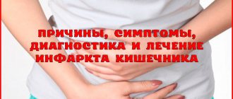
Intestinal necrosis is a condition in which tissue begins to die and lose its properties.
This process is most often not reversible and, if tissue necrosis has already occurred, then it will not be possible to restore the lost area. Therefore, such a pathology must be treated in the early stages in order to be able to save the person. The causes of necrosis are varied, and can be a consequence of a previous illness or an independent factor that has developed for its own reasons.
Types of necrosis
The intestines can be affected in different ways depending on what the necrotic area looks like, the location of necrosis, and the amount of dead tissue. Therefore, the following types of necrosis are distinguished:
| Classification | Examples |
| According to the degree of damage (how much space the necrotic area takes up) | Local - when only one part of any intestine is affected and necrosis does not spread to neighboring parts of the intestinal tract. Total - complete damage to the rectum, small and large intestines occurs, and part of the stomach may even be affected. |
| According to etiological factors (depending on what caused necrosis) | Ischemic - ischemia or infarction of the intestines occurs due to blockage of the vessels delivering blood to the intestines. If blood does not circulate for a long time, gangrene and even peritonitis can develop, when part of the small or large intestine is destroyed so much that all its contents enter the abdominal cavity, causing inflammation. Toxigenic - rotaviruses, coronaviruses, Candida fungi, clostridia infect the intestinal tract, causing necrosis of its tissues. Trophoneurotic - malfunctions in the functioning of the nervous system lead to improper innervation of the intestinal vessels, and therefore to necrotization of its areas. |
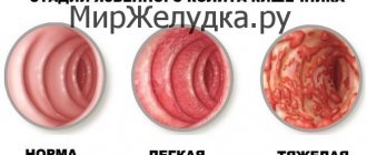
| According to clinical signs (how the disease manifests itself in development, each type can flow into the next, reflecting the degree of neglect of the disease) | Coagulative necrosis, or dry necrosis, develops due to dehydration associated with arterial insufficiency, which leads to the drying out of the intestinal mucosal wall and its separation from healthy areas. Liquation, or wet, is the next stage of dry necrosis. This stage is characterized by the proliferation of putrefactive microflora in those parts of the intestine that have already undergone necrosis. After it, gangrene often develops if medical assistance was not provided in a timely manner. Strangulation necrosis is most often caused by intestinal obstruction associated with obstruction of feces or the presence of a foreign body in the intestine. Also, the cause of this necrosis is a tumor that compresses the intestines from the outside, preventing blood from circulating normally. Thrombosis of mesenteric vessels and narrowing of the intestinal lumen can also cause this. Gangrene can form at any time during the development of necrosis. The dry form of gangrene is characterized only by impaired blood circulation, but the wet form leads to stasis of the veins and lymphatic capillaries, as well as the appearance of swelling. |
Video
How to determine intestinal necrosis: its symptoms and causes
Necrosis is the death of tissue of an organ.
This phenomenon is irreversible in the later stages of pathology development. What are the causes of the disease? Perhaps the most important of them is poor circulation in the body. The cause of stopping blood flow can be thrombosis; the vessels become clogged with a blood clot, which leads to cell death due to a complete lack of oxygen. The most common cause of clogged blood vessels is heart disease, which can develop in women aged seventy or older.
Currently, intestinal necrosis is found in 10% of patients under thirty years of age. The total form of the disease appears due to poor blood circulation in the intestine and in half of the cases can cause death.
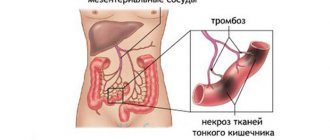
The mesentery supplies blood to both intestines, so when thrombosis of the mesenteric vessels occurs, the entire gastrointestinal tract suffers.
Do not forget that both necrosis of the large intestine and necrosis of the small intestine are possible.
Intestinal necrosis, causes
The cause may be infection with microbes. Most often, newborns have this disease.
The disease affects the intestinal mucosa; in this case, necrosis is not total, but if you ignore the symptoms of the disease, the disease will begin to damage the entire intestinal wall. Necrotizing enterocolitis is caused by intestinal fungi rotaviruses.
There is a rapid development of pneumatosis and intestinal gangrene. This disease can lead to the death of a person. It also affects people with nerve center disease.
Intestinal necrosis, symptoms
With this disease, the intestinal walls change smell and color. During a heart attack, the tissue walls become saturated with blood. As a result, the intestines take on a burgundy hue.
Symptoms of a disease directly depend on its cause.
Necrosis, which appears as a result of intestinal infarction, is characterized by initially intense pain in the abdominal area; with further development of the disease, the pain becomes constant.
When changing body position, the pain does not stop. The number of leukocytes in the blood increases, and when examining the abdomen, patients complain of severe pain in the necrotic zone. By palpation, the surgeon detects the affected part of the intestine.
If disturbances occur in the veins, then the symptom will be an increase in the temperature center, as well as discomfort in the abdominal area. Specialized diagnostic methods are prescribed:
- X-ray;
- laparoscopy;
- aortography.
Symptoms may include pain in the abdomen, a gag reflex, and penetration of intestinal contents into the stomach. There is no stool, but gas is present, and bloating occurs.
With symptoms of peritonitis, the patient’s general condition worsens:
- the skin takes on a grayish color;
- tachycardia appears;
- pressure decreases.
There are three stages of the disease
- Pre-necrosis. It is possible to detect the disease at the initial stage, in which case the cells can be restored.
- The process of tissue cell death. The color of the gastrointestinal tract changes. The cells gradually die.
- Tissues disintegrate.
At the initial stage, the disease is detected during a radioisotope study. A special substance is injected into the blood vessels, and after some time a study is carried out. In the area of the intestine affected by necrosis, blood circulation is stopped, which is why in the image the affected area takes on the appearance of a “cold” spot.
Treatment of intestinal necrosis
You can get rid of intestinal necrosis if you consult a doctor at an early stage. Regardless of the cause of cell death, urgent consultation with a surgeon is required. The patient is sent for an x-ray of the gastrointestinal tract or irriography (radiography, which is introduced into the intestines using an enema).
If the patient has no symptoms of peritonitis, the course of treatment is carried out by administering antibiotics and protein solutions. Prescribe washing of the digestive tract using probes. If there is no effect of antibiotic treatment, the patient is prescribed surgery. The part of the intestine that is affected by necrosis is removed.
Resection of the small intestine is performed much less frequently, but it is necessary for necrosis resulting from obstruction. During the operation, a part of the artificial passage is placed, which is necessary to unload the colon. Surgery should occur within the first 24 hours of intestinal damage.
Patients whose lives were saved by surgeons face severe difficulties during the recovery period after surgery due to the consequences of the disease.
With a disease such as intestinal necrosis, the prognosis after surgery is disappointing; every patient, despite the operation being performed after the control period, dies.
To eliminate intoxication after surgery, an anesthetic drug and antibiotics are injected into the patient’s blood to prevent complications.
Prevention
Recommendations are made when the causes of necrosis are identified. You need to try not to get food and drug poisoning, eat right, add more fruits and vegetables to your diet, and exclude solid foods. Avoid fatty and sweet foods. Quit smoking and drinking alcohol. Start treatment for various diseases of the digestive tract and central nervous system in a timely manner.
To deliver missing nutrients to the body, doctors prescribe parenteral nutrition.
Pay special attention to your health, pay attention to painful sensations, do not treat at home and do not delay seeking help from medical specialists.
Everyone should know what intestinal necrosis is, its symptoms and principles. This could save the life of your loved one or yourself. Take care of your health and be happy!
, please select a piece of text and press Ctrl+Enter.
Source: https://lechimzhivot.ru/kishechnik/zabolevaniya-lechenie/kak-opredelit-nekroz-kishechnika-ego-simptomy-i-prichiny.html
Causes
The following factors can cause intestinal necrosis:
- Intestinal obstruction, which is caused by prolonged accumulation of feces due to twisting of the intestines. The small intestine is less likely to suffer from such pathology than the large intestine. With significant physical activity, the large intestine can be severely compressed, which will block blood flow.
- Disturbances in the functioning of the central nervous system, which cause destruction of the intestinal walls.
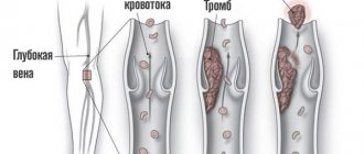
- Poor circulation in the intestinal walls can be caused by thrombosis (thrombi form in the intestinal vessels themselves, or migrate from other organs) or embolism (air entering the bloodstream).
- Damage to the intestinal tract by pathogenic microorganisms often causes necrosis in babies (especially infants). Their weakened body cannot fight infection, and therefore bacteria and viruses begin to destroy the intestinal walls very quickly.
- The body's allergic response to the presence of foreign bodies can cause necrosis.
- Chemical poisoning can also provoke necrosis of the tissues of the intestinal tract.
- When operations are performed on the stomach, the consequence (complication) may be that the part of the intestine closest to the stomach begins to die.
Causes of necrosis and symptoms of damage to the intestinal walls
Intestinal necrosis is a surgical pathology, that is, requiring immediate surgery. The danger lies in the fact that this anomaly can often be mistaken for poisoning with low-quality products, an upset stomach, or an exacerbation of chronic diseases.
Intestinal necrosis is a dangerous condition that can be fatal
What is pathology
Intestinal necrosis is the progressive destruction and death of organ cells, accompanied by gangrene. Decomposition products affect well-being, and inflammation spreads to neighboring organs. The pathology is secondary.
Strangulation type of necrosis develops due to acute intestinal obstruction
The process is irreversible, the entire functioning of the gastrointestinal tract gradually deteriorates, and dysfunction of the atrophied area is observed. Which is manifested by characteristic symptoms. There are three generally accepted classifications of the disease, as reflected in the table.
SignTypeDescription
| By area | Local | Limited localization of one section of the intestine, the lesion does not affect other sections. |
| Total | Complete damage to the large and small intestines gradually spreads to the stomach. | |
| By stage of development | Dry | Drying and separation of the mucous membrane due to dehydration. |
| Wet | Bacteria are actively multiplying. Without treatment, gangrene begins. | |
| Strangulational | Due to the resulting dysfunction, an obstruction forms, the deformation puts pressure on the organ, pinching the arteries. |
Another classification is due to the development of pathology: ischemic, toxigenic, trophoneurotic.
Reasons for development
The cause of intestinal necrosis is a heart attack, that is, a stop in the flow of blood, and therefore oxygen and nutrients.
This in turn may be caused by:
- volvulus and obstruction (the large intestine is more susceptible);
- a disorder of the central nervous system (the enteral system is directly subordinate to it);
- thrombosis or penetration of air into the vessels (can migrate from other organs);
- the activity of pathogenic microflora, necrotizing enterocolitis, provoked by candida, rotaviruses, coronaviruses (more common in children under one year of age, since due to weak immunity, bacteria multiply rapidly);
- foreign body (destruction - immune reaction);
- chemical poisoning;
- previous gastric surgery;
- mesenteric infarction (71% of deaths, since necrosis immediately affects the large and small intestines).
Volvulus can be caused by overeating, eating heavy (undercooked) food, or overexertion (lifting a load, jumping). Thrombosis can be provoked by peritonitis, malignant and benign neoplasms, obstruction, trauma, taking oral contraceptives, and cardiovascular disorders.
You will learn about intestinal obstruction by watching the video:
Symptoms of deviation
Symptoms often occur at a stage when the process has progressed greatly and immediate medical attention is needed. Symptoms of intestinal gangrene include:
- abdominal pain;
- sudden noticeable weakness;
- hyperthermia;
- hypotension combined with tachycardia;
- dry skin and mucous membranes;
- frequent trips to the toilet (not always with defecation);
- thirsty;
- loss of appetite and weight loss;
- vomiting (at a late stage with blood);
- nausea;
- streaks of blood in the stool.
Constant thirst accompanies the development of intestinal necrosis
Symptoms may not all appear at once (depending on the cause). Thus, with a heart attack that affects the arteries, acute pain occurs (constant, independent of body position), nausea, and vomiting. If the veins are blocked, then the pain is vague and hyperthermia is observed.
When volvulus occurs, pain, nausea, foul-smelling vomiting (the contents reflux inside), flatulence (gases easily escape), constipation, and asymmetric swelling of the abdomen are noted.
If the inflammation spreads to the peritoneum, then hypotension with tachycardia occurs, the skin acquires a gray tint.
For the first six hours, acute pain is observed, but a soft and painless abdomen, diarrhea, vomiting, and nausea remain.
Gradually, intestinal contractions decrease, the pain weakens, but overall health is rapidly depressed, the skin dries out and turns pale, and the tongue is dry and coated when examined. Fluid accumulates in the peritoneum.
MRI is one of the methods for diagnosing intestinal necrosis
The complete disappearance of pain is a bad sign, signaling the death of cells. Symptoms of dehydration and intoxication are rapidly increasing. Over time, the person becomes completely exhausted.
Complications and consequences
The most dangerous complication of rectal necrosis (large or small, as well as total type) is death. Without medical help it is inevitable. As a result of necrosis, severe intoxication and dehydration occur, which is dangerous due to shock, central nervous system depression, convulsions and fainting, and coma.
Diagnosis of the condition
Diagnosis begins with examination of the patient, palpation, and history taking. Dead areas are characterized by excessive softness and pain when pressed. The affected part is swollen, which can also be detected by palpation.
A blood test will help establish the correct diagnosis
After hospitalization, an X-ray is performed in the hospital (the picture shows a “cold” spot - the affected area), MRI, radioisotope scanning (the early stage can only be recognized in this way), ultrasound (allows you to evaluate neighboring organs and nearby tissues, for example, to identify necrosis of adipose tissue in the intestine, as well as blood flow speed), colonoscopy, irrigography (administration of contrast through an enema and subsequent x-ray).
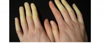
Instrumental studies make it possible to assess the condition of the intestine and visualize it. Dead tissue is usually white or yellowish in color.
Depending on the stage of the lesion, the tissue changes: from normal color, but structural disturbances, to discoloration and decay (usually accompanied by peritonitis).
In the early stages, blood thinning drugs are used for therapy
Selective mesentericography and angiography will help to identify the characteristics of the blood supply (relevant for identifying the disease at an early stage). Contrast is injected into the vascular bed, then the picture is studied using magnetic resonance imaging.
A general blood test is also taken, in which an increase in leukocytes and ESR is observed. Additionally, urine and stool tests are taken. Once the diagnosis is confirmed, the patient is sent for surgery.
You cannot delay contacting a specialist and self-medicate. Gangrene develops very quickly! Death can occur within a few hours.
Dead tissue can only be removed surgically
Treatment options
Identified at an early stage, intestinal necrosis (dry) or only a preceding cause can be treated with conservative therapy aimed at eliminating symptoms and provoking factors. Indicated in the absence of signs of inflammation of the peritoneum.
The goal of therapy is to thin the blood, eliminate and prevent blood clots, eradicate infection, and restore the body. Why are antibiotics (Aspirin), anticoagulants (Viatromb, Heparil), saline and protein solutions prescribed?
First, the intestines are cleaned of feces and food debris (foreign bodies). If necessary, intubation is performed, which allows stool to pass through an opening in the abdominal wall.
At the present stage of development of medicine, endoscopy and colonoscopy are not only diagnostic methods, but also treatment methods. With their help, a foreign object or other cause of intestinal blockage is removed.
After surgery, it is important to follow the diet prescribed by your doctor.
A sign of treatment success is that healthy tissue grows again in the dying areas.
When necrosis begins, there is only one option - surgery. The classic method is resection with anastomosis (open type of intervention). The dead part of the organ is cut out, the healthy remaining parts are sewn together. Sometimes a colostomy is fixed. Additionally, therapy is carried out to improve intestinal motility and prevent relapse.
In the early stages of death, it is permissible to use a minimally invasive method - laparoscopy on the vascular bed. It is appropriate to use only within 24 hours from the onset of necrosis.
Features of rehabilitation
After resection, intravenous nutrition is prescribed for the first two days. Then - liquid food. Gradually the patient is transferred to fractional meals (6-8 meals). The food is mainly protein. But preference is given to plant and dairy products.
The diet depends on the magnitude of the impact. But in any case, the digestive tract will have to adapt to a new mode of operation.
Let's take painkillers. After minimally invasive surgery - Diclofenac tablets. After resection, the first two days are given intramuscular narcotics (Droperidol), then Ketorolac. The duration depends on the patient's condition.
To prevent relapses, it is important to minimize the consumption of sweets
Respiratory and therapeutic exercises, early activity, physiotherapy (treatment with currents and heat, laser) are indicated. It is important to maintain suture hygiene to avoid infection and adhesions. The duration of rehabilitation depends on the scale of the operation and the initial condition of the patient.
To prevent relapse, it is recommended to avoid stress and change your diet. It is necessary to exclude rough foods, avoid excess fiber, fatty and fried foods, alcohol, and desserts.
There is no specific prevention. It is important to adhere to a balanced diet and an active healthy lifestyle. Go to the hospital in a timely manner, follow the doctor’s prescriptions, and monitor chronic cardiovascular and gastroenterological pathologies.
With early diagnosis and treatment of the disease, the patient has a greater chance of recovery
Disease prognosis
In general, the situation has a disappointing outlook. Approximately 50% of people survive surgery and return to normal life. However, 30% of them have complications.
With early detection of pathology and seeking help and conservative treatment, the prognosis is more favorable. The numbers exceed 50%. There are practically no complications.
Source: https://kishechnik.guru/zabolevaniya/prochie-bolezni/nekroz-kishechnika.html
Symptoms
Signs of intestinal necrosis often appear when the process is irreversible or barely reversible, and therefore you need to know the symptoms of necrosis and immediately call an ambulance, otherwise the consequences of delay can be fatal for a person.
Symptoms of necrosis are as follows:
- severe weakness, loss of strength;
- rise in temperature;
- the pulse quickens and the blood pressure drops;
- pale and dry skin;
- dry mouth;
- thirst;
- weight loss;
- appetite decreases;
- nausea and vomiting appears;
- in the later stages, abdominal pain occurs and blood appears in the stool.
Diagnostics
When seeking medical help, the patient will first undergo palpation of the abdomen.
With intestinal necrosis, there will be abnormally soft parts of the abdomen. To confirm the diagnosis, the following is prescribed:
- X-ray of the intestine;
- angiography or MRI;
- radioisotope scanning;
- Dopplerography (ultrasound examination of the intestinal arteries);
- colonoscopy;
- diagnostic laparoscopy.
According to the research results, if necrosis is detected, the patient is urgently sent to the surgical department for emergency care. If the cause of the pathology is not eliminated in time and intestinal function is not restored, the patient will die.
Features of rehabilitation
After resection, intravenous nutrition is prescribed for the first two days. Then - liquid food. Gradually the patient is transferred to fractional meals (6-8 meals). The food is mainly protein. But preference is given to plant and dairy products.
The diet depends on the magnitude of the impact. But in any case, the digestive tract will have to adapt to a new mode of operation.
Let's take painkillers. After minimally invasive surgery - Diclofenac tablets. After resection, the first two days are given intramuscular narcotics (Droperidol), then Ketorolac. The duration depends on the patient's condition.
To prevent relapses, it is important to minimize the consumption of sweets
Respiratory and therapeutic exercises, early activity, physiotherapy (treatment with currents and heat, laser) are indicated. It is important to maintain suture hygiene to avoid infection and adhesions. The duration of rehabilitation depends on the scale of the operation and the initial condition of the patient.
To prevent relapse, it is recommended to avoid stress and change your diet. It is necessary to exclude rough foods, avoid excess fiber, fatty and fried foods, alcohol, and desserts.
There is no specific prevention. It is important to adhere to a balanced diet and an active healthy lifestyle. Go to the hospital in a timely manner, follow the doctor’s prescriptions, and monitor chronic cardiovascular and gastroenterological pathologies.
With early diagnosis and treatment of the disease, the patient has a greater chance of recovery
Treatment
Treatment of intestinal necrosis is carried out in the following areas:
- Conservative therapy.
- Relieving therapy.
- Surgical intervention.
The first two directions are mandatory, but surgery is prescribed according to indications, but since necrosis at an early stage is detected only in small quantities, most patients will still need it.
Conservative therapy
A patient with necrosis is given:
- antibiotics;
- protein solutions;
- anticoagulants;
- electrolytes.
All this is done to reduce blood clotting, reduce the amount of thrombosis, eliminate infection and support the body.
Relief therapy
To reduce the load on the intestines, the patient’s stomach and entire intestinal tract are washed from all sides. If there is no accumulation of feces and undigested food, the likelihood of squeezing blood vessels will decrease. They can also, if necessary, intubate the large or small intestine, bringing the tube to the anterior wall of the abdomen, which will allow feces to be excreted through it in the future.
Surgical intervention
Most patients are indicated for intestinal resection (the necrotic part), but even this does not always provide a chance of survival. The patient has the damaged part of the intestine removed and the healthy ones stitched together; if this is not possible, then a colostomy is performed.
Laparoscopy may help if necrosis has just begun. Then such a small operation will eliminate the resulting defect without performing a full-fledged operation, which will significantly reduce the risk of infection.
Intestinal necrosis: classification, symptoms, treatment methods and prognosis
Intestinal necrosis is the death of organ tissue due to cessation of blood flow. Accompanied by severe intoxication and a sharp deterioration in general condition. Intestinal necrosis is irreversible and can be fatal. If pathology is detected, emergency surgical intervention is indicated.
By etiology
- Ischemic. It occurs due to blockage of the lumen of a large blood vessel responsible for the blood supply to the intestines (vein or artery).
- Toxigenic.
It develops when intestinal tissues are damaged by rotaviruses, coronaviruses, candida or clostridia. - Trophoneurotic.
Associated with circulatory disorders due to pathology of the central or peripheral nervous system.
According to clinical and morphological characteristics
- Dry (coagulation). Formed due to dehydration and coagulation of proteins in intestinal tissues.
- Wet (colliquation). Occurs when a bacterial infection joins the necrosis of cells.
- Strangulational.
It develops as a result of intestinal obstruction, which occurs due to obstruction by internal contents or compression of the intestine by adjacent formations. - Gangrene.
The last stage of necrosis, characterized by the spread of purulent inflammation to adjacent organs and tissues.
By prevalence
- Local. Necrosis affects only part of the intestine.
- Total. Tissue death spreads throughout the intestines.
Symptoms
The clinical picture of intestinal necrosis is caused by pain, severe intoxication of the body due to tissue breakdown and dehydration.
Specific manifestations
- intense, constant abdominal pain;
- bloating and passing gas in the absence of stool or bloody bowel movements;
- vomiting (possibly mixed with blood or a specific smell of intestinal contents);
- increased intestinal motility.
As the pathological process progresses, pain and peristalsis gradually weaken. The disappearance of pain in the abdomen is considered an extremely unfavorable sign, requiring immediate surgical intervention.
General manifestations
- sudden, increasing weakness;
- nausea;
- decreased blood pressure;
- sudden increase in heart rate;
- dizziness, sometimes loss of consciousness;
- dry mouth and thirst;
- increase in body temperature.
Reasons for the development of pathology
Factors predisposing to intestinal necrosis can be mechanical, infectious or toxic. The most common causes of the disease:
- Poor blood circulation in the intestinal area. This condition occurs as a result of thrombosis of an artery or embolism of a vein responsible for the blood supply to the intestinal wall. As a result of blood stagnation and oxygen deficiency, necrosis of organ tissue occurs, followed by intoxication of the body.
- Intestinal obstruction. Often the cause of necrosis is intestinal volvulus, which results in compression of the walls of the organ and its vessels. This condition can occur as a result of intestinal overfilling or sudden and strong tension in the walls of the abdominal cavity (high jump, lifting weights).
- Infectious intestinal lesions. Clinical manifestations of the disease may vary depending on the characteristics of the pathogen. The most dangerous is considered to be intestinal damage by clostridia. In this case, the necrotic process progresses intensively, which quickly turns into gangrene and causes peritonitis.
- Disturbances in the functioning of the central nervous system. Dysfunction of the central nervous system contributes to the development of dystrophy of the intestinal wall due to disruption of its innervation.
- Allergic reaction. This condition develops when a foreign body is present in the digestive organs, resulting in an immune response.
- Toxic effects. Intestinal necrosis can develop due to chemical poisoning or exposure to certain medications.
- Previous surgery on the stomach. If treatment of the stomach is insufficient, the pathological process moves to the intestines.
Laboratory research
- General blood analysis. ESR increases and leukocytosis occurs in the presence of areas of necrosis.
- Blood chemistry. The level of total protein, C-reactive protein, increases.
- Coagulogram. When the blood supply to the intestinal walls is disrupted, the D-dimer level increases.
Instrumental studies
- X-ray of the intestines. The study is informative in the last stages of necrosis.
- Radioisotope scanning. The method allows you to identify affected areas of the intestine, determine their location and extent of damage.
- Angiography.
The procedure allows you to detect blocked vessels using contrast MRI or CT. Contrast radiography of blood vessels is also used. - Dopplerography. An ultrasound research method that is used to identify disorders of the blood supply to an organ in the early stages.
- Diagnostic laparoscopy. An invasive research method that involves performing an operation to visually assess the organ and take samples of affected tissue for further research.
- Colonoscopy.
Endoscopic examination of the intestine, allowing you to assess the condition of the walls of the large intestine from the inside.
Conservative therapy
Drug treatment of intestinal necrosis is effective in the early stages of the disease. A prerequisite for such therapy is the absence of signs of peritonitis - inflammation of the walls of the abdominal cavity. Conservative therapy is used in a surgical hospital and includes the following drugs:
- antibiotics;
- electrolytes;
- protein solutions;
- anticoagulants.
Additional procedures:
- washing the digestive organs with probes (top and bottom);
- intestinal intubation (to remove intestinal contents).
Surgical therapy
Surgery is indicated in the absence of effect from conservative therapy. In the later stages of the disease, surgery is performed immediately. Intestinal resection is indicated - excision of the affected area within healthy tissue.
Surgery methods
Two types of surgery are used:
- Laparoscopy is an operation with minimal damage to the abdominal wall. To perform laparoscopy, the surgeon makes several small incisions, and removes necrotic tissue under the control of a video camera. Rehabilitation after such an intervention is easier. However, the method is advisable only in the first day of tissue death and with a limited pathological process.
- Laparotomy is an operation with extensive dissection of the anterior abdominal wall. The rehabilitation period after this operation is quite long and difficult. The main advantage of laparotomy is the possibility of a full inspection of all parts of the intestine and adjacent organs, and timely detection of changes in surrounding tissues.
Recovery period
The rehabilitation period after undergoing intestinal resection includes several points:
- Diet. For the first 24-48 hours, parenteral (intravenous) nutrition is prescribed, then the patient is transferred to liquid food. As the patient's general condition improves, the diet expands to include foods high in protein (mainly dairy and plant products). Fatty foods, rough foods, alcohol and sweets are excluded from the patient's diet. The patient is prescribed fractional meals with a frequency of meals 6-8 times a day.
- Physical exercise. For a speedy recovery of the body, therapeutic and breathing exercises are recommended.
- Physiotherapy. As an addition to treatment, therapy using laser, current, and heat is prescribed.
- Drug therapy during the rehabilitation period includes: antibiotics, painkillers, detoxification agents.
Forecast
The prognosis for intestinal necrosis depends on the timeliness of the patient seeking medical help. At the first stage of the disease, recovery is achieved in the vast majority of cases. The number of patients seen at this stage of the disease is minimal.
Surgical treatment of intestinal necrosis does not guarantee recovery. Only 50% of patients manage to return to their usual rhythm of life after surgery. A third of them have postoperative complications: adhesions, suppuration, bleeding.
Forecast
The prognosis after surgery is not very comforting; even intestinal resection does not save half of the patients. If conservative methods help and there is a chance to restore the damaged areas, then the survival rate is greater.
But this is only at the early stage of the disease, and only a few seek help during this period.
For everyone else, the chance of recovery is less than 50%, of which another 30% may develop complications.
Therapeutic measures
Treatment depends on the type and form of pathology, stage, and the presence of concomitant diseases. Complete healing is only possible if the patient presents with the problem in the early stages of the disease.
If there are no symptoms of peritonitis, then conservative treatment is possible. It involves the introduction of electrolytes, protein fluids, antibiotics and anticoagulants into the patient’s body. The lower and upper intestines are also washed using a special probe.
To reduce the load on the affected area, probing is performed. It involves inserting a thin tube into the intestinal lumen that sucks out the contents.
If conservative therapy does not have the desired effect or the patient’s condition is advanced, then surgery is performed. The patient undergoes a resection, that is, the part of the intestine that is affected by necrosis is removed. In some cases, a colostomy is required to help remove stool. To help the body recover faster, antibiotics, detoxification agents and a strict diet are prescribed.
The outcome depends on timely consultation with a doctor and the therapy performed. In more serious cases, death can occur.
Intestinal necrosis is a condition in which tissue begins to die and lose its properties. This process is most often not reversible and, if tissue necrosis has already occurred, then it will not be possible to restore the lost area. Therefore, such a pathology must be treated in the early stages in order to be able to save the person.
The causes of necrosis are varied, and can be a consequence of a previous illness or an independent factor that has developed for its own reasons.