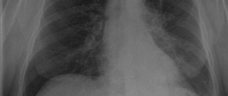
October 20, 2020 Admin Home page » MRI and CT scan of the chest Views: 1572
MRI diagnostics of the heart is a very important method for studying this organ. It allows you to very accurately estimate the speed of blood flow and the size of the heart chambers. Using MRI, clear images of the heart and adjacent soft tissues and blood vessels are obtained.
A distinctive feature of MRI of the heart and blood vessels is that they cannot be stationary. In addition, it affects the quality of visualization of changes in the chest when a person breathes. High-resolution MRI makes it possible to clearly distinguish the boundaries between soft tissues containing water. As a result, tomography is performed on devices with high field strength. What does MRI of the heart and blood vessels show? MRI of the heart and blood vessels makes it possible to study the features of the anatomical structure of the heart and coronary system, the functioning of the heart chambers, valves, and changes in the myocardium.
Thus, MRI of the heart and blood vessels makes it possible to identify various tumors, defects, signs of atherosclerosis, heart attack, obesity of the heart, and evaluate the results of their treatment. Sometimes a special contrast agent is injected into the blood vessels to provide clearer visualization and enhance the “visibility” of the vessels. A contrast agent, gadolinium, is usually administered. The method is called MR angiography
What does an MRI of the heart show?
The examination can provide valuable information about a number of parameters of the structure and functioning of the heart and surrounding tissues:
- congenital heart defects;
- general and local contractility of the left ventricle;
- ischemia and myocardial infarction;
- cardiomyopathy with definition of its type (restrictive, hypertrophic, dilated);
- congenital absence of the pericardium;
- constrictive pericarditis;
- the presence of effusion (free fluid) in the pericardial cavity;
- intracardiac thrombi;
- heart tumors;
- pericardial cysts;
- perivalvular abscesses and fistulas;
- valve insufficiency.
Tomography with contrast
MRI uses contrast for a clearer image and to analyze blood flow.
MRI of the heart with contrast takes a little longer. Contrast is administered intravenously. The drug used for contrast is neutral for humans, i.e. cannot be rejected by the body. In rare cases, allergic reactions are possible (less than 1% of all those studied). Therefore, people prone to allergies should notify their doctor before starting the study.
How do they do it? The tomograph records the speed of passage of the contrast agent through the bloodstream and shows the narrowing and expansion of vascular trunks. If narrowed or enlarged areas are detected, the image is saved to a computer and transmitted to the treating cardiologist for a final diagnosis.
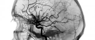
MRI scanner and heart scan result
What does MRI of the heart vessels show?
Significant vessels for diagnosis are the aorta, coronary arteries, pulmonary trunk and branches of the pulmonary arteries. Using MR imaging, the following types of pathological changes in the vessels listed above can be detected:
- aortic aneurysm, aortic dissection and thrombi;
- intramural hematoma of the aorta;
- aortic injury;
- aortitis;
- vascular atherosclerosis;
- thrombi and emboli of the pulmonary arteries.
Special MRI modes make it possible to determine the speed of blood flow in the vessels of the heart, to determine zones of normal and reduced blood supply to individual areas of the myocardium. In most cases, cardiac MRI with contrast is performed for this purpose.
Alternative Methods
In addition to MRI, there are alternative methods for studying internal organs. They are: ultrasound, radiography and angiography.
Vascular angiography is an X-ray examination of the vascular system using a contrast agent. Used for all arteries and veins of the body, including coronary ones. Using this method, it is possible to clarify the normal structure, as well as the speed of blood flow, and calculate a tumor entwined with blood vessels.
Radiography using contrast has more contraindications than similar tomography. However, it is quite possible to study using angiography for those who are allergic to MRI contrast. Contrast agents for the two different examination methods are also different. For tomography - based on gadolinium, for angiography - based on iodine.
Coronorography
A method for studying venous lumens for diagnosing coronary artery disease also uses contrast.
Indications for cardiac MRI
You can get a referral from your doctor for magnetic resonance imaging of the heart and blood vessels in the following cases::
- suspicion of a congenital malformation of the heart or blood vessels;
- control over thrombus dissolution during anticoagulant therapy;
- suspicion of benign or malignant neoplasms in the heart;
- germination of tumors from nearby tissues and organs (pericardial tumors);
- studying the operation of prosthetic valves;
- control of blood flow through shunts after coronary artery bypass grafting;
- detection of aneurysm, dissection or thrombosis of the aorta, determination of the rate of expansion of the aneurysm when the study is repeated;
- determination of the size of myocardial infarction, differential diagnosis of myocardial ischemia and necrosis, determination of the scar at the site of the infarction.
Indications
The main indication for the study is the need for detailed visualization of the circulatory organs in the event of an inaccurate result of cardiac ultrasound. The method can also be used as an alternative to multislice computed tomography (MSCT).
In cardiological practice, it is important to correctly calculate the mass of the myocardium, chamber volumes, ejection fraction, and also to establish the cause of the development and progression of heart failure, which is clearly shown by cardiac MRI.
The high resolution of tomography allows for optimal assessment of the contractile function of the heart. Breath-hold techniques help track the location of the coronary artery. The method makes it possible to evaluate the operation of the valve and indicate backflow of blood or stenosis. Tomography is the gold standard for assessing pericardial thickness. The method allows you to obtain clear images of the aorta, pulmonary arteries and veins, and assess myocardial perfusion.
Indications for cardiac MRI are formed if the following conditions are suspected:
- ventricular aneurysm and pseudoaneurysm;
- dilated and hypertrophic cardiomyopathy;
- congenital and acquired heart defects;
- myocarditis;
- cardiac fibrosis;
- arrhythmogenic myocardial dysplasia;
- hemochromatosis is an infiltrative cardiomyopathy in which iron accumulates in the myocardium;
- cardiac remodeling;
- amyloidosis;
- sarcoidosis;
- Chagas disease;
- Tumors in the heart are also easily detected, which helps to recognize the disease in the early stages.
Information content
The information content of the method directly depends on the type of device used, its power and settings.
An open (low-field) tomograph is recommended for examining patients with claustrophobia and other conditions that complicate the process. The power of the device usually ranges from 0.23 to 0.5 Tesla. Whether to do cardiac MRI with such low settings is a question of rationality, because the quality of the images is low.
High-field installations have a power of 1-1.5-3 Tesla, which provides thin slices and, therefore, a more detailed picture. In addition, this type of device works much faster.
Contraindications for MRI of the heart and blood vessels
No examination is carried out in two cases:
- the patient’s body contains foreign bodies made of metal, with the exception of those made of titanium;
- the patient has implanted electronic devices (pacemaker, etc.).
Administration of gadolinium-based contrast is contraindicated.:
- pregnant and lactating women;
- patients with chronic renal failure;
- persons intolerant to gadolinium-based drugs.
Contraindications
The examination procedure itself is usually well tolerated by patients and does not cause much discomfort. However, there are a number of contraindications, absolute and relative, that may interfere with its implementation.
The first group of contraindications makes the procedure impossible. These include:
- the presence in the patient’s body of various devices and metal products for medical purposes (pacemakers, intracardiac catheters, insulin pumps, vascular stents, clips, steel endoprostheses, etc.) - the magnetic field can lead to heating of individual elements, causing device failure or displacement, which will cause injury to surrounding tissues;
- increased body weight (over 120 kg) – the patient’s waist is incommensurate with the diameter of the device.
The second group of contraindications makes the procedure possible subject to strict adherence to a number of conditions and recommendations of the attending physician:
- fear of confined spaces;
- pregnancy (first trimester);
- nervous and mental disorders;
- serious condition due to any illness;
- heart failure in the stage of decompensation (usually III, IV functional classes);
- the patient's inability to remain completely still;
- the presence of tattoos with metal-containing dyes.
Preparation for the procedure
MRI of the heart and blood vessels does not require special preparation. It is recommended to come to the examination in loose, comfortable clothing without metal fasteners made from natural fabrics. Metallic elements and electrostatic discharge from synthetic fabrics may reduce image quality.
It is advisable to take with you to the clinic the results of previously conducted examinations, doctors’ opinions, extracts or epicrisis about the treatment performed, if any. Any medical information related to the problem for which MRI of the heart and blood vessels is performed can make the functional diagnostic doctor’s conclusion more complete and meaningful.
Preparation and conduct of the study
MRI without contrast does not require any preparation. If you are planning an examination with contrast, you are required to fast for 6 hours. Before entering the room with the tomograph, you should:
- talk in detail with your doctor about past illnesses and operations, not forgetting to tell about any allergic reactions;
- remove all metal objects from yourself - jewelry, watches, clothes with metal buttons, buckles, buttons and locks.
When the patient is ready for the examination, he is helped to lie down on the tomograph bed. It is necessary to wear headphones, as the device makes a lot of noise when operating. During the procedure, the doctor is in the adjacent room. There is a special button inside the tomograph to communicate with it. A series of photographs are taken before and after intravenous administration of a contrast agent. Contrast allows the doctor to see blood vessels and assess their patency.
How to do a heart MRI
To carry out the procedure, the patient is invited to the magnetic resonance imaging room. The largest machine in the office is a magnetic resonance imaging scanner. The patient is placed inside the device, lying on a special retractable table in a supine position. In order to protect your hearing from the noise that the device makes during operation, it is recommended to use earplugs or headphones.
During the examination, the doctor may ask the patient to hold his breath using voice communication. The patient can use voice communication to inform the doctor about any discomfort that he may experience during the procedure.
If an MRI is performed with contrast enhancement, a contrast agent is injected intravenously into the patient. To administer the drug, a special catheter can be used, which is inserted into a vein in the patient’s arm. The catheter can deliver contrast into the blood throughout the entire MRI procedure. After the procedure, the catheter is removed. In its place only a small puncture of the skin remains.
Interpretation of MRI results
After the scan is completed, medical specialists begin to decipher the data obtained and draw up a conclusion, so the patient needs to wait in the clinic. The conclusion and photographs are handed over to the patient. They can also be recorded on digital media. With the received conclusion and photographs, the patient must immediately contact the attending physician. If the patient comes to the clinic on his own initiative, he will be advised which specialist he should contact after the examination: a therapist, a cardiologist, an oncologist.
Magnetic resonance imaging of the heart has been used in medicine since the 90s of the 20th century, but the widespread use of the method is hampered by the high cost compared to ultrasound or CT. MRI uses more expensive equipment and requires highly qualified medical personnel, and contrast will also require additional costs. The initial cost of cardiac MRI ranges from 5 to 8 thousand rubles. The cost, of course, depends on the region, the level of the clinic and specialists, the type of MRI scanners and the contrast used. Prices for contrast agents range from 5 thousand rubles.
But if it is impossible to make a correct diagnosis using other methods or to clarify it, MRI of the heart and coronary vessels will help. This method is absolutely harmless, which allows it to be prescribed repeatedly within a short period of time, without side effects for the patient. MRI eliminates the need for invasive angiography, which can cause complications for the patient. If cardiac surgery is planned, then the role of the MRI procedure will also play a significant role in order for doctors to compile a complete picture for the operation.
MRI, unlike CT, shows soft tissue more clearly, allows you to examine in detail the narrowing of the coronary vessels, and determine the localization of dying areas of the heart muscle suffering from ischemia.
Decoding the received data
Deciphering means recognizing pathological changes in photographs and three-dimensional images of organs and vascular networks and correlating these changes with a particular disease. The medical records that a patient can provide to a doctor about his or her illness are very helpful in making a diagnosis and can be used to determine the dynamics of the pathological process over time and under the influence of treatment.
A radiologist or a functional diagnostics doctor is engaged in decoding. Decryption takes about an hour. The patient receives photographs on film, paper or digital media, as well as a doctor’s report.
How much does the procedure cost?
Prices for examinations in Moscow clinics:
| Clinic name | Price (rubles) |
| European Medical Center | 31340 |
| Medical Clinic "Medicine" | 15360 |
| Scandinavian "Health Center" | 14990 |
| Clinical Hospital on Yauza | 12950 |
| Medical | 12000 |
| Diagnostic | 11490 |
How often can you do an MRI of the heart and blood vessels?
There are no restrictions on the scope of the study and the frequency of procedures performed, since the magnetic resonance imaging method does not involve the use of ionizing radiation and other harmful effects on the body. The examination can be performed at the diagnostic stage, before surgery to clarify the localization and extent of pathological changes, after surgery to monitor the healing process, and some time after treatment to monitor the state of the cardiovascular system. And all this without harm to the human body.
Contraindications for the study
Due to the specific design of the tomograph, some patients cannot be admitted to the procedure:
- if there are metal implants in the patient’s body (with the exception of titanium structures, since titanium is not ferromagnetic), the metal heats up and shifts under the influence of magnetic radiation, which can pose a threat to the patient’s life;
- suffering from panic attacks, claustrophobia, mental disorders;
- women in the first trimester of pregnancy;
- patients in extremely serious condition requiring constant monitoring of vital functions;
- people with body weight exceeding the maximum allowable value for the tomograph model;
- for contrast studies – liver and kidney failure, pregnancy at any stage.
How is it carried out?
Before starting the procedure, the patient fills out a questionnaire where he answers questions about his health, previous operations, the presence of tattoos, implants, etc. Sometimes this survey is conducted orally. When answering questions, you should not be shy and speak honestly. There are diseases for which MRI is contraindicated. These include:
- Presence of ferrocontaining implants.
- Previously installed pacemaker.
- Renal and heart failure (with MRI with contrast).
Cardiac MRI is done with caution in people with claustrophobia, mental disorders, and alcoholism.
Main stages:
- The patient is placed on the table, the head is fixed with a special pillow of medium softness, and headphones are put on. The table slides into the tomograph capsule, the ring begins to rotate - the study has begun.
- You need to lie still. For this purpose, some clinics prefer to secure the arms and legs of the subject with straps before the study begins.
- During the tomography, the doctor will monitor the patient from an adjacent room. It is necessary to talk about all unpleasant sensations; the tomograph is equipped with a built-in microphone. If necessary, the procedure can be interrupted.
- After the scan is completed, you can go home immediately.
Decoding magnetic resonance imaging of the heart takes time, it ranges from 2 to 6 hours. Most often, you can receive decrypted results and a recording disc in your hands the next day. In high-traffic clinics, the waiting time for results may be longer.
The final diagnosis is made by the attending physician. Taking into account the medical history, he evaluates the tomograph readings and compares them with the results of other studies.