What does expansion of the heart to the left mean?
The average weight of this cone-shaped hollow muscular organ in an adult man is approximately 300 grams, in a woman - 250 grams. Its dimensional characteristics are comparable to the size of a clenched fist: longitudinal size - 12 centimeters, transverse - 8-9 centimeters, anteroposterior - 6 centimeters.
An expansion or increase in the volume of an organ is called cardiomegaly. If it is expanded on the left, this is a symptom of transformation of the left ventricle, which means hypertrophy of the heart muscle. This sign means that it has become larger in diameter.
As a rule, the increase in volume occurs on the left side. This may be a manifestation of hypertension (hypertension), blood stagnation in the systemic circulation, and other pathologies. Sometimes this phenomenon is called “bull’s heart”.
Symptoms of what diseases are visible in the image?
When the patient is given the results of the study, he is explained what disorder was discovered and which doctor should be contacted in the future. For example, cardiovascular pathology is often visualized on fluorography as a displacement or expansion of the mediastinal shadow - what kind of disease it is can be found out using echocardiography (EchoCG) or other diagnostic methods. An expanded mediastinal shadow is one of the signs of an increase in the size and mass of the heart.
We suggest that you familiarize yourself in more detail with the essence of changes in the chest organs that fluorography can detect.
Heaviness of the roots of the lungs
Many patients are interested in the question: “On fluorography, the roots are stringy - what is it?” Heaviness often means thickening and nodularity. The roots of the lungs consist of the main bronchi, pulmonary arteries and veins, bronchial arteries and veins, lymph nodes and vessels. If these anatomical structures are compacted, it means that there is an acute or chronic inflammatory process in the patient’s lungs (for example, pneumonia or bronchitis).
Changes of this nature are also found in smokers with many years of “experience”. The roots are often compacted in people who work in hazardous chemical industries (for example, when inhaling fumes of gasoline, ammonia, asbestos and other hazardous substances). Notice how the roots of the lungs look heavy in the image.
Pronounced vascular pattern
The vascular picture is formed from the shadows of the pulmonary arteries and veins. If it is clearly visible on the image, this means that the blood supply to the vessels has increased significantly compared to normal. This fluorography conclusion indicates an inflammatory process in the lungs: pneumonia, bronchitis, pneumonitis (inflammation of the walls of the alveoli - the end elements of the respiratory apparatus in the lungs, shaped like bubbles).
An overly pronounced vascular pattern appears even in the initial stages of development of a malignant neoplasm in the lungs. To rule out cancer, the patient will need to be tested for cancer markers and undergo a more thorough examination.
A pronounced vascular pattern (see the image below) in the lungs can also be a sign of cardiovascular pathology (in particular, congenital heart defects).
Presence of focal spots
If the results of fluorography indicate the presence of focal spots, this means that small areas of darkening of the pulmonary field are detected in the image. As a rule, the diameter of the spots does not exceed 10 mm. If they are located in the middle or lower part of the lungs, this is a sign of pneumonia, but if in the upper part, this is an x-ray symptom of tuberculosis.
If the inflammatory process is in an active stage, the contours of the darkening zones are uneven, in some places the merging of several spots into one is noticeable, the vascular pattern is too pronounced. When inflammation subsides, the contours of the focal spots are smooth. A small darkened area may indicate the development of a pathological neoplasm in the organ. Below is a picture in which focal spots are clearly visible.
Blackout zones
The large area of darkening in the lungs, which is visible in the image below, can have different sizes and shapes, have thinned or thickened, smooth or uneven edges. When answering the question of how to decipher fluorography, which contains darkened zones, it should be pointed out that pneumonia and tuberculosis most often manifest themselves this way.
Presence of fibrous tissue in the lungs
A type of connective tissue that is made up of collagen fibers and has high strength is called fibrous tissue. It appears as a result of injuries suffered by a person, acute or chronic inflammatory diseases of the lungs, or surgical intervention.
Fibrous tissue replaces damaged cells and protects healthy tissue from the source of pathology. Therefore, such a fluorography result can be detected in a recovered person. What fibrosis looks like in the picture, see below.
Calcifications in the lungs
Fluorography with a description containing the word “calcifications” means that small round shadows have been identified in the lungs, comparable in density to bone tissue. Their presence indicates that the person had previously had contact with patients with pneumonia or tuberculosis, but he himself did not develop the disease (pay attention to the picture below).
Calcifications consist of calcium salts that surrounded the infection that had entered the body and, thus, completely isolated it (“preserved”).
Adhesions and pleuroapical layers
Interpretation of fluorography of the lungs may contain an indication of the presence of adhesions. They are a proliferation of connective tissue and appear as a result of an inflammatory process suffered by the patient previously. An adhesive process detected on fluorography in most cases does not require medical intervention.
Sometimes connective tissue growths cause significant discomfort to a person, causing chest pain and breathing problems. In this case, it is necessary to seek medical help and undergo a more thorough diagnosis.
Pleuroapical layers are thickenings of the apices of the lungs. Like adhesions, they are signs of previous inflammation that has spread to the pleura. Most often, pleuroapical layers appear after pulmonary tuberculosis. The extensive adhesive process is shown in the photo below.
Deformity of the thoracic spine
Decoding the numbers 21 according to the results of fluorography is a deformation of the spine in the thoracic region. Since the bones are clearly visible in the image, the radiologist will be able to notice scoliosis. Curvature occurs as a result of displacement of the vertebrae to the left or right side relative to their axis.
Scoliotic deformity can be C-, S- and Z-shaped (with one, two and three arcs of curvature, respectively). By measuring the angle of the spinal deformity (as shown in the picture below), the specialist will be able to indicate the stage of development at which the detected scoliosis is located on fluorography.
Signs of pneumosclerosis
Pneumosclerosis is a pathological process consisting of the replacement of lung tissue with connective tissue and is accompanied by a decrease in the level of elasticity and intensity of gas exchange in the affected areas. Due to the proliferation of non-functioning connective tissue, the bronchi become deformed, the lung tissue shrinks and thickens, and the lungs decrease in size.
Most often, pneumosclerosis develops at the final stage of inflammatory diseases or degenerative processes in the lungs, and sometimes it can be triggered by improper treatment. In some patients, this disease becomes a consequence of hemodynamic abnormalities (that is, disturbances in the movement of blood through the vessels due to different pressures in different zones of the circulatory system) that occur in the pulmonary circulation.
Often, pneumosclerosis is diagnosed in patients with mitral valve stenosis (narrowing of the left atrioventricular orifice), heart failure (inability of the heart to pump blood in the required volume), thromboembolism (acute blockage) of the pulmonary artery.
Changes in aperture
The diaphragm is a muscular partition that separates the thoracic and abdominal cavities. Fluorography visualizes both deformations of its shape and changes in its location. Most often they are caused by excess weight, pleurisy (inflammation of the pleura), diseases of the gastrointestinal tract, and deformation of the adhesions.
To determine the cause of changes in the diaphragm, the patient is prescribed additional diagnostics. The deformed muscular septum is shown in the image.
Causes of pathology in adults
Provoking factors for cardiomegaly can be:
- diabetes;
- hypertension;
- long-term treatment with antibiotics;
- the onset of pregnancy;
- anemia (anemia) – a decrease in the level of hemoglobin protein in the blood, which is responsible for transporting oxygen in the blood;
- renal failure;
- alcohol abuse;
- rheumatism – inflammation of the connective tissue structures of the cardiovascular and musculoskeletal systems;
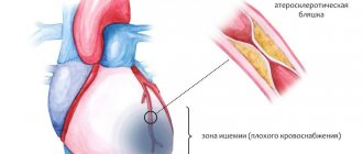
- ischemia – impaired blood supply to the myocardium;
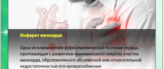
- Myocardial infarction – damage to the heart muscle. It develops with an acute lack of oxygen. Occurs due to blockage of a blood artery by a blood clot. Subsequently, the damaged areas die off. The process of their death is called necrosis;
- athletes with enlarged heart muscle undergoing irregular training;
- genetic predisposition;
- cardiomyopathy - thickening of the walls of the ventricles, stretching of the cavities of the heart.
The described pathology does not manifest itself in any way at first, but over time, with increasing loads, the following symptoms may appear:
- a condition characterized by shortness of breath, fatigue;
- swelling in the lower extremities;
- the appearance of a feeling of heaviness on the right under the ribs;
- cephalalgia, tinnitus;
- causeless cough.
Enlarged heart in children
An increased volume of an organ in children can be a congenital pathology, which will subsequently cause serious health problems. It is important to identify such deviations from the norm immediately - from the first days of a child’s life to six months.
Cardiomegaly in children can occur due to the presence of an inflammatory process of connective tissue in the body - rheumatism. This disease, in turn, can appear as a complication after a sore throat, exacerbation of chronic tonsillitis, or with some types of caries. It is acquired and develops regardless of the child’s age.
Signs of the described pathology in children may be:
- weakness;
- dyspnea;
- pain in the sternum area;
- fever 1-2 weeks after recovery from tonsillitis;
- sweating;
- increased fatigue;
- pain in muscles, joints.
The reasons for the appearance of signs of an enlarged heart on a fluorography image may also be:
- hypertrophic cardiomyopathy - manifests itself in thickening of the wall of the ventricle, most often the septum between the ventricles;
- myocarditis is an inflammatory process in the heart muscle. It can be of viral, fungal, bacteriological origin, or may appear due to autoimmune problems or improper use of medications;
- endocarditis – inflammation of the endocardium (inner lining);
- pericarditis is an inflammatory process in the serous membrane.
The last two pathologies in minor patients are quite rare.
If the described symptoms occur, you should consult a doctor; it is highly undesirable to leave this situation without proper attention.
What it is
The diagnosis of aortic thickening is made by identifying a cholesterol plaque or growth on the wall of the vessel, which can be seen using radiography, fluorography, and ultrasound.
Normal vessel and with a sealed wall
The doctor should immediately begin examining the patient to identify the underlying cause of the defect. If this is not done, the pathology will soon develop into more serious diseases, for example, dissection or rupture of the aorta.
It is believed that atherosclerosis itself is not fatal, but a person can easily die from the consequences of not being treated.
Compaction in the aorta can occur anywhere along the entire vessel, depending on its location:
- At the root of the aorta;
- On the arc;
- In the ascending part;
- In the downward direction.
What additional diagnostic measures are taken?
To make an accurate diagnosis and prescribe appropriate treatment, the following types of examinations are required.
- X-ray of the sternum area. You can notice an expanded shadow of the heart group and determine congestion in the circulatory system.
- Electrocardiography (ECG).
- Echocardiography (EchoCG). Allows you to determine the parameters of the heart muscle, the size of the chambers, identify necrosis (death of cells/tissue areas) or ischemic disease (impaired blood supply to the myocardium due to damage to the arteries).
- Ultrasound examination (ultrasound) of the heart muscle.
- Computed tomography (CT).
- Magnetic resonance imaging (MRI).
- Immunological and biochemical blood tests. They determine the levels of hemoglobin, bilirubin, protein, hormones and urea.
The doctor will be able to prescribe adequate treatment only after making a diagnosis. And to determine the disease, the doctor carefully examines not only the data of hardware examinations, but also the results of clinical tests, as well as the clinical picture.
Reasons for the formation of compactions in the lungs
Pulmonary compactions can form under the influence of various factors, among which the most prominent are the following conditions:
- pneumonia of a bacterial nature;
- damage to lung tissue by tubercle bacilli;
- syphilis manifests itself in the form of the formation of painless nodes;
- fungal lung damage;
- cancer formation in lung tissue;
- manifestation of metastasis from affected nearby organs;
- grounding of various parts of the lungs;
- pulmonary infarction.
In some cases, the formation of compactions in the lungs is a consequence of exposure to ionizing radiation, systemic diseases, leukemia, the use of certain groups of drugs, and the result of constant inhalation of dust particles. Sometimes pulmonary infiltrate is confused with enlarged lymph nodes located in the thoracic area, a diaphragmatic hernia, pleural adhesions, and pathologies of the vascular system.
Treatment
For almost any disease with a symptom of heart enlargement to the left on fluorography, doctors recommend leading a healthier lifestyle, adhering to a special diet, which means avoiding spicy, fatty and salty foods. Patients are advised to give up bad habits and may be prescribed special exercises.
As a rule, medications from the following groups are prescribed for cardiomegaly:
- angiotensin-converting enzyme (ACE) inhibitors;
- angiotensin receptor blockers;
- beta blockers;
- diuretics.
Angiotensin-converting enzyme inhibitors
Such drugs dilate blood vessels and lower blood pressure, reducing the workload on the cardiovascular system. The drugs have the following names: Enalapril, Captopril, Lisinopril. A side effect is often a dry cough.
Angiotensin receptor blockers
Being analogous to ACE inhibitors in terms of pharmacological effect, these drugs do not cause a dry cough as a side effect.
Beta blockers
The use of beta blockers helps reduce heart rate and normalize blood pressure. However, such drugs are only auxiliary in treatment.
Diuretics
Diuretics (diuretics) can improve blood flow to the heart and help lower blood pressure. The names of these drugs: Chlorthalidone, Hydrochlorothiazide.
It is not recommended to resort to self-medication; only a doctor can make a diagnosis and determine an effective drug therapy regimen.
Surgery may also be required. It consists of restoring or completely replacing the arterial valve. In severe cases, a whole organ transplant is necessary.
Prevention
Prevention of cardiomegaly can be a change in lifestyle. One of the main factors in the occurrence of heart pathologies is obesity. It is recommended to maintain a normal body mass index. Losing weight if you are overweight, against the background of an already enlarged heart, helps stabilize the condition.
A special diet is recommended to prevent cardiovascular diseases. It is advisable to consume fish, soybean, flaxseed, corn or olive oil at least twice a week. To strengthen the heart muscle, you need to eat cranberries, cabbage, viburnum, eggplants, peaches, dried apricots, apples, pomegranates, walnuts, melon. To normalize blood pressure, you need to avoid excessive consumption of salt and salt-containing foods. In addition, it is advisable to reduce the consumption of alcoholic beverages or completely abandon them.
Although an increase in heart volume may be caused by intense physical activity, in moderate doses, sports activities can only benefit the patient. Thus, yoga, Pilates, and walking will help strengthen the heart. If hypertrophy has already been identified, it is better to consult a physiotherapist. He will help you choose a set of therapeutic exercises, designed for about half an hour. Regularly performing this set of physical activities will reduce the volume of the heart muscle.
Eight hours of sleep and avoidance of excessive physical and emotional stress are helpful. In addition, regular walks outside are recommended.
Thus, a healthy lifestyle and proper nutrition will help minimize the risk of complications with an enlarged left ventricle.
Coronary diseases
Hypertension is the most common cause of heart enlargement.
This is explained by the fact that due to increased blood pressure, the organ is forced to pump large volumes of it and work in enhanced mode.
This causes the heart muscles to enlarge and the organ itself to expand.
Video:
If a person has ischemia, the heart muscle cells constantly do not receive enough nutrients, as a result of which they degenerate, and connective tissue appears in their place.
The latter, unlike muscle tissue, is not capable of contraction; as a result, the organ cavities become deformed and increase in size.
READ Age at which a child can have fluorography
What to do if an x-ray showed that the organ is enlarged, and the cause of this phenomenon is a disease of the cardiovascular system?
The answer to this question is simple and obvious - treat the root cause and return the organ to normal limits.
If a patient is diagnosed with hypertension, he is usually prescribed pharmaceuticals that lower blood pressure. The latter helps restore the normal size of the organ.
A patient with hypertension or coronary artery disease who has been diagnosed with an enlarged heart must take medications.
Video:
The fact is that despite the increased size of the organ, a large heart performs its most important function - pumping blood - much worse, which means that human organs and systems do not receive the nutrients they need - heart failure develops, and the whole body suffers.
That is, returning the organ to its normal size helps prevent heart failure, which in some cases can simply save a person’s life.
Risks and possible complications
There are various prognoses that are directly dependent on the diagnosis made when the heart expands to the left.
- For arterial hypertension (hypertension, high blood pressure), the prognosis is considered favorable. After taking the medications prescribed by the doctor, the patient’s condition returns to normal, the heart returns to its normal parameters and does not enlarge again.
- If a ventricular septal defect is observed, surgery is necessary. Otherwise, the pathology will lead to aortic valve insufficiency, serious rhythm disturbances will occur, and left ventricular dysfunction is possible. All of the above can cause death. After the operation, the patient’s condition returns to normal, and he no longer faces such heart problems.
- Dilated cardiomyopathy (stretching of the heart cavities) is characterized by a poor prognosis. Full recovery becomes possible only after a heart transplant.
- Hypertrophic cardiomyopathy (thickening of the walls of the ventricles) - the prognosis is relatively unfavorable, the disease may be asymptomatic. Without proper and timely treatment, there is a risk of death. When undergoing treatment procedures, the likelihood of death becomes lower.
- Metabolic cardiomyopathy is a non-inflammatory myocardial lesion of various etiologies. It can cause insufficiency of cardiac functions, in particular contractile function. The prognosis is favorable. If you establish proper metabolism, complete recovery will occur.
- Myocarditis. Characterized by a favorable prognosis. Complete recovery is possible within 1-2 months in nine out of ten cases, after 1 year in the remaining patients.
- Aortic stenosis (the mouth of the aorta becomes narrow, as a result of which blood flow from the left ventricle to the aorta is impaired) - without treatment, the life expectancy will be from 1 to 4 years from the time the disease is detected. With proper treatment, the prognosis is relatively favorable.
- Exudative pericarditis (inflammation of the serous membrane of the heart) has a favorable prognosis, all patients recover.
How can you easily recognize a smoker?
You can determine whether a person smokes or not in different ways; no special procedures are needed for this.
- A smoker's skin has a yellowish or grayish tint.
- Even at a very young age, people who smoke regularly show fine facial wrinkles.
- An unpleasant odor of tobacco from your breath during a conversation also indicates that you smoke.
- The peculiar smell of clothes also gives away a smoker. Even the skin has a specific smell when smoked regularly.
- Yellow plaque and frequent inflammation of the gums, sometimes accompanied by bleeding.
- Prolonged coughing attacks, which sometimes lead to vomiting.
- The tips of the thumb and index finger of the right hand are yellowish from regular smoking.
Fluorography: indications, procedure and interpretation
Tuberculosis is a very dangerous and very common disease. In order to identify it, a procedure is performed - fluorography. This is a rapid diagnostic method that allows you to determine the presence of structural and other changes in the lungs, as well as other chest organs and blood vessels.
The procedure does not require special preparation and takes minimal time. The study is recommended for every adult and is carried out at least once a year. For people with existing diseases, the study is performed more often to monitor the dynamics of the development of the disease and evaluate the effectiveness of treatment.
After reading the information below, you will learn how the procedure works, what its advantages and disadvantages are, as well as a breakdown of the indicators.
Indications for fluorography
Lung fluorography is a diagnostic method for examining the chest organs, which is based on X-ray radiation. Typically, a procedure is performed to detect the development of tuberculosis.
Fluorography refers to mass research, since it allows you to examine thousands of people per day, provided that the device operates normally. Conducting research has its advantages and disadvantages.
The latter include: high dose of radiation; Old devices do not always allow you to obtain the highest quality and accurate images, as well as timely identify film defects.
The positive aspects of the procedure include:
- Minimum time, labor and material costs.
- High information content during mass screening of people for tuberculosis.
- Modern devices have the ability to send images over the Internet, as well as compare them with previous studies of the patient.
Fluorography is mandatory in the following cases:
- For an annual preventive examination to detect tuberculosis. Once a year, every person over 18 years of age must undergo the procedure to ensure that they do not have the disease. Fluorography is mandatory for: future students; workers of medical, educational institutions and public catering establishments; women preparing for motherhood and everyone who lives with them; visitors to sports clubs and swimming pools; conscripts of the military registration and enlistment office.
- To identify inflammatory processes in the lungs of a fungal or bacterial nature.
- In order to determine the presence of tumor formations, not only in the lungs, but also on the heart, large blood vessels.
- To identify foreign bodies in the chest area.
- To determine structural and dimensional changes in lung tissue, the formation of cavities, the presence of air accumulation in the lungs.
Fluorography helps determine the presence of changes in the lungs and helps in making a diagnosis. If negative processes are identified during the study, then additional studies are prescribed, for example, X-rays, CT, MRI. Fluorography is not performed for the following categories of persons:
- Women during pregnancy (especially up to 25 weeks).
- Children under 15 years of age (from 16 to 18 only if there are serious indications).
- Lying patients who cannot take a vertical position even for a short time.
- People with respiratory failure.
- Patients with claustrophobia (fear of closed spaces).
How to prepare for fluorography and the procedure for conducting it
Fluorography does not require special preparation; the only requirement is to abstain from smoking for several hours before the procedure. Basic principles of the study:
- The procedure is carried out at any time and does not require special preparation.
- Before conducting the study, you should stop smoking for several hours - this will allow you to get a clearer and clearer picture.
- Before starting the procedure, you should expose your upper body and remove jewelry (chains, necklaces, etc.).
- The study is carried out only in an upright position, so it is not prescribed for bedridden patients.
- During fluorography, you should strictly follow all the recommendations of the radiologist. The picture is taken while inhaling as deeply as possible - during this period the lungs open well, which allows you to get a more complete picture.
- During the study, X-rays pass through the body from behind and fall on a cassette with ultra-sensitive film to form a pattern, which is subject to further study and research.
Interpretation of fluorography results
The procedure allows us to identify the following changes in the lung tissue and other organs of the chest:
- Strong pulmonary pattern. This phenomenon indicates the presence of inflammation, sclerotic changes or a tumor disease. In some cases, this may indicate the presence of a heart disease or pathological changes in blood vessels, so additional diagnostics are required to confirm the diagnosis.
- Focal shadows. Such manifestations are typical for inflammation, in particular tuberculosis. There can be one or many lesions; as a rule, the size does not exceed one centimeter.
- Pathological changes in the roots of the lungs (compaction, expansion, weighting).
- Calcifications are specific traces (shadows) of an infection or disease that did not develop, but was suppressed by the human immune system.
- Fibrous changes. These indicators indicate the occurrence of an inflammatory process in the lungs.
- Accumulation of fluid in the pleural cavity. This phenomenon indicates pleurisy. The fluorographic image clearly shows the amount of liquid.
- Displacement of organs (large blood vessels, heart, lymph nodes, bronchi, esophagus, trachea). This is usually observed with heart disease, accumulation of fluid or air bubbles in the pleural cavities. If such a phenomenon is detected in the image, the patient is prescribed additional procedures and research methods.
- Aperture changes. As a rule, it indicates an abnormal structure of the organ, the presence of adhesions, and obesity. Often the change is observed after surgery or chest trauma.
Fluorography is a quick and simple (although not entirely safe) method for detecting tuberculosis and other diseases or pathological changes in the lungs and other organs of the chest. It must be carried out once a year, does not require specific preparation, and informative results are ready within a few days.
Page 5
Source: https://24doctor.info/medical_tests/flyuorografiya/