Disruption of blood flow (arterial or venous) poses a huge danger to human health and life. As practice shows, death occurs in 25% of clinical situations; in approximately 50% of cases, severe disability occurs, associated with the need for mutilation.
Thromboembolism is an acute circulatory disorder in tissues that develops as a result of blockage of a vessel by a clot of formed cells (platelets, layers of other structures plus fibrin protein fibers).
It is more of a consequence than an independent disease. Although doctors use the term to describe the disorder.
Diagnosis is urgent, in a hospital setting. If necessary, resuscitation measures are carried out to restore the person’s normal condition.
Mixed therapy. Conservative and operational. Alone, they don’t make much sense, except in the mildest cases.
Forecasts are vague and depend on many factors: from the age and gender of the patient to hormonal status, the presence of somatic diseases and other points that are assessed by doctors.
Development mechanism
As the name of the pathological process suggests, thromboembolic syndrome consists of two components.
The first is the formation of a blood clot, which in the future will play a major role in the development of the deviation.
The second stage consists of embolism, that is, the separation of a blood clot from the site of development and its movement along the bloodstream.
The initial phase is formed as a result of the influence of three factors at once.
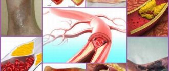
- Changes in the rheological properties of blood. It becomes less fluid. Hence hypercoagulability - excessive coagulation. Liquid tissue begins to coagulate directly in the vessels. This does not happen to everyone; conditions such as hematological diseases or endocrine disorders are necessary.
- Reduced blood flow rate. It develops against the background of the same change in its properties, since the liquid tissue becomes thick, it moves worse, and accordingly, third-party complications appear, including arterial hypertension, stroke, and heart attack against the background of tissue ischemia.
- Violation of the anatomical integrity of blood vessels. As a result of injuries, diagnostic and therapeutic interventions (iatrogenic factor), or as a result of spontaneous tissue destruction of hollow structures against the background of increased vascular fragility, taking non-steroidal anti-inflammatory drugs.
In the system, these factors cause the formation of blood clots. Often the formation is found far from the vessel, where it then settles.
The vast majority of cases involve damage to the limbs or heart walls. At this stage, the anomalous structure is attached to the site of its own formation. But this won't last long.
The second stage is called embolism. The thrombus breaks off (usually as a result of the influence of a mechanical factor) and moves along the bloodstream. It is impossible to say exactly where he will stop.
Typically, relatively small arteries, areas of anatomical bends where the blood supply structures are initially thinner, are affected.
Then the process moves quickly. A thrombus clogs a vessel to a certain extent:
- If we are talking about obstruction at a level of less than 20%, the symptoms are sluggish.
- Up to 60% - moderate.
- Up to 70% - critically severe, focal.
As for more than 80%, rapid tissue death and necrosis occurs.
If the pulmonary artery, important centrally located vessels, is damaged, the patient will soon die from critical complications.
In the opposite situation, when the limbs are affected, rapid necrosis begins. Amputation required.
You need to know the mechanism in order to correctly assess the situation, carry out high-quality, timely treatment to eliminate the symptoms, the root cause, and also prevent relapses.
Definition
Deep vein thrombosis (DVT) occurs when blood clots form in the deep veins, usually the legs , although the arms, abdomen, or area around the brain can also be affected. A blood clot forms as a result of damage to a vein, thickening of the blood, or slowing of blood flow . PE, a potentially fatal complication of DVT , occurs when a blood clot breaks off and travels through the bloodstream and into the lungs.
Between 10% and 30% of people diagnosed with DVT/PE die within one month3. Many people with DVT develop long-term complications such as pain, swelling, skin discoloration, peeling or ulcers in the affected limb, and up to 40% have recurrent disease within 10 years2,3.
Because signs and symptoms are often nonspecific and similar to other conditions, DVT may be undiagnosed or misdiagnosed 3,4. DVT affects men and women of all ages and races. Many known risk factors (see chart) are increasing, resulting in an increasing public health importance of DVT/PE 3 .
Risk factors for DVT/PE1,2,4
- Damage to a vein (for example, from a fracture or surgery)
- Immobility (for example, during long travel or hospitalization)
- Surgical intervention
- Increased estrogen levels (taking birth control, hormone replacement therapy, or pregnancy)
- Certain medical conditions and/or treatments (eg, cancer/chemotherapy, heart failure, inflammatory diseases, or kidney disease)
- Genetic predisposition
- Elderly age
- Obesity
- Smoking
Symptoms depending on location
Thromboembolic disease mainly affects the arteries of the body, creating a colossal danger at the moment, since there is practically no time left to respond.
Death is likely in a matter of minutes or the development of dangerous complications.
The signs of thromboembolism are not the same and depend on where exactly the blockage occurred and are characterized by ischemic and hypoxic phenomena.
Damage to the abdominal arteries
They are also called mesenteric. The pathological process is accompanied by the development of a clinical picture of acute abdomen.
Among the typical points:
- Pain in the abdominal area. Intense, pressing, burning, cutting. Accompanied by movement back and forth, wandering, localization is not clear, so the patient cannot say exactly where the discomfort is concentrated.
- Nausea and severe vomiting. Possibly with blood if the disorder progresses.
- Flatulence, increased gas formation.
- Against the background of bloating, defecation disturbance is detected. A person may experience painful urges (tenesmus) with an inability to relieve themselves. It is possible to develop intestinal obstruction (probability about 70%).
- Collaptoid state. An increase in blood pressure, and then its rapid drop, increased sweating, tachycardia, increased heart rate, pallor of the skin, shallow breathing, depression of consciousness. An extremely worrying sign.
- Increase in body temperature (hyperthermia at a level slightly above 37.5 degrees).
Thromboembolism of the abdominal arteries is extremely dangerous. The development of intestinal necrosis and death from peritonitis in a short time is possible.
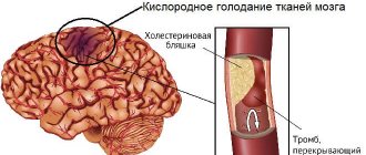
Blockage of arteries in the brain
Classified as ischemic or hemorrhagic stroke. Both forms carry great danger. Accompanied by general and focal neurological symptoms.
The first ones are almost always the same, with different intensities:
- Unbearable headache. Baling, pressing. Localized in the back of the head, crown. The temples or has a diffuse, diffuse character; it is impossible to determine exactly where it is located.
- Nausea, vomiting. Reflex, short-term.
- Impaired consciousness. Syncope.
- Dizziness. Vertigo. With the inability to navigate in space.
- Weakness, drowsiness, asthenia. The opposite effect is possible with the development of psychomotor agitation.
Focal signs depend on the specific location of tissue necrosis. If the occipital region is involved, vision suffers, the temporal region suffers from hearing, consciousness and memory, the frontal region affects behavior, intelligence, the parietal region suffers from smell, cognitive abilities, and so on.
Symptoms of the pre-stroke condition, depending on the location, are described in this article.
MRI helps to put an end to the question. Although not in all cases, survey data are sufficiently informative. Especially with the development of massive damage to brain structures (major stroke).
Involvement of the arteries of the extremities in the process
Accompanied by a critical malnutrition of the arms or, much more often, legs.
The clinical picture is typical, so it is almost always possible to detect the problem directly during the initial examination.
Among the symptoms:
- Pain in the leg area on the affected side.
- Severe numbness up to complete loss of sensitivity.
- General critical condition. Collapse.
- Paleness, bluishness of the skin layers, a feeling of goosebumps. The appearance of a vascular pattern on marble-colored skin.
In the absence of high-quality immediate surgical treatment, gangrene cannot be avoided. And then death (especially if the femoral artery is blocked).
Pulmonary artery damage
The most dangerous option in terms of the likelihood of death of the patient. Affects a key vessel of the pulmonary circulation.
When the blockage is more than 80%, sudden death occurs, without preliminary symptoms. The person does not have time to understand anything.
In less complex situations, the clinical picture develops in a matter of minutes. Doctors have about half an hour to provide assistance.
The following points indicate an emergency:
- Chest pain. Expressed. Unbearable. Bursting.
- Paleness of the skin.
- Dyspnea. Reaching asphyxia. When the body position changes to lying from sitting, threatening complications develop. A person may suffocate and die.
- Cough. At first unproductive, then with the release of bloody, foamy sputum of a pinkish hue, with scarlet streaks. Negative sign. Indicates the development of cardiac asthma.
- Impaired consciousness.
- Collaptoid state. Coma in difficult cases.
- An increase in body temperature to significant levels of more than 38 degrees.
Fatal complications, respiratory failure, increase quite quickly. Doctors have very little time to provide assistance and even transport them to the hospital.
Renal artery disease
It develops slowly and also progresses slowly over several days.
Among the typical manifestations:
- Lower back pain. From the side of the lesion, movement to the back and spine is possible.
- Discomfort when urinating.
- Delayed urine output.
- Increasing phenomena of oliguria. Reducing daily diuresis to 300-500 ml, and in critical cases nothing is separated at all.
- Blood in urine. Macrohematuria. The shade changes from straw yellow to pinkish and even red.
- Increase in body temperature.
- Collapathoid state.
Malignant hypertension may develop within a few days. With critical indicators of blood pressure, destruction of target organs (heart, brain, etc.).
Attention:
The progression time from the first manifestation to the death of the patient is 2-3 days.
Involvement of the peritoneal veins in the process
The symptoms of the lesion are approximately the same as described earlier (mesenteric arteries). The difference is that the time frame for progression of the pathological process is higher.
On the one hand, this is good, since there is much more time to provide medical care. On the other hand, blockage of veins poses a significant danger.
It is this type of pathological process that most often ends in necrosis of intestinal loops and the need for mutilating surgery to remove dead areas.
There are other, unnamed forms of the disorder. Thus, when the coronary arteries are damaged, an extensive infarction develops, most often ending in the death of the patient. Other veins in the body may be involved.
Symptoms of thromboembolism are specific, which is why doctors suspect the disease during the initial examination. The clinic plays an important diagnostic role.
Symptoms of venous thromboembolism
The clinical picture of thromboembolism depends on which organ was supplied with blood through the blocked vessel.
Leg vein embolism
At first, the signs of venous thromboembolism of the lower extremities are similar to the symptoms of phlebothrombosis - the leg becomes swollen, the skin color changes, intermittent claudication and pain appear. The second and third stages are manifested by more specific changes:
- loss of sensation;
- severe swelling;
- decrease in muscle volume;
- the limb becomes cold;
- signs of gangrene appear;
- ischemia affects deep tissues.
An external sign is a change in skin color when the position of the leg changes. In an elevated position, it becomes pale, and if lowered, the skin turns red. At the third stage, gangrene begins, requiring emergency medical care.
Blockage of the pelvic veins
Embolism of the pelvic veins leads to disruption of the outflow from the internal and external genital organs. Blockage of the lumen of the pelvic veins is manifested by sharp pain and swelling in the groin area. Swelling may spread to the buttocks, abdomen and legs. Due to the overflow of blood in the superficial veins, a vascular pattern appears on the skin, and the skin acquires a bluish or purple tint. There may be disturbances in urination and bowel movements.
Violation of the blood supply to the pelvic organs also occurs with thromboembolism of the femoral artery and its vessels. Signs of blockage: sudden acute pain, pallor and marbling of the skin, decreased sensitivity and local temperature.
Signs of portal vein embolism
Portal vein embolism blocks the access of venous blood from the stomach, pancreas and intestines to the liver. When its main trunk is completely blocked, the patient dies due to rapid ischemia of the organs connected to it. If the embolus enters one of the branches of the vein, the patient experiences: lack of stool and bloating, bleeding from the veins of the stomach and esophagus, severe abdominal pain, ascites (accumulation of fluid in the abdominal cavity).
Due to hemodynamic disturbances, portal hypertension develops. Restricting blood flow to the heart reduces minute and stroke volume and blood pressure. As a result, shortness of breath develops and respiratory arrest may occur. Developing liver failure is expressed in yellowing of the eyeballs.
Causes
Factors in the development of the pathological process are varied. Among the possible ones are the following:
- Atherosclerosis. Indirectly causes the onset of the disease. Although not always. Depends on the characteristics of the human body.
- The presence of endocrine disorders that artificially provoke changes in the rheological properties of blood.
- Vascular abnormalities. Vasculitis, phlebitis.
- Aneurysms (wall arterial bulges).
- Chronic heart failure and dangerous types of arrhythmias (ventricular/atrial fibrillation).
- Suffered injuries, with violation of the integrity of blood vessels and the development of hematomas.
- High hemoglobin level due to tumors and other diagnoses.
Risk factors:
- Age over 50 years.
- Being male. Female hormones have a natural thrombolytic effect, so the risks are lower before menopause. Then they are roughly compared.
- History of arterial hypertension.
- Smoking, alcohol abuse, drug addiction.
- Excessive or insufficient physical activity.
- Poor diet with large amounts of animal fat and salt.
- Recent surgeries.
The causes are assessed in the system for early detection of etiological issues that need to be urgently addressed.
Causes of thrombosis and vascular embolism
Doctors identify the following prerequisites for thrombosis and emboli:
Diabetes mellitus causes the patient's blood to thicken.
- Violation of the integrity of the endothelial lining. If the internal vascular wall is damaged under the influence of mechanical or chemical factors, platelets adhere to this place in order to restore its structure, and a blood clot is formed.
- Changes in the coagulation system. This is a complex physiological cascade of blood coagulation and anticoagulation properties, which is disrupted by genetic abnormalities and the influence of external factors.
- Slowing blood circulation. It occurs due to blood thickening and a sedentary lifestyle, which is often observed in elderly people or in bedridden patients.
- Artificial heart valves. Clots form on them, so patients after endoprosthetics are advised to take blood-thinning medications.
- Atherosclerosis. If there is a lot of cholesterol and harmful lipids in the blood, atherosclerotic plaques form on the walls of blood vessels, accumulating platelets on their surface.
- Diabetes. With severe endocrinological disease, metabolic disorders occur in the blood.
- Mechanical injuries of blood vessels. Their direct impact provokes the release of platelets to restore tissue integrity.
- Smoking and other bad habits.
- Sedentary lifestyle.
Some medications (glucocorticosteroids, combined oral contraceptives) can also thicken the blood.
Diagnostics
It is carried out in a hospital. Regardless of the form of the process. Almost nothing can be done on site.
The specialized specialist is a vascular surgeon. But not all hospitals, even in regional centers of Russia, are staffed with such doctors.
The problem is the qualifications and necessary knowledge. Therefore, patients are usually transported to general surgery.
List of events:
- Oral questioning regarding complaints, medical history and visual examination with palpation and functional tests. Detects a probable disease almost immediately.
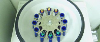
- Ultrasonography of structures.
- Angiography. Along with the previous named method, it is considered the gold standard for urgent diagnosis. Done immediately.
- MRI if necessary.
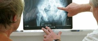
- X-ray of the chest and abdomen. Including to determine probable complications and current consequences of thromboembolism.
- Measurement of blood pressure, heart rate.
Attention:
Laboratory methods do not play a big role, except in cases of heart attack, when special marker proteins are released into the blood.
Preventive actions
Venous thromboembolism is a preventable disease. Prevention involves following the following medical recommendations:
- the use of elastic compression of the legs for varicose veins, before and during any medical interventions or during hormone therapy;
- getting up as early as possible after surgery;
- performing therapeutic exercises;
- to give up smoking;
- physical education and sports with mandatory weight loss;
- drug correction of diseases affecting blood clotting.
The most dangerous variant of VTE is pulmonary embolism. The risk of early death from leg vein thrombosis gradually increases as vascular disorders progress. Venous thromboembolism, as a factor in sudden cessation of blood circulation, requires timely identification and implementation of all necessary treatment and preventive measures.
Treatment
Mixed therapy. Conservative and operational. The use of tablets alone is possible only for mild and relatively harmless forms of thromboembolism, and even then not always. It is necessary to take into account the specifics of the situation.
Several groups of drugs are urgently used:
- Thrombolytics. Streptokinase, Urokinase. Dissolve clots.
- Anticoagulants. Aspirin, including Cardio modification, Heparin. Thin the blood.
- Anti-inflammatory non-steroidal origin in small dosages. Nimesulide, Nise, Ketorolac. If used incorrectly, it will provoke the formation of new blood clots, so you need to exercise maximum caution.
- Antispasmodics. Restore normal vascular tone, relieve excess tension and narrowing. Papaverine, Drotaverine.
The operation is mandatory. Its essence is to eliminate the blood clot mechanically, by removing part of the vessels, amputating a fragment of the intestine, the affected limb, and installing a special filter device to prevent further movement of blood clots.
Regular examinations by a hematologist (every 3 months) for 3 years or more are indicated. The other methods don't make sense.
Forecast and consequences
The outcome in most cases of arterial damage is conditionally unfavorable, even with timely treatment.
30% die, another 50% acquire permanent disability after surgery.
Only 20% of people can expect relatively good results. Discrepancies in case of vein damage are 10-15% in the direction of improved performance. And that's not always the case.
The consequences are terrible, deadly:
- Major heart attack, stroke.
- Necrosis (gangrene) of the intestines and limbs.
- Sepsis, blood poisoning.
The result is death or disability. Not all complications are named, but the most obvious ones in terms of the prospect of early death.
Stages of the disease
Thromboembolism of the great vessels is divided into stages according to the degree of damage to organs and tissues:
| Stage | Duration and prognosis | Degree of damage | Signs |
| First | The first 2 hours after an embolism, when blood circulation is restored, the functions of the organ are completely restored | Functional disorders | sharp pain |
| cold extremities | |||
| absence of pulse in peripheral arteries | |||
| minor limitation of joint mobility | |||
| Second | From 12 hours to 24 hours, assistance provided at this stage restores function by 85% | Organic changes | lack of pain and tactile sensitivity |
| traffic restrictions | |||
| bluish skin color | |||
| Third | 1–2 days after embolism, restoration of the main blood flow affects only the level of limb amputation | Necrosis | all types of sensitivity and mobility are lost |
| gangrene develops |
In rare cases, at the necrotic stage of embolism, rapid restoration of blood flow leaves a chance to save the leg, but in no more than 25% of cases.
Prevention
No specific methods have been developed. But you can minimize the risks if you adhere to certain points:
- Avoid stress. If it is impossible to master relaxation techniques. In order not to cause a hormonal “explosion” in every situation at work, study, etc.
- Eat consuming less animal fat and salt (no more than 7 grams).
- Sleep at least 7 hours per night.
- Adhere to an adequate individual physical activity regimen. Don't overexert yourself and don't sit still.
- Treat all pathologies in a timely manner, regardless of type and location.
- Regularly visit at least a therapist if you have problems with the cardiovascular system - a cardiologist. At least once a year for preventive examinations. Then the doctors will advise you on what to do if necessary.
Venous thromboembolism is a dangerous immediate or gradually increasing pathological process of partial or complete blockage of a blood vessel.
Requires urgent medical attention. Otherwise, dangerous complications cannot be avoided.
How to get rid of thrombosis and embolism?
It is better to prevent thrombus formation. The tendency to thrombosis and embolism is high in pregnant women. Hormonal changes in their body provoke an increase in blood clotting ability. The period of birth of a child is also dangerous, especially if a caesarean section is used. Expectant mothers should regularly attend antenatal clinics and follow the doctor's recommendations. A sedentary lifestyle is a factor that a person can modify, in contrast to metabolic and functional disorders in the body. If, due to the nature of his work, he remains in a standing or sitting position for a long time, the blood in his vessels stagnates, which contributes to the formation of blood clots. Warm-ups, moderate exercise, and swimming help a lot. Getting rid of bad habits, a healthy lifestyle and regular medical examinations will help avoid thrombosis and embolism.