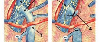
The disease is congenital in nature. The anomaly is a capsule with a cavity filled with liquid. A coelomic pericardial cyst can be located on a small stalk (pedicle) or be fused to the structures of the outer pericardial sac.
The initial stages of minor dysplasia are latent; in more complex cases, shortness of breath, cough, discomfort in the left side of the chest and heartbeat disturbances develop. This benign formation is more often diagnosed in females aged 20 to 45 years. Men encounter these types of cysts three times less often. Treatment consists of surgical removal of the neoplasia.
Pericardial cyst (coelomic pericardial cyst)
What is a cyst and how can it be dangerous?
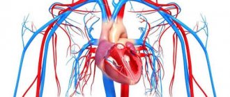
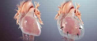
The pericardium or cardiac sac is a kind of bag that separates the heart from other organs and is its outer shell.
Between the pericardium and epicardium there is a small layer containing a specific colorless liquid. This layer allows the heart muscle to work without friction between the cardiac sac and the lining of the heart.
Sometimes a benign formation with a separate membrane appears on the outer shell of the heart. Inside it contains serous fluid - this is a coelomic cyst.
The tumor originates from the parietal layer of the pericardium and has a structure similar to it.
The International Classification of Diseases (ICD 10) encrypts this disease with code I31.
A coelomic cyst can be dangerous if it increases in size, if it is not diagnosed in a timely manner and if there is no treatment. In such cases, it can cause not only disturbances in the functioning of the heart, but also in other organs and systems.
Brief epidemiology:
- in the structure of heart diseases it is quite rare, only 4-10% of the total;
- women are more susceptible to its appearance;
- occurs in people of working age.
When and how to treat
If the pericardial formation is small in size and does not cause clinical symptoms or discomfort, then treatment is not prescribed. The person is registered and his condition is checked from time to time. Measures are taken in case of the following problems:
- rapid cyst growth;
- big sizes;
- symptoms of dysfunction of the heart and mediastinal organs as a result of compression;
- high probability of complications (rupture, infection, internal hemorrhage).
Conservative therapy does not help in this case. The only possible cure is surgery.
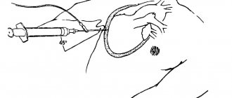
Previously, the use of therapeutic puncture was practiced, when the contents were removed and a sclerosant was introduced into the cavity. This method is almost never used anymore, since the secretion is produced again, and the chemical can cause constrictive pericarditis if it gets past the cyst.
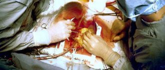
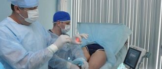
Removal is performed by thoracotomy or thoracoscopy. During an open operation, the formation is removed from the lining of the heart along with the walls that form it, and the area of violation of the integrity of the pericardium is sutured without harm to the functioning of the organ. Endoscopic techniques are more modern and minimally invasive methods. For this purpose, special equipment is used, which, through small incisions, allows a person to get rid of a cyst in the pericardium. The entire process is displayed on the screen, helping the surgeon to carry it out, control and correct it.
You can learn more about the method of removing a pericardial cyst using thoracoscopy from this video:
Usually such operations are successful, complications after them are extremely rare. Cases of relapses as a result of excision of the cystic cavity are not described. The prognosis is favorable; with timely intervention, complete restoration of the patient's working capacity is possible.
Case from practice
A 39-year-old patient was admitted to my hospital. Since childhood, he had problems with his heart, which recently began to bother him almost constantly, his pulse quickened, and shortness of breath appeared. A month before treatment, he was treated at the clinic for episodic ventricular tachycardia against the background of monomorphic single extrasystoles. I took Allapinin and Amiodarone. I did not notice any particular improvement while taking the medications.
After an echocardiography, a rounded cavity formation with clear walls was discovered. MRI confirmed the presence of a pericardial cyst. Treatment included radiofrequency ablation to restore normal pulses and removal of the cavity using thoracoscopy. A month after complete rehabilitation, Holter monitoring was performed. No rhythm disturbances were found.
In this case, a double pathology was observed. The source of impulse in the left ventricle caused rhythm disturbances and episodes of tachycardia. The cause of the rapid pulse was a coelomic cyst, and it also acted as a provoking factor for arrhythmia and extrasystole.
The following sources of information were used to prepare the material.
Most cardiac pathologies develop in the pericardial sac. Inside the walls of the pericardium there is not only the heart, but also numerous vessels. Pathologies of this tissue can lead to a variety of disorders. What features does a pericardial cyst have, and does it pose a danger to the patient?
Classification of pericardial tumors
According to the presence of association with the cavity of the outer shell of the heart:
- extraparacardial - laced cysts;
- pericardial (communicating) cysts or diverticula;
- parapericardial - cysts on a thin stalk or connected through a planar fusion.
By number of cameras:
- single-chamber;
- multi-chamber.
According to the course of the disease:
- asymptomatic;
- having complications;
- without complications.
Due to appearance:
- congenital (occurring during the intrauterine life of the fetus);
- acquired (arising as a result of inflammatory heart diseases or injuries).
By location on the heart:
- cardiophrenic node on the right 6%;
- cardiophrenic node on the left 3%;
- other departments 1%.
Causes
According to one theory, a pericardial cyst can develop against the background of inflammatory heart diseases - myocarditis, pericarditis or endocarditis.
While scientists are considering two theories about the causes of coelomic pericardial cysts.
I theory
According to the first theory, the formation of such cystic formations is associated with a violation of embryoogenesis. It is believed that cysts form in areas of pericardial weakness that bulge like a diverticulum. They communicate with the pericardial cavity and in the future may separate (unlace) from it and become isolated.
In addition, scientists suggest that a coelomic cyst may be a consequence of improper fusion of embryonic lacunae. Normally, these structures unite and form the pericardial sac. However, if one of the lacunae develops unevenly, a true diverticulum or a cyst isolated from the pericardium can form. So far, this is the theory that most scientists adhere to.
II theory
A number of experts believe that the formation of a coelomic cyst can be provoked not only by disturbances of embryoogenesis, but also by some factors affecting the pericardium after birth:
- inflammatory diseases (myocarditis, pericarditis, endocarditis);
- post-traumatic hematomas of the heart;
- tumor processes;
- parasitic diseases.
Symptoms of the disease
In most cases, at the onset of the disease, the pericardial cyst on the heart is asymptomatic. But as the tumor grows, surrounding tissues and organs are compressed.
Discomfort and aching pain in the heart area, pressure behind the sternum, lack of air, palpitations - this is not a complete list of manifestations of the neoplasm.
Symptoms mimic angina pectoris (even to changes on the ECG).
At the stage of differential diagnosis, it is important to conduct other research methods to prevent the cyst from rupturing and fluid entering the heart.
Depending on the direction of tumor growth, symptoms of compression of certain organs and systems predominate:
- Esophagus: - dysphagia (difficulty swallowing food and water).
- Bronchi: - asthma attacks; - the occurrence of a dry cough during exertion, shortness of breath; - difficulty breathing; - shortness of breath; - dry cough when changing body position.
- Atrium: - heartbeat; - pain in the chest, radiating to the shoulder or shoulder blade; - tachycardia; - heart rhythm disturbance.
- Circulatory systems: swelling of the neck veins.
What influences the formation of a pericardial cyst?
In recent years, cardiologists have quite often discovered this pathology in their patients. But what causes the appearance of pericardial tumors? According to doctors, this is influenced by several factors:
- Inflammation of cardiac tissue. These include pericarditis, endocarditis and myocarditis.
- Diseases of a parasitic nature.
- Myocardial injury. A cyst may occur due to the formation of a hematoma in the cardiac region.
- Pathological formation of the pleura at the stage of embryonic development.
- The appearance of a pericardial tumor. The formation of a coelomic cyst may relate to one of the stages of the tumor.
Causes of pericardial cysts
Diagnosis of mediastinal tumors
Depending on the prevalence of certain symptoms, the patient is initially admitted to the cardiology or pulmonology department.
Diagnosis of coelomic pericardial cysts includes several types of examination:
- Physical examination. It will help to identify nonspecific disturbances of the heart, which will determine further research tactics: - changes in tones during percussion (dullness of sound); - heart murmurs; - weakening of breathing.
- Electrocardiography (ECG). – Specific changes are recorded during adhesive processes in the pericardial area.
- X-ray of the chest, as well as fluoroscopy of the heart (this uses contrast, which is injected into the esophagus). — Allows you to identify a spherical shadow in the cardiophragmatic sinus (coelomic cyst). — Its outer contour will be smooth, clear, the inner contour in most cases merges with the shadow of the heart.
- Echocardiography (EchoCG). – Used for visualization and differential diagnosis of formation.
- Computed tomography (CT of the mediastinum). – Allowing a detailed examination of the characteristics of the tumor, its distribution in the mediastinum.
- MRI of the mediastinum and chest organs. — The most informative method of non-invasive studies. — Helps to make a differential diagnosis between benign and malignant processes.
- In the most difficult cases, diagnostic thoracoscopy (invasive method) is used. Used if the patient has: - diaphragm hernia; - malignant neoplasm of the heart muscle; - parasitic cyst (echinococcus).
Prevalence
Coelomic pericardial cyst
A pericardial cyst is a protrusion of the outer layer of the pericardium, which has the shape of a sac with thin walls. Inside the bag, as a rule, there is a colorless or slightly yellow liquid. It is called coelomic because in the process of embryonic development there are sections of the coelom - the embryonic tissue from which the pericardium is formed. Prevalence data differs among different authors. On average, coelomic pericardial cyst occurs in 6-15% of cases of all mediastinal tumors.
The mediastinum is an anatomical space in the chest filled with a complex of organs, including the heart. In women, coelomic pericardial cysts are more common. Its favorite location is the right cardiophrenic sinus. More than half of all cases are found here. A less common site of origin for coelomic pericardial cysts is the left cardiophrenic sinus. And the most rare localization of a pericardial cyst is the middle and upper parts of the mediastinum. Most often, this pathology is observed in middle-aged people; in more rare cases, it affects children and the elderly.
Types of treatment
The choice of treatment method determines the size of the tumor.
- Puncture of coelomic cyst. - Removing fluid from the cavity using a puncture needle. — This method has a temporary effect and the fluid can build up again, often increasing the formation in size and returning the symptoms of the disease.
- Sclerosis. – The introduction of a special substance into the cyst cavity can often cause complications (for example, constrictive pericarditis).
- Surgical method.
Surgery
The most effective method to get rid of the tumor is surgical treatment.
Surgical intervention involves removing the tumor, followed by suturing the pericardium and eliminating the complications that it caused.
Operations are performed in several ways:
- thoracotomy: classic open approach;
- Thoracoscopy: is less invasive and reduces the risk of side effects. — During thoracoscopic surgery, only a few main incisions are made on the chest. — Special endoscopic equipment is introduced, which allows access to the cystic cavity with minimal damage to surrounding tissues. — Removal of fluid from the neoplasm is carried out if the cyst is large or contains air. — During thoracoscopy, the tumor is completely enucleated, the risk of recurrence is minimal.
Any surgical treatment method (both thoracoscopy and thoracotomy) can lead to common complications:
- bleeding during surgery and in the postoperative period;
- vascular thrombosis;
- effect on the brain, since the operation is performed under general anesthesia (dizziness, short-term memory impairment).
What it is
A similar pericardial cyst is a kind of cavity formation, inside of which there is a colorless liquid. In this case, the pathology is connected structurally, as well as anatomically, with the pericardium itself. This anomaly was first described in the 19th century, when it was called “pericardial diverticulum.” It was then assumed that this formation was a protrusion of the parietal layer.
However, in the 40s of the 20th century, scientists proved that the appearance of such a pathology is a defect in the development of the coelom (the cavity in which the organ develops independently of other organs), due to which the disease received its current name. According to statistics, the share of such entities is only 4% of all. It often affects people under 40 years of age, and the anomaly is several times more common in women.
Complications
Neoplasms of the outer layer of the heart are rarely complicated. But such cases can still happen.
An increase in fluid volume leads to cyst growth and compression of internal organs.
Rupture of a pericardial cyst is a rare but very serious complication. It leads to a violation of the integrity of the cyst and fluid gets into the walls of the heart and nearby organs.
Manifested by symptoms of pleuropulmonary shock, hydrothorax, and may lead to malignancy.
Pleuropulmonary shock is a life-threatening complication that requires urgent treatment.
Manifested by the following symptoms:
- increase in severe shortness of breath;
- pale skin;
- drop in blood pressure and tachycardia;
- acrocyanosis;
- persistent cough;
- difficulty breathing, up to periods of apnea.
Rehabilitation after surgical treatment
In modern medicine, rehabilitation is of great importance. This is explained by the fact that diseases are getting younger and are increasingly affecting people of working age.
There can be risks of complications after any operation, even a relatively minor one. Rehabilitation is aimed at helping the patient adapt after the surgical intervention itself or after its complications, if any.
Restoring physical activity and ability to work is its main goal.
Recovery methods:
- Physical therapy (physical therapy) is a set of exercises developed for each patient.
- Metered walking - a gradual increase in the distance traveled.
- Diet: - reducing the amount of salt, foods containing large amounts of fat and cholesterol; - increasing the consumption of foods containing polyunsaturated fats (fish, seafood, olive oil); - introduce foods containing large amounts of fiber into your diet (bran, fruits, vegetables).
- Breathing exercises.
- Drug therapy: - lipid-lowering drugs (statins) - to correct blood cholesterol levels. These drugs also help stabilize cholesterol plaques and reduce the risk of postoperative vascular thrombosis. - antiplatelet therapy (drugs of the Acetylsalicylic acid group) - affect platelet aggregation, which reduces their deposition on the surface of the plaque, and inhibits the growth of atherosclerotic plaque.
Forecasts
After surgery, recovery is individual. Much depends on the age of the patient and how the intervention proceeded. Thoracoscopy is a minimally invasive technique. The risk of serious injury to the heart muscle is virtually eliminated. After excision of a cyst in the pericardium, many patients get out of bed within 24 hours. Rapid recovery is ensured by analgesics that relieve pain. Sometimes your doctor will prescribe antibiotics to prevent infection.
The patient can be discharged from the hospital 5-7 days after removal of the pericardial cyst. Usually, before discharge, additional examination is carried out to exclude postoperative complications.
In order to eliminate the risk of complications before and after treatment, experts recommend seeking help for the slightest ailments and pain in the heart area.
Prevention measures
There are no specific measures to help avoid the appearance of benign heart tumors in adulthood. A nonspecific measure is timely detection and therapy.
For this purpose, there are preventive examinations and medical examinations that need to be completed once a year.
The following studies will help find a tumor in the early stages:
- chest x-ray;
- ECG;
- EchoKg;
- CT scan of the mediastinum and chest.
Diagnostic tests
If a patient is suspected of having pericardial cysts, the doctor prescribes a whole range of instrumental studies:
- MRI is one of the most modern and unique methods. It allows you to see new growths. With its help, it is possible to understand whether these are really cysts or tumor or inflammatory diseases;
- EchoCG will also help determine the presence of neoplasms. Additionally, ultrasound makes it possible to see and evaluate the condition of the heart and its functionality;
- Catheterization. Performed by a professional surgeon. Indeed, during this invasive technique, penetration into the cavities of the heart occurs. Thus, the atria and ventricles are examined. All this allows you to see how intact the heart walls are at the moment;
- CT is an instrumental diagnostic method that allows the surgeon to see all the contours of the cysts. You can also understand how they are located, as well as what their features are.
- Thoracoscopy is a variant of endoscopic examination. It will visually show all pericardial neoplasms (what they represent, what their shapes and characteristics are).
To identify the location of the cyst, the following steps are taken:
- Multiplanar radiography, used as one of the first methods for identifying tumors. This is the first stage in the examination.
- Computer diagnostics makes it clear what kind of pathology is present in the body. Shows the location and radius of growth of the neoplasm.
- Echocardiography - determining the presence of pericardial cysts, as well as the condition of the heart.
- Thoracoscopy is a study using an endoscope that displays an image of formations.
- Magnetic resonance imaging is a modern method for diagnosing the disease, which determines the presence of a cyst and simultaneously helps to identify its nature.
- Catheterization refers to invasive methods that allow you to see the condition of the heart.
- Pericardial cyst on CT scan. In this case, all the outlines of the formations and their location are visible.
An experienced doctor, when visiting his patient for the first time, will be able to notice some protrusion of the chest, which indicates the presence of pathology.
Self-medication in all areas of the human body is fraught with adverse consequences, but in our world there are people who are categorically against surgical intervention. And they turn to traditional medicine for help.
Herbal medicine for any type of cyst can be said to be the same, and it consists of the following:
- Burdock juice, also called burdock. The composition of its leaves and roots includes alkaloids, about 50 percent inulin, resin, and some vitamins. Its action lies in its blood-purifying properties, which is why burdock is a means to combat neoplasms. To prepare burdock juice, you need to wash it thoroughly, dry it and grind it in a meat grinder, squeeze it and leave it for 5 days in a cool and dark place. It should be taken 2 times a day before meals for two months. However, it is imperative to undergo an ultrasound scan before and after the start of such therapy.
- Elecampane, which also contains natural inulin. Most often, this particular plant helps in the fight against small cystic formations. A decoction of elecampane is prepared as follows: yeast is diluted in 3 liters of boiled water that has cooled, then 40 grams of dried herb are added and left to infuse for 2 days. This infusion should be taken for 21 days, 2 times a day.
- Acacia with its excellent composition, which includes alkaloids, glycoside vitamins, quercetin, kaempferods, is an excellent remedy for treating cysts of various kinds. 5 tablespoons of flowers and leaves of the plant are poured with half a liter of vodka. Infuses for a week. This medication should be taken with caution due to the alcohol content. 1 teaspoon half an hour before meals, 2 times a day, course of therapy - 2 months.
Such methods of combating cyst formation should be carried out only under strict control and in accordance with the prescribed regimen, without avoiding a medical examination and consultation with doctors.