© Author: Sazykina Oksana Yuryevna, cardiologist, especially for SosudInfo.ru (about the authors)
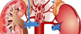
Thromboembolic complications are a rather serious problem in surgery, because they not only cause a severe course of the postoperative period, but can also lead to the sudden death of the patient. According to statistics, about 100 thousand patients in Russia die annually from sudden pulmonary embolism (PE). Mortality due to the development of massive pulmonary embolism is about 5%. Thromboembolism is represented by blood clots that form in the lumen of blood vessels and spread with the blood flow throughout the body. More often, blood clots form in the lumen of the veins of the lower extremities, and then enter the right half of the heart and then into the arteries of the lungs.
Regardless of the size of the blood clot, an artery of one diameter or another is blocked, as a result of which the area of the lung tissue supplied with blood by the branches extending from the blocked artery does not receive proper nutrition, and the tissue dies. The larger the thrombus, the wider the lumen of the blocked artery, the more branches do not receive blood, the more cells die in a larger area of the lung. The death, or necrosis, of cells is called pulmonary infarction. This is a pathognomonic morphological sign for pulmonary embolism (PE).
pulmonary embolism followed by pulmonary infarction
If a pulmonary infarction due to obstruction of the pulmonary artery by a thrombus leads to damage to a large amount of lung tissue, acute cardiopulmonary and respiratory failure occurs, which without treatment leads to death. That is why the prevention of thromboembolism in the postoperative period is one of the pressing problems of surgery.
But pulmonary embolism poses a danger to not only surgical patients, but also urological, traumatological, gynecological and obstetric patients. That is, for all patients who are planning or have already undergone surgery.
In addition to pulmonary embolism, thromboembolic complications include thrombosis of the inferior vena cava and acute phlebothrombosis of the lower extremities. These thromboses not only provide a direct background for the development of pulmonary embolism, but also pose a threat to the patient’s health.
thrombosis of deep veins of the leg (left) and inferior vena cava (right)
Causes of thromboembolism
The causative factors of venous thromboembolic complications (VTEC) can be divided into disturbances of normal blood flow in the veins of the lower extremities, as well as predisposing factors.
The first group of reasons includes all factors that contribute to the activation of the so-called Virchow triad, the essence of which is as follows. The formation of a blood clot in the lumen of a vessel is possible if the blood flow in the vein slows down, there is a violation of the integrity of the vascular wall, and there is also a tendency to blood hypercoagulation. All of these conditions occur during the early postoperative period in patients with conditions requiring emergency or elective surgery.
Thus, the development of VTEC is possible in the following conditions (the percentage of patients with venous thromboembolic complications out of the total number of those operated on is indicated in parentheses):
- Surgeries on the abdominal organs, including therapeutic or diagnostic laparoscopy (19)%,
- Gynecological operations, including therapeutic and diagnostic curettage of the uterine cavity and cesarean section (11.2%),
- Urological operations, including resection of prostate adenoma (7.1%),
- Neurosurgical operations (24%),
- Surgeries for malignant tumors of various locations (30%),
- Prosthetics of the knee or hip joints, as well as combined trauma and fractures requiring surgery or long-term immobilization (immobilization) of the patient (84%).
Predisposing factors include:
- Gender – in women, blood clots form in the veins more often due to hormonal characteristics,
- Age - the older the person, the higher the likelihood of blood clots in the veins,
- Lifestyle – “sedentary” and sedentary work contribute to stagnation of blood in the veins,
- The presence of varicose veins in the lower extremities - the more nodes and the higher the incompetence of the vein valves, the slower the blood flow through the vessel and the higher the tendency to platelet aggregation,
- Taking hormonal contraceptives (COCs - combined oral contraceptives), which significantly change the rheological properties of the blood,
- Hereditary disorders of the blood coagulation system - thrombophilia, or a tendency to increased thrombosis.
Basic prevention of pulmonary embolism in various situations
Blockage of the pulmonary artery by a thrombus (PE) most often occurs with thrombosis of the deep veins of the leg.
Therefore, patients at high risk of pulmonary thromboembolism are treated with elastic compression and anticoagulants.
It is indicated before and after surgical interventions, especially long and extensive operations, in traumatology practice, for ischemic stroke, and in pregnant women.
Order of the Ministry of Health on the prevention of pulmonary embolism
The special role of pulmonary artery blockage in complications of the postoperative period is reflected in regulatory documents. This is due to the fact that PE is the third leading cause of death after myocardial infarction and stroke, and 90% of deaths are caused by delayed diagnosis and treatment. Therefore, all patients considered at risk are recommended to:
- the earliest possible physical activity after surgery, childbirth - standing up and active walking;
- avoiding prolonged standing or sitting with legs down;
- wearing compression hosiery (stockings) of compression class 1 - 2, bandaging with elastic bandages before and during surgery (except for operations on the lower extremities);
- in the postoperative period, administration of anticoagulants or pneumatic compression if Heparin and its analogues are contraindicated;
- wearing elastic medical knitwear during inpatient and outpatient treatment.
We recommend reading the article on the prevention of thrombosis. From it you will learn about what thrombophilia is, symptoms, diagnosis and treatment of thrombophilia.
And here is more information about how to prevent thrombophlebitis after surgery.
Risk groups for pulmonary embolism
Antithrombotic therapy is indicated for the following categories of patients:
- with prolonged bed rest - physical inactivity leads to the formation of unstable blood clots, which are prone to destruction and detachment from the wall;
- in the case of a planned operation, stress and pain stimulate the formation of blood clots; a long operation, especially in the pelvic cavity, lower extremities increases the risk of venous thrombosis. Any tissue damage also triggers the release of clotting factors. Blood loss and centralization of blood circulation increase blood viscosity and thrombophilia;
- when using general anesthesia, too strong or prolonged anesthesia causes hypercoagulation and provokes stagnation of blood in the legs;
- after administration of whole blood or red blood cells;
- elderly - after 40 years, anticoagulant activity decreases, the structure of the vascular wall is disrupted, cardiac output and the return of venous blood to the heart decrease, the risk of thrombosis increases with previous physical inactivity;
- with malignant neoplasms – blood clotting is activated;
- with excess body weight – thrombosis is associated with impaired fat metabolism;
- history of venous thrombosis, thromboembolism, varicose veins.
Additional risk factors include heart failure, atrial fibrillation, bacterial endocarditis, myocarditis, and cardiomyopathy. PE is often detected in patients with diabetes mellitus, sepsis, congenital thrombophilia, and antiphospholipid syndrome.
Pregnancy, especially complicated by gestosis, the labor and postpartum periods are also included in the list of risk factors. Accelerated formation of blood clots can be provoked by the use of hormones for contraception, treatment of menopause, kidney and intestinal diseases, heart attack and stroke, antitumor radiation or chemotherapy.
How is prevention carried out?
In addition to the general principles of preventing pulmonary embolism (activity, compression, heparin therapy), there are features in various clinical situations.
Before surgery
If a high risk of developing pulmonary thromboembolism is detected, then it is necessary:
- shorten the preoperative hospital period - examine on an outpatient basis;
- use gymnastics, massage, physiotherapy, if there are no contraindications;
- avoid overeating;
- use elastic bandaging of the lower extremities or compression hosiery;
- take tranquilizers and psychotherapy to reduce the release of stress hormones.
Watch the video about the development of pulmonary embolism:
Before surgery, the blood volume deficit is eliminated and the hematocrit is normalized. In case of increased coagulation activity, Refortan is used intravenously, as well as Heparin or Fragmin with great caution so as not to cause bleeding; any anticoagulants are canceled 24-12 hours before.
In traumatology and postoperative period
Direct anticoagulant therapy is carried out with the help of Heparin for at least 3 days and low molecular weight analogues - Tsibor, Fragmin, Clexane.
They are recommended to be used 2 times a day, first together with Heparin, reducing its dose, and then independently.
The minimum period of heparin therapy is 2 weeks, and in case of cancer it is often extended to 3 months. It is extremely important to prevent bleeding.
In addition to these drugs, after major orthopedic operations the following are used:
- antiplatelet agents for elevated platelet levels - Cardiomagnyl, Plavix;
- solutions to maintain hematocrit at 30 - 33%;
- early transfer to a normal diet, since protein and especially fat emulsions increase the formation of blood clots;
- painkillers and anti-inflammatory drugs;
- transfusion of fresh frozen plasma for deficiency of anticoagulant factors.
In patients with ischemic stroke
In the acute period of a stroke, almost every seventh patient experiences pulmonary embolism. The difficulty in identifying this complication is due to the fact that often a blood clot forms in a paralyzed limb, the blockage of the vessel is asymptomatic, and due to impaired consciousness there are no complaints. If prophylactic anticoagulant therapy is carried out, the probability of thromboembolism is reduced to 0.7%.
In this regard, patients who have obesity, varicose veins of the lower extremities, congestive heart failure, pulmonary lesions, and an indwelling venous catheter are indicated for the prevention of pulmonary embolism. For this purpose the following is used:
- adequate administration of solutions or drinking regimen to maintain high blood fluidity;
- development of paralyzed limbs from the first days of illness;
- bandaging or compression with medicinal knitwear throughout the entire period of bed rest;
- Fraxiparine, Clexane subcutaneously once a day;
- Thrombo Ass, Aspirin cardio at a dose of 150 - 300 mg per day.
In pregnant women
PE is considered the most common cause of maternal death. Its risk during pregnancy increases 5-6 times, and in the postpartum period, especially after cesarean section, the probability is even higher.
The most susceptible to pulmonary thromboembolism are patients over 35 years of age, as well as those who have:
Heparin is used to prevent pulmonary embolism in pregnant women. It is administered for up to 2 months with a moderate risk of thromboembolism and at least three months with a high risk. After childbirth, women can be switched to Warfarin, Pradaxa, Xarelto. Most often, breastfeeding is not canceled during anticoagulant therapy.
Types of PE prevention
Depending on the goals, primary and secondary measures to prevent pulmonary thromboembolism are distinguished, and in addition to traditional medication, a surgical method is also used.
Primary and secondary
To prevent the formation of blood clots in the venous network, primary prevention is carried out. It includes:
- identification of risk groups;
- wearing compression stockings;
- the earliest possible physical activity after childbirth, operations;
- the use of massage and therapeutic exercises;
- maintaining normal platelet counts and blood viscosity;
- the use of Heparin, low molecular weight analogues with a transition to Warfarin during operations and injuries;
- normalization of body weight;
- when traveling - sufficient drinking regime, compression stockings, prohibition of alcohol, movement;
- avoiding smoking;
- blood pressure control, in case of hereditary predisposition - additionally coagulograms and D-dimer;
- choosing contraception without hormonal drugs.
Secondary prevention is aimed at preventing relapse in cases of already developed pulmonary embolism. It involves the administration of anticoagulants in therapeutic doses and surgical treatment. For long-term use, Warfarin is most often recommended.
How to assess the risk of VTEC?
Any surgical doctor planning surgical intervention for his patient must be able to assess the risks of thromboembolic complications, and in particular, the risk of developing pulmonary embolism.
The risk assessment of VTEC is determined based on the nature of the surgical intervention:
- by a low risk of thromboembolic complications in the postoperative period in surgical patients. The risk of pulmonary embolism during surgery is less than 0.2% of all those operated on, including 0.002% of deaths as a result of massive thromboembolism. These include laparoscopic interventions and transurethral urological manipulations on the prostate.
- An average degree of risk with the incidence of thrombosis in less than 5% of operated patients is typical for major operations. These include removal of the gallbladder, appendectomy with complications (phlegmonous, gangrenous appendicitis), cesarean section or amputation of the uterus, removal of part of the stomach or intestines, removal of prostate adenoma with transvesical access.
- Interventions that are accompanied by a high incidence of VTEC (more than 80% of thrombosis in the deep veins of the legs, more than 40% of thrombosis in the inferior vena cava and more than 10% of pulmonary embolism, including fatal ones) include extended operations - removal of malignant neoplasms, traumatological and orthopedic operations with joint replacement, as well as neurosurgical interventions.
In this regard, the first group of operations implies a low degree of risk of VTEC, the second group - a moderate degree of risk, and the third group - a high degree of risk of VTEC.
Prevention, diagnosis and treatment of postoperative pulmonary embolism
Key words: postoperative pulmonary embolism, heparin prophylaxis, bemiparin. Pulmonary embolism (PE) is one of the fairly common and dangerous emergency situations, incl. in the postoperative period. The cause of pulmonary embolism is the occlusion of the trunk or small branches of the arteries of the pulmonary circulation by thromboembolism with the development of right ventricular and then biventricular heart failure. In the structure of mortality from cardiovascular diseases, pulmonary embolism ranks third after myocardial infarction and stroke. In economically developed countries, 0.1% of the population die annually from this pathology [1, 3, 9]. It is not possible to give an accurate estimate of the incidence of postoperative pulmonary embolism, since this condition is often asymptomatic. During life, the diagnosis of pulmonary embolism is established in less than 70% of cases. Mortality among patients without pathogenetic therapy, according to various authors, is 40% or more, with massive pulmonary embolism it reaches 70%, and with timely started therapy it ranges from 2 to 8% [1, 9]. According to a number of authors, pulmonary embolism is the cause of 5% of deaths after general surgery and 23.7% after orthopedic operations. Pulmonary embolism occupies one of the leading places in obstetric practice: mortality from this complication ranges from 1.5 to 2.7% per 10 thousand births, and in the structure of maternal mortality it is 2.8–9.2% [2]. In European countries, in particular in France, the incidence of pulmonary embolism is up to 100 thousand cases per year, in England and Scotland 65 thousand are hospitalized with pulmonary embolism, and in Italy – 60 thousand patients annually [8]. In the United States, up to 150 thousand patients are diagnosed annually with pulmonary embolism as a complication of various diseases. Among hospitalized patients, 70% are therapeutic patients [8]. According to the Framingham study, death from pulmonary embolism accounts for 15.6% of all in-hospital mortality, with surgical patients accounting for 18% of cases, and 82% were patients with therapeutic pathology [1, 9]. It should be understood that the source of the thrombus that reaches the pulmonary circulation is mainly located in the vessels of the inferior vena cava basin, namely in the veins of the lower extremities and pelvis. Most often, the primary thrombus is located in the ileocaval segments or proximal veins of the lower extremities (popliteus-femoral segment). Venous thrombosis localized in the distal deep veins of the lower extremities (legs) is complicated by pulmonary embolism in 1–5% of cases. Recently, there have been reports of an increase in cases of pulmonary embolism from the superior vena cava (up to 3.5%) as a result of the placement of venous catheters in intensive care units and intensive care units [1, 9]. The most dangerous for the development of pulmonary embolism are “floating thrombi”, which have a fixation point in the distal part of the venous bed; the rest of them are located freely and throughout their entire length are not connected to the walls of the vein, and their length can vary from 5 to 20 cm. “Floating thrombus” usually forms in veins of smaller caliber, and the process of thrombus formation spreads proximally to larger ones: from the deep veins of the leg – into the popliteal vein, then into the deep and common femoral artery, from the internal – into the common iliac, from the common iliac – into the inferior vena cava [1]. The size of thromboemboli determines their localization in the vessels of the pulmonary artery; they are usually fixed at the sites of division of the pulmonary vessels. According to various authors, embolization of the trunk and main branches of the pulmonary artery occurs in 50%, lobar and segmental - in 22%, small branches - in 30% of cases. Simultaneous damage to the arteries of both lungs reaches 65% of all cases of pulmonary embolism, in 20% only the right lung is affected, in 10% only the left lung is affected, the lower lobes are affected 4 times more often than the upper ones [9]. Risk factors for venous thromboembolism (PE in particular) are the following: old age, prolonged immobility (due to paresis of the limbs, after injuries, in the postoperative period, with frequent and long flights on planes or trips in cars, etc.), cancer, trauma (especially fractures of large bones), surgical interventions and intravascular invasive procedures (subclavian catheter, etc.), taking certain medications (hormone replacement therapy, use of oral contraceptives, chemotherapy), chronic cardiac or respiratory failure, pregnancy and the postpartum period, thrombophilia. There is evidence that PE is also associated with problems such as obesity, metabolic syndrome, hypertension, smoking, and cardiovascular events (myocardial infarction, stroke). Thus, thromboembolic complications, according to various authors, occur in 30–60% of patients with strokes that caused paralysis of the lower extremities, in 5–35% of patients after myocardial infarction, in more than 12% of people with congestive heart failure. A serious risk factor is a history of venous disease of the lower extremities, especially deep vein thrombosis. The incidence of pulmonary embolism increases with age: the average age of patients with pulmonary embolism is 62 years, while the majority (at least 65%) of patients are over 60 years of age, and in patients over 80 years of age, pulmonary embolism occurs 8 times more often than in persons under 50 years of age [1, 9]. Thrombi from the veins of the lower extremities and pelvis with the blood flow enter the right atrium, then into the right ventricle, where they fragment. From the right ventricle, blood clots enter the pulmonary circulation. Massive pulmonary embolism is accompanied by an increase in pressure in the pulmonary artery, and this leads to an increase in overall vascular resistance in the lungs. Overload of the right ventricle occurs, a drop in cardiac output and the development of acute cardiovascular failure. Diagnosis of postoperative pulmonary embolism is not an easy task. Symptoms of pulmonary embolism do not have clear specificity; the clinical picture is associated with exacerbation of diseases such as coronary heart disease, chronic heart failure, lung diseases, or is one of the complications of cancer, trauma, or extensive surgical interventions. The most characteristic symptoms of PE are “silent” shortness of breath of the inspiratory type with a frequency of up to 24 beats/min. and higher, tachycardia more than 90 beats/min., pallor with an ashen tint of the skin. With massive pulmonary embolism, there is (in 20% of cases) severe cyanosis of the face, neck and upper half of the body. Substernal pain during embolism of the main trunk of the pulmonary artery is tearing in nature (irritation of the afferent endings embedded in the artery wall). A sharp decrease in coronary blood flow due to a decrease in stroke and cardiac output can cause angina pain. Acute liver swelling with right ventricular failure causes severe pain in the right hypochondrium, often combined with symptoms of peritoneal irritation and intestinal paresis. The development of acute cor pulmonale is manifested by swelling of the neck veins and pathological pulsation in the epigastrium. An accent of the second tone is heard on the aorta, a systolic murmur is heard under the xiphoid process, and a gallop rhythm is heard at Botkin’s point. Central venous pressure may be significantly elevated. Severe arterial hypotension, up to the development of shock, indicates massive pulmonary embolism. Hemoptysis, observed in 30% of patients, is caused by the development of pulmonary infarction [1, 7, 9]. Instrumental diagnostic methods require special procedures that have limited distribution. These include, first of all, angiography of the pulmonary circulation (the “gold standard” for diagnosing pulmonary embolism) and lung scintigraphy. With angiography, direct signs of PE are a filling defect in the lumen of the vessel and “amputation” (i.e., breakage) of the vessel, and indirect signs are dilation of the main pulmonary arteries, a decrease in the number of contrasted peripheral branches, and deformation of the pulmonary pattern. X-ray studies are not very informative. The most characteristic symptoms of acute cor pulmonale (in 15% of cases) as manifestations of pulmonary embolism are expansion of the superior vena cava and the shadow of the heart to the right, as well as swelling of the conus of the pulmonary artery, which is manifested by flattening of the waist of the heart and bulging of the second arch beyond the left contour. Expansion of the lung root may be observed (in 4–16% of cases), its “chopping off” and deformation on the affected side. With embolism in one of the main branches of the pulmonary artery, in the lobar or segmental branches in the absence of background bronchopulmonary pathology, depletion (“clearing”) of the pulmonary pattern is observed (Westermarck’s symptom). Discoid atelectasis, observed in 3–8% of cases, usually precedes the development of pulmonary infarction and is caused by bronchial obstruction with hemorrhagic secretions or an increased amount of bronchial mucus, as well as a decrease in the production of alveolar surfactant. Pulmonary infarction develops 2–3 days after embolization and is clinically manifested by pain in the chest when breathing and coughing, shortness of breath, tachycardia, crepitus, moist rales in the lungs, and hyperthermia. Currently, the algorithm for diagnosing PE is based primarily on the use of computed tomography (CT), a sensitive, non-invasive and relatively accessible method of examination, but it is first necessary to exclude patients who will not require CT (for high-risk patients - using an echocardiogram (EchoCG), for patients low risk - by assessing the likelihood of pulmonary embolism and determining the level of D-dimer). CT is currently recognized by experts as the most acceptable method for confirming the presence of a thrombus in the pulmonary vessels, recommended for routine clinical practice. In high-risk patients, conventional (single-detector) CT is sufficient to confirm or exclude PE, but in low-risk patients it is recommended to use multi-detector CT, which allows more clearly visualization of the segmental and subsegmental branches of the pulmonary artery and reliably confirm or exclude PE. If multidetector CT is not available, these patients are either diagnosed with PE when single-detector CT is positive, or double follow-up is required to rule out PE when negative single-detector CT is compared with venous compression ultrasonography, and if both tests do not detect thrombi, only then the diagnosis of pulmonary embolism can be removed. On the ECG, acute overload of the right ventricle due to high pressure in the pulmonary circulation leads to the appearance of SI and QIII waves (the so-called SI-QIII type). In leads V1, V2, the amplitude of the R wave increases. An S wave may appear in leads V4–V6. The ST segment shifts discordantly downward in leads I, II, aVL and upward in leads III, aVF, and sometimes V1 and V2. At the same time, a pronounced negative T wave appears in leads V1–V4, as well as in leads II and aVF. Overload of the right atrium can lead to the appearance of a high P wave in openings II and III (P-pulmonale). Signs of acute overload of the right ventricle are more often observed with embolism of the trunk and main branches of the pulmonary artery than with damage to the lobar and segmental branches. Using echocardiography, it is possible to assess the parameters of hemodynamics and the structural state of the myocardium, the severity of hypertension in the pulmonary circulation, the presence of thrombotic masses in the cavities of the heart, exclude heart defects and evaluate the results of treatment. The most common findings include dilatation of the right heart and pulmonary artery, paradoxical movement of the interventricular septum, tricuspid regurgitation, lack of collapse of the inferior vena cava, and a patent foramen ovale, which is rare. In the results of laboratory tests, a high titer of D-dimer may indicate the presence of venous thrombosis and PE in a patient only if other conditions that occur with the formation of fibrin are excluded, for example, foci of necrosis and inflammation (in diseases of the abdominal and thoracic organs, abscesses, after recent surgical interventions and injuries). At the same time, this indicator can be used as a screening method due to its high sensitivity. However, there are no indicators that clearly indicate the occurrence of pulmonary embolism. The probability of pulmonary embolism can be preliminary assessed using the MW Roges and PS Wells scale (2001) [7]: • clinical symptoms of deep vein thrombosis of the lower extremities – 3 points; • when carrying out differential diagnosis, pulmonary embolism is more likely than other diseases – 3 points; • tachycardia >100 beats/min. – 1.5 points; • immobilization or surgery during the last 3 days – 1.5 points; • history of deep vein thrombosis of the lower extremities or pulmonary embolism – 1.5 points; • hemoptysis – 1 point; • oncological pathology currently or up to 6 months ago. – 1 point. If the sum does not exceed 2 points, the probability of pulmonary embolism is low; with a score of 2–6 – moderate; more than 6 points – high. In addition, in recent years in Europe the so-called Geneva scale is often used [4]: • tachycardia ≥95 beats/min. - 5 points; • tachycardia 75–94 beats/min. – 3 points; • clinical signs of deep vein thrombosis of the lower extremities (pain on palpation of the vein + swelling of one limb) – 4 points; • suspicion of deep vein thrombosis of the lower extremities (pain in one limb) – 3 points; • confirmed deep vein thrombosis of the lower extremities or a history of pulmonary embolism – 3 points; • surgery or fracture within the last month – 2 points; • hemoptysis – 2 points; • oncological pathology – 2 points; • age over 65 years – 1 point. If the sum does not exceed 3 points, the probability of pulmonary embolism is low; with a score of 4–10 – moderate; ≥11 points – high. The main areas of treatment for PE are hemodynamic and respiratory support, reperfusion (thrombolysis or surgical removal of emboli from the pulmonary arteries), and anticoagulant therapy. Moreover, the treatment strategy significantly depends on the degree of risk [1, 7]. Anticoagulant therapy should be started immediately in patients with a high or moderate probability of PE while still in the diagnostic process, without waiting for final confirmation of the diagnosis. Considering the high risk of rapid development of life-threatening complications, the need for aggressive therapy and its careful clinical and laboratory monitoring, it is advisable to carry out all therapeutic and diagnostic measures in patients with suspected acute pulmonary embolism in intensive care units. Treatment of pulmonary embolism in high-risk patients: 1. Begin treatment with fast-acting direct anticoagulants, followed by as early a transition to indirect anticoagulants as possible. 2. To prevent further progression of right ventricular failure, it is necessary to eliminate systemic arterial hypotension. For this purpose, it is recommended to use vasopressors. In patients with low cardiac output and normal blood pressure, inotropes such as dobutamine and dopamine may be used. 3. Aggressive fluid resuscitation is not recommended. 4. Patients with hypoxemia require oxygen therapy. 5. In high-risk patients with pulmonary embolism accompanied by cardiogenic shock and/or arterial hypotension, thrombolytic therapy is indicated. 6. If thrombolysis is absolutely contraindicated or has proven ineffective, surgical embolectomy is an alternative method of reperfusion and percutaneous catheter embolectomy or thrombus fragmentation may also be considered as an alternative method of reperfusion. Treatment of pulmonary embolism in patients at low (moderate or low) risk: 1. Anticoagulant therapy. 2. Routine use of thrombolytic therapy in low-risk patients is not recommended, but may be considered in some moderate-risk patients. Thrombolysis is most effective in patients who have undergone reperfusion within 48 hours of the onset of PE, but thrombolytic therapy may also be successful in those patients whose first symptoms of PE appeared 6–14 days ago. Thrombolytic therapy is not indicated in low-risk patients. For thrombolytic therapy for pulmonary embolism, the use of three drugs is currently approved [1]: 1. Streptokinase or according to the usual regimen - a loading dose of 250 thousand IU (over 30 minutes), then 100 thousand IU per hour for 12–24 h, or accelerated - 1.5 million IU for 2 hours. 2. Urokinase or according to the usual regimen - loading dose of 4400 IU/kg body weight (for 10 minutes), then 4400 IU/kg body weight per hour in for 12–24 hours or according to an accelerated regimen - 3 million IU for 2 hours. 3. Tissue plasminogen activator (alteplase): 100 mg for 2 hours or 0.6 mg/kg body weight for 15 minutes. (maximum dose 50 mg). In modern conditions, a special role is given to the prevention of pulmonary embolism, which, according to the order of the Ministry of Health of the Russian Federation dated 06/09/2003 No. 233 “On approval of the industry standard “Protocol for the management of patients. Prevention of pulmonary thromboembolism in surgical and other invasive interventions ””, consists in the following events: • detection of patients with a high degree of risk of pulmonary pulmonas. • The most early activation of the patient in the postoperative period: lifting and active walking (however, a long static load is contraindicated in the standing position, sitting). • Elastic compression of the lower extremities (elastic bandages or stockings 1 or 2 compression classes). Elastic bandages or stockings are used before surgery, during surgery (except for operations on the lower extremities) and after surgery up to discharge from the hospital. • interspersing pneumatic compression (recommended for use for contraindications to heparinoprophylaxis). • The use of drugs. The prophylaxis algorithm with direct anticoagulants is the prescription of sodium heparin in a daily dose of 15,000 IU, with body weight below 50 kg, the daily dose of heparin is reduced to 10,000 IU. Heparin is introduced under the skin of the abdomen, the interval between injections is 8 hours. The duration of preventive heparinization is at least 3 days. Prolonged bleeding serves as an absolute contraindication to the appointment of anticoagulants. The prophylaxis algorithm with indirect coagulants includes the use of low -molecular heparins of the first generation: Dalteparin, Obrooparin, Enoxyparin. These drugs are convenient in that they can be administered subcutaneously once a day in doses of 0.6 ml/day (0.8 ml/day with body weight above 120 kg), 5000 units/day (7500 units with body weight from above 120 kg) or 40 mg/day (60 mg/day with body weight above 120 kg), respectively. Antiplatelet agents. At the level of platelets exceeding the norm, the purpose of antiplatelets is shown: acetylsalicylic acid, tyklopidine, clopidrogeel in general metal doses. Despite the fact that Order No. 233 regulates the use of Nefractionated heparin and low -molecular heparins of the first generation, it should be remembered that in our arsenal there is already a drug of low -molecular second -generation heparins. Currently, it is actively studied, and the amount of data on its clinical application is limited. Nevertheless, studies have already been conducted that characterize its effectiveness in patients after orthopedic, gynecological and other types of operations. Bemiparin (Tsibor®) is a low -molecular heparin of the second generation. It has the lowest molecular mass equal to 3600 and the longest period of half -life (5.3 hours). Preliminary results of recent studies suggest that low molecular weight heparins, in particular Bemiparin, can be used to treat and prevent thromboembolic complications, including And outside the hospital, in patients with mild fat and in patients with cancer [5]. Such an opportunity was investigated in a double blind multicenter study of Canbesure (n = 703) [6]. This study included patients over 40 years old who conducted surgical intervention over previously diagnosed malignant tumors (primary or metastatic) gastrointestinal tracts, urinary tract or female reproductive system. The study included only patients with a life expectancy of at least 3 months, in which the operation was carried out under general anesthesia or spinal anesthesia and lasted at least 30 minutes. Before randomization, all patients received Bemiparin subcutaneously at a dose of 3500 IU 1 r/day for 8 ± 2 days (the first dose was administered 6 hours after surgery). Then patients randomly were divided into a group of continuation of Bromyparin thromboprophylaxis in the same mode for an additional 20 ± 2 days and a placebo-control group. 28 days after the operation, bilateral ascending venography was performed. Distant results were observed for 3 months. The main end point of efficiency was the totality of symptomatic and asymptomatic thrombosis of deep veins (TGV), non -phalual fat, as well as cases of death from any reason. A prospective record of an additional combined endpoint was also carried out, which included proximal TGV, symptomatic non -fatal pulp and deaths due to fatla. The main final point of efficiency was registered in 10.1% of Bemiparin group patients and 13.3% of patients in the placebo group. Although the differences between the groups did not reach a statistically reliable level (p = 0.263), the level of relative risk of developing pulmonary fiber in the Bemiparin group was lower by 24.4%. The frequency of the onset of the additional final point was significantly lower in the Bemiparin group (0.8% versus 4.6%, p = 0.016), and the relative risk of developing the body in the Bemiparin group was 82.4% lower than the placebo group. The risk of hemorrhagic complications in the extension of thromboprophylaxis of Bemiparin by 3 weeks. Reliably did not increase. Serious bleeding developed in 0.6% of Bemiparin’s patients against 0.3% of the placebo group (inaccurate differences, p = 0.572). The results of the Canbesure study allow us to conclude that extended to 4 weeks. Brombiparine thrombopalance at a dose of 3500 IU/day after abdominal or pelvic operations to remove malignant tumors reliably reduces the risk of severe thromboembolism without increasing the risk of hemorrhagic complications, compared with the standard weekly course of thromboprophylaxis with direct anticoagulants. In conclusion, it should be said that postoperative fat remains an urgent modern problem. Despite the presence of a large volume of literary data, the choice of treatment regimen and prevention of postoperative fella lies on the shoulders of the attending physician and depends on the security of the medical institution.
Literature 1. Kotelnikov M.V. Pulmonary embolism (modern approaches to diagnosis and treatment). M., 2002. 2. Makarov O.V., Ozolinya L.A., Parkhomenko T.V., Kerchelaeva S.B. Prevention of thromboembolic complications in obstetric practice // Ros. honey. magazine 1998. No. 1. P. 28–32. 3. Darryl Y. Sue, MD Pulmonary Disease // Frederic S. Dongard (ed.): Current: Critical Care Diagnosis & Treatment. US – a language medical book. First Edition. P. 496. 4. Le Gal G., Perrier A. Contemporary approach to the diagnosis of non-massive pulmonary embolism // Curr. Opin. Pulm. Med. 2006..Vol. 12(5). P. 291–298. 5. Martanez-Gonzilez J., Vila L., Rodriguez C. Bemiparin: second-generation, low-molecular-weight heparin for treatment and prophylaxis of venous thromboembolism // Expert. Rev. Cardiovasc. Ther. 2008. Vol. 6(6). P. 793–802. 6. Monreal BM, Vignoli A, Lecumberri VR et al. Bemiparin in oncology // Drugs. 2010. Vol. 70 (Suppl. 2). P. 35–42. 7. Rodger M., Wells PS Diagnosis of Pulmonary Embolism // Thromb. Res. 2001. Vol. 103. P. 225–238. 8. Sharma GVRK, Schoolman M., Sasahara AA Diagnosis and Treatment of Pulmonary Embolism // Melvin M., Sheinman, MD (eds.) Cardiac Emergencies. WB Saunders Company, 1984. P. 349. 9. Task Force Report. Guidelines on diagnosis and management of acute pulmonary embolism. European Society of Cardiology // Eur. Heart J. 2000. Vol. 21. P. 1301–1336.
What are the symptoms of thromboembolic complications?
Complications such as deep vein thrombosis of the lower extremities are characterized by severe pain in the lower leg and foot, accompanied by a blue or purple color of the skin below the site of thrombosis. These symptoms are due to the fact that when a vein is blocked, blood does not flow from the limb, leading to bursting pain. Even minor discomfort in one or both limbs after surgery should not go unnoticed by the doctor.
acute thrombosis of leg veins
Pulmonary embolism has manifestations of varying severity. Sometimes, due to the insignificance of symptoms, pulmonary embolism of small branches may remain unrecognized, leading to complications from the lungs and heart, for example, to the development of chronic thromboembolic pulmonary hypertension.
patient with pulmonary embolism
Typically, pulmonary embolism of small branches is accompanied by attacks of dry cough or hemoptysis with pain in the chest of various localizations. Often the patient experiences attacks of sudden shortness of breath and a feeling of lack of air. Loss of consciousness may be present.
Massive pulmonary embolism is characterized by severe pain in the chest, shortness of breath, hemoptysis and cyanosis (blue discoloration) of the skin of the face, neck, earlobes and chest strictly up to the horizontal line between the nipples. Clinical death can occur instantly, which without treatment turns into biological death. In some cases, the patient may simply get up and die.
Diagnosis of VTE
The diagnosis of deep vein thrombosis of the leg or inferior vena cava can be confirmed using vascular ultrasound.
X-ray signs of pulmonary embolism (Fig.: NSC “Institute of Cardiology N.D. Strazhesko”)
PE is confirmed by chest x-ray, but the absence of characteristic x-ray signs does not justify excluding the diagnosis. In other words, the diagnosis of thromboembolism, even with a normal chest x-ray, can be established on the basis of clinical data.
A mandatory study for suspected VTE is a blood test for D-dimer, as well as a study of the blood coagulation system (INR, fibrin, clotting time, associated partial thrombin time - APTT, prothrombin time - PTT and prothrombin index - PTI).
After a comprehensive assessment of the data obtained, treatment begins.
Treatment of thromboembolic complications
Any doctor needs to remember that the mortality rate for massive pulmonary embolism without treatment is more than 90%, so therapy should be started as early as possible.
The main principle of treatment is to dissolve the blood clot and correct the blood clotting disorder. In this regard, in the intensive care unit the following drugs are administered intravenously to the patient:
- Low molecular weight heparins - heparin at a dose of 31-33,000 units/day for 5-7 days or enoxaparin at a dose of 180 mg/day for 5-7 days,
- Drugs for thrombolysis - streptokinase at a dose of 250,000 units in the first 30 minutes, then 100,000 units on the first day or alteplase at a dose of 100 mg on the first day.
Of the tablet medications, warfarin is used at a dose of 10 mg for 5-7 days.
vena cava filter that catches blood clots
If there are indications, the patient can undergo surgical treatment of thrombosis - installation of a vena cava filter in the lumen of the inferior vena cava or embolectomy of the inferior vena cava.
The indications for surgery are the following:
- Recurrent pulmonary embolism during adequate anticoagulant therapy,
- Extensive or progressive thrombosis of the inferior vena cava,
- Planned or performed surgery in a patient with a history of pulmonary embolism.
How to prevent thrombophlebitis after surgery
Thrombophlebitis occupies one of the first places in the structure of morbidity after surgical intervention. Often it is asymptomatic, and its manifestation is only a sudden pulmonary embolism. Therefore, it is necessary to prevent thrombophlebitis in postoperative patients using non-drug methods and medications.
What is thrombophlebitis
Thrombophlebitis is a combination of inflammation of the venous wall and the formation of a blood clot. Its development is provoked by a combination of two factors: increased blood clotting and venous stagnation.
| ⇐ Previous12345678Next ⇒ Preventive measures against venous thrombosis should take into account etiopathogenetic factors. In this regard, special attention should be paid to patients with an increased risk of venous thrombosis. The highest percentage of vein thrombosis and thromboembolic complications, as noted above, is observed in surgical patients. In the postoperative period, prevention includes early mobilization of patients, elevated position of the limbs in bed 20-30° above the bed level, elastic bandaging of the limbs during surgery and during the entire period of bed and semi-bed rest. In patients who have suffered acute venous thrombosis or after surgery for venous disease, the duration of elastic bandaging of the limb is determined by the clinical picture. Elastic compression, squeezing the subcutaneous and muscular veins, significantly accelerates blood flow through the deep veins (5-7 times at rest). Physical therapy and breathing exercises are prescribed in the preoperative period and in the first hours or days after surgery. Intermittent pneumatic compression, performed using special devices, significantly reduces the incidence of postoperative thrombosis and embolism. Currently, various types of pneumatic massagers are proposed and used in practice. For the same purpose, passive dorsiflexion of the foot is used using a pedal driven by a motor. The method of electrical stimulation of the lower leg muscles is as follows: the muscles are stimulated every 2 s using a galvanic current transmitted by two electrodes, one of which is placed on the back surface of the upper third of the lower leg, the other on the dorsum of the foot. The intensity of the current is adjusted in such a way as to obtain rapid, gentle flexion of the foot, which ensures intermittent pushing of blood through the veins of the leg. Specific prevention includes antithrombotic therapy and is carried out for patients with various risk factors. Other preventive measures for surgical and postoperative thrombosis include: elimination of tissue hypoxia and water-electrolyte disturbances, timely restoration of normal values of blood volume, prevention of hypotension, adequate pain relief during surgery. 117. DIC syndrome in surgical patients. Prevention measures, basic principles of treatment of DIC syndrome. DIC syndrome is the most common type of hemostasis pathology. Its basis is generalized blood coagulation in the microvasculature with the formation of a large number of microthrombi and blood cell aggregates. In this case, normal blood circulation is blocked in most organs and systems, leading to the development of deep dystrophic changes in them. Following intense blood clotting, hypocoagulation (decreased blood clotting ability), thrombocytopenia (decrease in the number of platelets per unit volume of blood) and hemorrhage (bleeding) develop.
At the same time, the severity, prevalence and rate of development of DIC syndrome are very diverse - from fulminant fatal forms to latent (hidden) and protracted, from generalized blood coagulation to regional and organ thrombohemorrhages. Treatment of DIC syndrome: Treatment of DIC syndrome depends on the stage of the process . First of all, it is necessary to eliminate the cause that caused the activation of thrombokinase (thromboplastin). If there is no visible cause, then it is necessary to begin syndromic therapy aimed at restoring adequate hemodynamics, microcirculation, respiratory function of the lungs, and correction of metabolic disorders. Stage I of DIC syndrome is recommended (mandatory!) to use heparin at 100-400 U/kg (daily dose) subcutaneously 3-6 times a day or intravenously by continuous infusion (from 400-500 to 2000 U/h or more) through a dispenser .
Heparin administered directly into the plasma at a rate of 0.1-0.25 U/ml increases the activity of AT-III in relation to factors Xa and 1Xa by 1000(!) times, interrupts the process of intravascular coagulation and thereby prevents the development of consumptive coagulopathy. To normalize the rheological properties of blood and improve microcirculation, the use of 6% or 10% ethoxylated starch up to 10 ml/kg or rheopolyglucin in the same dosage has recently been widely recommended. For the same purpose, the following are prescribed: intravenous chirantil 10-20 mg per day, papaverine 3-5 mg/kg (per day), complamin 10-20 mg/kg per day. In stage II of DIC syndrome, the dose of FFP is increased to 10-15 ml/kg per day. Heparin in microdoses is added only to FFP. Transfusion of dry plasma and fibrinogen is contraindicated(!). The introduction of FFP should be carried out as a jet. In stage III (hypocoagulation), the administration of protease inhibitors (contrical, trasylol - 1000-2000 ATP units/kg body weight) is indicated. One of the most promising and safe methods of treating DIC syndrome is therapeutic plasmapheresis. Its effect is due to the unblocking of the mononuclear phagocyte system and the removal of products from the bloodstream coagulation and fibrinolysis, proteins of the “acute phase” of inflammation, circulating immune complexes and other compounds that cause the development of endotoxicosis syndrome.At least 70-90% of the TCP are exfused with full replacement of FFP. ⇐ Previous12345678Next ⇒ Does this page violate copyright? | |
| Reasons for increased clotting: | Causes of venous stagnation: |
|
|
When these factors combine, platelets accumulate on the damaged vascular wall under conditions of slow blood flow, which become enveloped in a dense network of fibrin and form a blood clot.
Such a clot can break away from its base and enter the heart, and through its chambers into the pulmonary artery system.
This is how pulmonary embolism develops, a life-threatening condition.
Why is the likelihood of developing the disease higher after surgery?
Most patients after surgery have several of the listed factors for the development of thrombophlebitis. The most important of them is immobilization of the patient.
The likelihood of thrombophlebitis is highest in patients after joint replacement, since in this case there is direct injury to the blood vessels. In non-orthopedic operations, the risk of thrombosis is determined by the combination of blood clotting factors and the type of operation.
Criteria have been developed according to which the likelihood of thrombophlebitis is divided into three degrees:
- low (up to 10%) – with an operation duration of up to 30 minutes and the absence of unfavorable factors other than age, as well as with an intervention duration of more than 30 minutes in patients under 40 years of age without other risk conditions;
- average (10 – 40%) – lengthy operations on internal organs, the patient’s age over 40 years or other negative factors, operations shorter than 30 minutes in patients with previous thrombophlebitis;
- high (40 – 80%) – fractures or orthopedic operations on the lower extremities, operations for cancer, paralysis of the lower extremities, amputation of extremities.
One of the factors of thrombosis is damage to the veins. Thus, the course of the postoperative period can be complicated by thrombophlebitis after a catheter.
Measures to prevent thrombophlebitis
Depending on the risk group for the disease, non-drug and medicinal methods of preventing it are used.
Prevention of venous thrombosis of the lower extremities
The methods listed below are indicated not only for orthopedic, but also for any other interventions associated with the risk of thrombosis:
- early activation of the patient, sitting up in bed on the first day after surgery;
- elastic bandaging or the use of compression stockings before and after surgery;
- pneumatic compression using cuffs placed on the lower limbs into which air is periodically pumped;
- at a very high risk - implantation of a vena cava filter - a “clot trap”, surgically installed into the lumen of the inferior vena cava.
Prevention after surgery
In addition to the non-drug prevention measures listed above, doctors use medications:
- unfractionated heparin is used for moderate risk of thrombosis;
- low molecular weight heparins (Fraxiparin, Clexane or Fragmin) are the drugs of choice for the prevention of thrombophlebitis;
- fondaparinux (Arixtra) is indicated for high thromboembolic risk during orthopedic operations;
- "Warfarin".
These drugs are prescribed for a week, some of them require laboratory monitoring of blood clotting parameters (heparin, warfarin).
Cancer patients are recommended to continue the administration of low molecular weight heparins for at least a month after the intervention.
Thrombophlebitis after a catheter: treatment and prevention consist of lubricating the skin over the catheterization site with heparin-containing drugs, washing the catheter with a heparinized solution, and regular inspection of the catheterization site.
Prevention of exacerbation of the disease
To avoid exacerbation of thrombophlebitis, it is necessary to carry out its prevention. This is especially true for people who are undergoing surgery. They are recommended:
- avoid complete immobility;
- when in bed, keep your feet on a bolster 15–20 cm high;
- drink more fluids;
- do not expose your feet to overheating;
- use compression stockings;
- as prescribed by a doctor, take venotonic drugs and agents that improve blood flow.
We recommend reading the article on thrombolytic therapy. From it you will learn about the indications for thrombolysis, the procedure, and possible complications.
Traditional methods of combating thrombophlebitis after surgery
None of the traditional methods will be as effective as drug prevention of blood clots. However, you can use the following methods:
- compress on shins made from cabbage leaves;
- rubbing tincture of lilac and chestnut flowers in alcohol;
- soda compresses;
- performing exercises with contractions of the calf muscles (tiptoeing, flexion and extension of the feet);
- ingesting an infusion of string, St. John's wort, coriander, chamomile, 100 ml twice a day.
Prevention of thrombus formation after surgery is based on early activation of the patient, the use of compression stockings, medications, and physical therapy. Additionally, folk recipes can be used to improve blood flow in the veins of the lower extremities.
Source: https://CardioBook.ru/profilaktika-tromboflebita-u-posleoperacionnyx-bolnyx/