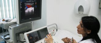
What is a stethoscope?
A stethoscope is a medical device with which a doctor can listen to noise in internal organs, thereby identifying deviations in their functioning. Such an examination is carried out at every appointment with a pediatrician or therapist.
The configuration of the device is as follows:
- Head. This is exactly the part that is applied to the area where the organ is located and picks up auscultatory sounds, that is, noises. It can be one-sided, having a membrane or funnel, or two-sided, which has both.
- Flexible tube. She connects the head to the arms and makes noises so that the doctor can hear them.
- Temples. They are metal tubes that are a continuation of a flexible tube. They are equipped with a spring that allows the headphones to fit more deeply and correctly into the ear canals.
- Olives. These are soft attachments that are located on the arms.
Components of a stethoscope
There are several types of stethoscopes, for example, obstetric, pediatric, cardiological.
Stethoscope or phonendoscope?
The stethoscope is the first version of a device for listening to body sounds. To obtain sound, a sound-collecting funnel was used - a “bell”.
The phonendoscope appeared later (by the way, the term was proposed by the Russian scientist Nikolai Sergeevich Korotkov). In a phonendoscope, the sound-collecting funnel is covered with a membrane - a resonator, to enhance a certain frequency spectrum.
Often the device combines both a “bell” and a membrane—a resonator. It turns out to be a stethophonendoscope. In most stethophonendoscopes, switching between the funnel and the membrane is carried out using axial rotation of the head, but other systems are also found: KaWe Planet - longitudinal rotation of the head
Dimeda - switching using a lever (discontinued model).
As a rule, high frequencies (heart sounds) are better heard with a funnel, while low frequencies (lung sounds) are better heard with a membrane. However, a lot depends on the manufacturer and the specific model of the stethoscope. For example, the 3M Littmann “Master” series has a dual-range membrane, which allows you to tune to high or low frequencies depending on the force of pressing the head.
Characteristics of a phonendoscope
A stethoscope and a phonendoscope are similar in appearance. A phonendoscope is also intended for listening to noise in internal organs. It comes with the same components as a stethoscope. However, there is also an additional membrane, thanks to which sounds are captured much better.
The most effective in diagnostics are phonendoscopes, which are equipped with a flat head. They are used to listen to the lungs.
The flat head of a phonendoscope is more sensitive than the head of a stethoscope
What is cardiac auscultation
“Auscultation” is the scientific name for listening. It is most often used to recognize sounds made by the heart or lungs. At the same time, a qualitative assessment of what is heard is made, that is, compliance with the norms of frequency, volume, and nature of sounds.
This technique is used not only in diagnostics. It is also actively used to monitor the patient’s condition with heart defects, which are already known.
The “music” of the heart muscle consists of alternating two tones:
- The tones are short and intermittent.
- The noises are continuous.
The heart sounds like water flowing in a pipe. The nature of the noise is influenced by a number of factors:
- blood flow rate;
- roughness of the walls of blood vessels;
- obstacles encountered in the path of the flow;
- listening location, proximity to the site of narrowing of the vessel.
At the same time, sound effects can be of a different nature. They can be:
The flow rate depends on the viscosity of the blood. Its movement through the vessels creates noise. This means that if any changes occur in the structure of the blood, this will affect the tone of the heart.
A phonendoscope is used for auscultation. This procedure is familiar to everyone since childhood: the doctor, with the words “breathe, don’t breathe,” applies the device to the chest.
What is a stethophonendoscope?
Nowadays in medical practice such devices as stethophonendoscopes are used. They are essentially a mixture of a stethoscope and a phonendoscope. That is why they are highly effective in detecting noise and can be used to examine various internal organs.
According to external data, such a device is practically no different from its predecessors. It comes with the following:
- A capsule equipped with a convex or flat membrane. This part of the device is responsible for capturing sounds. The head is covered with metal or plastic. Metal capsules are most preferred because they last a long time.
- Flexible tubes that connect the head to the olives and conduct noise signals. They are made from rubber or vinyl. Doctors like the latter material more, as it more effectively protects against external sounds.
- Headphones located at the end of the tubes. They can be plastic or rubber. The latter are the most comfortable because they fit tightly to the ears.
A stethoscope has more advanced functionality. Therefore, it is in great demand among doctors.
A stethoscope is a device that combines the advantages of a stethoscope and a phonendoscope.
Main components of a stethoscope:
Ear olives
There are several options:
- Hard (made of plastic, rarely used in modern stethoscopes, because they put painful pressure on the ear canal);
- Semi-rigid (material like thick PVC);
- Soft anatomical - made of soft PVC with thin walls.
The latter type is preferable - in addition to the fact that the anatomical soft olives do not put much pressure on the doctor’s external auditory canal, they improve sound insulation from external noise.
In “top” models of stethoscopes, the olives can rotate around their axis, which allows you to use the stethoscope with greater comfort - when moving the stethoscope up and down, the olives do not rub your ears and allow you to avoid acoustic artifacts arising from friction. On the left in the photo is a new ergonomic Riester earpiece with the ability to rotate around its own axis. On the right is a standard soft ear olive.
Metal arcs
Reputable manufacturers set the correct angle of the arches at the factory in the most comfortable position for the doctor. However, it is possible to adjust the rotation of the tubes, a kind of “fine tuning” to the individual characteristics of the user’s ear.
Spring. Happens:
- External. The most “budget” option;
- External folding. It is convenient because it adds compactness to the stethoscope, allowing you to store the device in a narrow pocket and the ability to quickly adjust the tilt of the arches;
- Internal hidden. Has a positive effect on ergonomics - does not cling to hair.
Some manufacturers (for example, Rudolf Riester) use a double hidden spring - this design allows you to correctly distribute the pressure on the ear canal and press the olives tightly but gently.
Acoustic tubes
Their quality determines how long your stethoscope will last you. Unfortunately, it is only possible to find out the quality of the material used during operation (especially if you purchased an inexpensive Asian device). In any case, it is worth considering that:
- Stethoscope tubes do not like strong heat and severe frost. Do not store the stethoscope on the dashboard of a car (they may fade) or in an unheated car interior in winter (cracks may appear).
- They do not like contact with oil and aggressive antiseptics such as chlorine. If necessary, wipe the tubes with 96.5% alcohol.
Membrane rings
- Conventional plastic ones are used in budget models from Asian manufacturers. They reduce the cost of the device, but cause discomfort to the patient;
- Uncooled. The so-called “warm plastic” does not cause an unpleasant reaction in the patient when the stethoscope head is applied to the skin;
- Metal. They have a positive effect on the overall “indestructibility” of the stethoscope, however, like any metal, they are cold. Before the examination, it is necessary to either warm the head of the stethoscope in the palm of your hand, or ask the patient to be patient.
Head.
The most common type is a stethophonendoscope: on one side there is a “bell”, on the other there is a membrane. The acoustic data of the stethoscope depend on the internal shape and design of the head, which manufacturers use their own know-how. Reputable manufacturers announce the characteristics in numbers or provide an acoustic graph: And the smoother and higher the number in db (decibels), the more noise you will hear and be able to differentiate without straining your hearing.
There is a membrane on the head of the phonendoscope. In addition to sensitivity, membranes are:
- flat,
- convex,
- convex along).
What type of membrane should I choose? There is no definite answer to this question; it all depends on the design features as a whole. In any case, the membrane must be sensitive in the range from 10 hertz to 1 kilohertz and made of high-quality materials.
What types of devices are there?
There are several types of stethoscopes and phonendoscopes:
- Cardiological. They are used to listen to murmurs in the heart and blood vessels. Such devices are distinguished by the fact that they are able to more effectively capture sound signals and have increased sensitivity.
- Pediatric. Such devices are used to listen to organs in children. In terms of functionality, they do not differ from those used for adults. The only difference is that the head is smaller. This allows the doctor to better detect the noises in the child and pinpoint the location where they are coming from.
- Obstetrics. Such devices are used to listen to the fetal heartbeat during pregnancy.
- Electronic. This is a modern type of phonendoscope that not only helps in capturing sound signals but also has additional functions. This may be the ability to record auscultation results, a Bluetooth option, or save patient information.
There are also training devices. Their difference is that two people can listen to noise at once. This allows you to objectively assess whether the student has correctly picked up the noises.
The training device allows two people to use it simultaneously
Types of wheezing
There are several types of wheezing that can be heard during pneumonia:
Crepitus
During pneumonia, the alveoli fill with fluid. When the breathing process occurs, they periodically stick together and come apart, making a quiet sound. This phenomenon often occurs at the very beginning of the development of pneumonia, as well as during recovery. This sound resembles a light crackling sound and is heard only when inhaling.
Crepitation can be detected by listening to the lung using a phonendoscope. The doctor presses it tightly against the patient’s skin, thereby reducing the audibility of low-frequency sounds. If the patient is a man and has hair on his chest, it is necessary to lubricate this area with fat so that imitation crepitus does not occur when rubbing dry hair.
Crepitation can be congestive and inflammatory. The first type is usually observed in the lower pulmonary regions. This kind of crepitation is less sonorous than inflammatory. In the latter case, compacted tissue is found around the alveoli, which is better able to conduct sound.
Wet wheezing
This type of wheezing can be fine-bubble, large-bubble or medium-bubble. It all depends on the involvement of small, medium or large bronchi in the process. They accumulate fluid that forms during inflammation. It is called exudate. When you breathe, the liquid gurgles. Moist rales are heard in both phases of breathing.
If pneumonia goes away without complications, fine bubble noises are often observed. They sound like small bubbles bursting. When pneumonia is complicated or advanced, coarse wheezing occurs. The sound can be heard not with the help of a special device, but even at a short distance from the patient. Medium-bubble noises occur when the lung edema, fluid enters the small or medium bronchi. They sound like crackling.
Dry wheezing
This type of noise occurs when the air passing through the bronchi does not find an obstacle, which is liquid.
Dry wheezing appears at the beginning of the development of pneumonia, which occurs against the background of other diseases of the respiratory system, such as, for example, bronchitis. They are observed in both phases of breathing and sound like rustling. During the course of the disease, bronchial obstruction sometimes occurs. This often occurs in patients suffering from bronchial asthma. At the same time, a whistling sound is heard. The air stream passes through the bronchi, as if through a pipe. This sound is easy to hear without special equipment.
Dry noises indicate a narrowing of the lumen in the bronchi.
This occurs due to tumors, swelling of the mucous membrane, and the presence of lumps of viscous sputum.
Pleural friction rub
If another disease, dry pleurisy, is associated with pneumonia, a pleural friction noise appears. It resembles scraping sounds and is similar to crepitus. However, such noise is heard constantly, in both phases of breathing. It appears when the inflamed layers of the pleura rub against each other under the influence of air flow.
Pleural friction noise is characterized by the following properties:
- dry intermittent sound; superficiality of noise felt close to the ear; variability of sound (can appear and disappear) - the exception is the chronic form of the disease; low sound prevalence; heard in both phases of breathing; presence of pain.
Typically, a pleural friction rub is found in the lower part of the chest, on the side. Sometimes it is difficult to distinguish it from wet wheezing. In this case, you need to know some nuances. First, when applying pressure with a stethoscope, the pleural noise becomes louder. As for coughing and deep breaths, the sound does not change or disappear.
Bronchophony
Bronchophony is the strengthening of the patient's head when listening to the lungs. At the same time, he pronounces the word in a whisper, and the doctor hears him perfectly. If bronchophony is pronounced, there is also a metallic tint to the sound. This type of noise indicates compaction in the lungs, which appeared as a result of inflammatory infiltration or for other reasons. With bronchophonia, vocal tremors are often detected.
The essence and history of the method
A phonendoscope in the hands of a doctor is so familiar that it does not evoke any emotions. However, by historical standards, it appeared recently - back in the 19th century, doctors listened to the patient’s heart and lungs directly with the ear, applying it to the patient’s body. Rene Laennec was the first to improve a procedure that was not entirely pleasant for a doctor; in order to exclude direct contact with the patient’s body, he listened to the heart using a sheet of music rolled up into a tube. And I was very sincerely surprised when I heard the heart sounds better and more clearly. Later, the doctor invented a primitive stethoscope, and even later, the Russian scientist P. N. Korotkov invented a phonendoscope, which doctors still use today.
The heart is a constantly working organ. When it contracts, peculiar sounds are formed that are well transmitted through tissue structures. It is these sounds that the doctor listens to using a phonendoscope.
The heart auscultation algorithm is quite simple, but only a doctor with practical experience can evaluate it correctly. The method has no contraindications; it can be used to examine patients of any age group.
Auscultation of the heart also has listening points - certain areas on the chest where various parts of the myocardium are projected in detail. Through auscultation you can:
- Assess heart rhythm.
- Analyze the contractile force of the myocardium.
- Assess the timbre coloring of sounds that are heard when using a phonendoscope.
- Identify extraneous noise.
Using a phonendoscope, the doctor can preliminarily determine the presence of the following pathologies:
- ischemia (CHD);
- heart muscle defects;
- ventricular hypertrophy;
- arrhythmias;
Arrhythmia on ECG
- inflammatory processes in the myocardium.
The results of auscultation determine further tactics for patient management. Having suspected a pathology, the doctor will refer the patient for additional examinations, after which he will be able to accurately establish the diagnosis.
Historical background and features of the methodology
A stethoscope is a device for performing cardiac auscultation. It was invented by the French doctor Rene Laennec. This significant event took place in 1816. Externally, this device is a wooden tube with funnel-shaped extensions of different diameters at the ends.
Literally a year later, R. Laennec published the work “Mediated Auscultation”. It describes the experience itself and the practical application of this technique. It was this French doctor who identified and systematized the main symptoms that health workers rely on when performing auscultations.
The stethoscope was actively used for a century. Even at the beginning of the 20th century, rural paramedics continued to use this particular instrument, although at that time this model had undergone modernization.
This medical device was replaced by a binaural instrument with a bell-shaped head . Afterwards the membrane structure was put into operation. During the active use of these devices, experts noticed a number of features:
- Low-frequency sounds are best listened to with a bell-shaped stethoscope. For example, murmurs of mitral stenosis.
- For high-frequency instruments, a membrane tip is more suitable, that is, aortic insufficiency will be diagnosed with this instrument.
In 1926, the phonendoscope was invented. Its advantage over its predecessors is the combination of two listening technologies and the presence of a membrane-bell-shaped head. This medical device is more universal and equally clearly detects heart sounds regardless of their frequency.
Today, the requirements for devices are more stringent, so stethoscopes with noise filtering and signal amplification functions are being developed. In the meantime, the procedure is carried out in silence. Sometimes, in order to listen to the heart more clearly, the patient is asked to squat. For comparison, auscultation is performed in the supine and sitting positions.
How to choose the “right” device
Stethoscopes are distinguished by the purity of the sound conducted, its volume, design and operating principle. When choosing, you need to pay attention to several parameters:
- The material used to make the head and rigid tubes
is aluminum, steel or plastic. Polymer products are the cheapest, but their conductivity is the lowest; wheezing is difficult to hear through such a stethoscope. Aluminum models are more practical, steel models are the most accurate, but also expensive. - The membrane of the head
of the device must be flexible; the degree of fit to the body and audibility, respectively, depend on this. Its size is also important: the larger the diameter, the wider range of sounds in the body can be heard. - The tube conducting the sound
must be thick so that the waves do not dissipate on the way to the ear. The larger its internal diameter, the better. The minimum required length is 30 cm, but it is more convenient to use 50-55 cm. - Ear tips
(tips) made of plastic do not change shape, they fit worse to the auricle, and accordingly, audibility suffers, in contrast to using a stethoscope with soft tips. - The type of sound transmission
of the device is mechanical, based on the transmission of noise according to the laws of physics, or electronic, which has a sound amplifier and speakers for the ears.
Stethoscopes are divided into 3 types depending on the specific application. They differ in the degree of sound conductivity and its accuracy.
- Pediatric
(therapeutic) for listening to heart rhythms and respiratory tract conditions. The membrane and its rim do not cause a feeling of cold when touched, for the comfort of patients of different ages. - Cardiac
sounds transmit sounds of different frequencies for a more accurate auditory determination of the work of the heart. - Obstetrics
for listening to the fetal heartbeat in the womb.
Many doctors, especially in private clinics, prefer universal devices suitable for diagnosing all the problems described - phonendoscopes.
Rating of the best stethoscopes
| Nomination | place | Name of product | price |
| Best Mechanical Stethoscopes | 1 | KAWE Planet Cardiology (43920) | 16 000 ₽ |
| 2 | ADC ADSCOPE 602 | 6 000 ₽ | |
| 3 | Riester Cardiophon 2.0 | 6 500 ₽ | |
| 4 | AMRUS 04-AM507 | 400 ₽ | |
| Best Electronic Stethoscopes | 1 | LITTMANN Electronic Stethoscopes 3100BK27 | 34 320 ₽ |
| 2 | Switel BH170 | 3 890 ₽ |
Best Mechanical Stethoscopes
The simplest devices are mechanical stethoscopes, they are also the most reliable, but they do not always transmit sounds perfectly. As we have already said, it is important to choose the “right” model.
KAWE Planet Cardiology (43920)
Rating: 4.9
The leader in the rating is the KAWE Planet Cardiology stethoscope (43920) with an adjustable arm and a floating membrane with a diameter of 42 mm. The original production instrument can be identified by the serial number printed on the golden metal door head, which, by the way, can be switched from low to high frequencies to diagnose problems in different organs.
The total length from the massive head to the adjustable ear olive is 75 cm; it is convenient to carry out diagnostics while maintaining a comfortable distance between the patient and the doctor. At the same time, the sound quality is outstanding, the noise from the fingers is leveled out, like other background sounds from the outside. Good audibility is also facilitated by a movable membrane that adapts to body contours as accurately as possible; it is capable of transmitting noise under the skin up to 120 mm. The stethoscope system is two-tube for better sound conduction and increased diagnostic accuracy.
The KAWE Planet Cardiology stethoscope (43920) is widely used by doctors of various specializations and receives only positive reviews, so it will not be superfluous in your home medicine cabinet; if handled skillfully, it will help diagnose various diseases in the early stages. The price of the device is about 14,000 rubles.
Advantages
- Two-pipe sound transmission system;
- Excellent sound quality up to 120mm depth.
- Rotating head mechanism and adjustable arms;
- Gold-plated head with a “warm” rim;
- Adjustable massive head with frequency switching;
- Light weight 147 grams;
Flaws
- High price.
ADC ADSCOPE 602
Rating: 4.8
In second place in the rating is the ADC ADSCOPE 602 cardiac stethoscope made in the USA. The stylish device, black in color from the arm to the head, 71 cm long, is easy to use for diagnosing low and high frequency noise by switching the head accordingly, the touch of the rim of which does not cause a feeling of cold.
A special feature of the device is a high-tech AFD membrane with an adjustable frequency of high sensitivity. It transmits the slightest noise, which the stainless steel olives help the ear to catch - they do not lose sound thanks to Adsoft™ PVC ear tips. A cardiological headband with binaural arms fixed at an angle of 150 ensures a comfortable fit of the stethoscope in the doctor’s ears and better audibility.
The tube is double, it is made of flexible PVC, which maintains mobility and elasticity throughout the life of the stethoscope. The total weight of the medical device is 215 grams (diaphragm for adults) and 243 grams with pediatric diaphragm. The set includes a bell, ear tips, and an identification plate.
The average price of ADC ADSCOPE 602 is 10,300 rubles.
Advantages
- Frequency switching;
- The headband is not cold;
- Anatomically located arches;
- Sealed Adsoft™ liners;
- Double conductive tube;
Flaws
- High price.
Riester Cardiophon 2.0
Rating: 4.7
In third place is a simpler model of the Riester Cardiophon 2.0 stethoscope with an updated head design with improved conductivity of noise in the body up to 3 times in a wide range of 10…100 Hz. The device has soft adjustable olives that fit snugly to the auricle; sound insulation is also improved in the conductive tube - they are made of elastic material. The rings of the double-diaphragm head do not cool down and do not cause discomfort.
The stethoscope is equipped with two membranes with a diameter of 44 and 32 mm with the perception of low and high frequencies, respectively. A Y-shaped tube separated by acoustic valves for each ear of the doctor will help you hear all the slightest noise.
The Riester Cardiophon 2.0 package includes a box for storing the device, 2 replaceable membranes and a badge. The total weight of the stethoscope is 204 grams, and the average price is 6,500 rubles.
Advantages
- Improved sound sensitivity;
- Optimal price;
- Head rings are not cooled;
- Sound valves in the tube for each ear;
- Elastic olives;
- Membranes for listening to low and high frequencies;
Flaws
- Minimum equipment, no replacement olives.
AMRUS 04-AM507
Rating: 4.6
The model AMRUS 04-AM507 closes the rating in the category of mechanical stethoscopes, ideal for pediatric diagnostics due to the bright colors of the tubes - they can be green (aqua), pink or red. The rotating double-sided head is designed to work with children: it is smaller, equipped with an elastic membrane, and the rim does not get cold when listening. A conductive tube 55 cm long with thick walls does not dampen noise, conical vinyl olives in the auricle contribute to their preservation, but the fit of the chrome tubes is too strong - the spring is elastic, but puts too much pressure on the ears, this is noted by all ordinary people and doctors who use the AMRUS stethoscope 04-AM507. The device has one membrane, its diameter of 35 mm contributes to sensitivity to a narrow spectrum of sounds.
The average price of 400 rubles is the lowest in our rating.
Advantages
- The lowest price;
- Does not cool the skin;
- Convenient olives;
- Choice of tube colors;
Flaws
- Narrow spectrum of sound conductivity;
- The tight spring on the arms puts pressure on the ears.
Steps
1 Selecting and setting up a stethoscope
- 1 Get a high quality stethoscope.
A high quality stethoscope is important. The higher the quality of your stethoscope, the easier it will be for you to listen to your patient's body.- A single-sided stethoscope is better than a double-sided one. The channels in a double-ended stethoscope may rub against each other. This noise can make it difficult to hear heart sounds.
- A thick, short, and relatively rigid bore is the best option if you don't plan on wearing the stethoscope around your neck. In this case, a long channel is preferable.
- Make sure there are no holes in the canal by tapping the diaphragm (the flat part of the stethoscope head). Use headphones as a tapping aid to listen to sounds. If you don't hear anything, then there are holes.
- 2 Check the ear tips of your stethoscope.
It is important to make sure that the ear tips point in a frontal direction away from you and that they fit well. Otherwise, you may not be able to hear anything with your stethoscope.- Make sure the ear tips are facing in front of you. If they look the other way, you won't be able to hear anything.
- Make sure the ear tips fit snugly and provide good isolation to block out external noise. If the ear tips do not fit tightly, many stethoscopes have replaceable tips. Visit a medical supply store to purchase the tips you need.
- With some stethoscopes, you can also tilt the ear tips forward to provide a better fit.
- 3 Check the tension of the ear tips of your stethoscope.
In other words, make sure the ear tips are close to your head, but not too close. If your ear tips are too tight or too loose, readjust them.- If the ear tips are too loose, you may not hear anything. To tighten the tension, apply pressure to the ear ties near the ear tips.
- If the ear tips are too tight, you may damage your ears and make it difficult to use your stethoscope. To release the tension, stretch the ties very carefully.
- 4 Select the appropriate head for your stethoscope.
There are many different types of stethoscope heads. Choose the one that suits your needs. Stethoscope heads come in a variety of sizes for adults and children.
2 Preparing to use the stethoscope
- 1 Choose a quiet place to use the stethoscope.
Use your stethoscope in a quiet place. Find a quiet place to ensure that the body sounds you intend to hear will not be drowned out by background sounds. - 2 Position the patient correctly.
To hear the heart and abdomen, you will need your patient to be in a supine position. To hear the lungs, you will need to have your patient sitting up. In other words, ask your patient to lie down. Heart, lung and bowel sounds vary depending on the patient's position: that is, sitting, standing, lying on either side, and so on. - 3 Decide whether you will use a diaphragm or a bell.
The diaphragm, or flat side of the tip, is better suited for listening to mid- and high-frequency sounds. The bell, or round part of the tip, is better suited for listening to low-frequency sounds.- If you want a stethoscope with really high sound quality, you may need to consider an electronic stethoscope. An electronic stethoscope provides amplification so that it is easier to listen to the sounds of the heart and lungs. Using an electronic stethoscope can make it easier to listen to your patient's heart or lung, but be aware that they are expensive.
- 4 Have your patient wear a hospital gown or lift their shirt to expose skin.
Use the stethoscope on bare skin to avoid picking up the sounds of rustling fabric. If your patient is a man with a hairy chest, hold the stethoscope still to avoid any rustling sounds.- To ensure patient comfort, warm the stethoscope by rubbing against the sleeve, or consider purchasing a stethoscope warmer.
3 Listening to the heart
- 1 Hold the diaphragm over the patient's heart.
The position of the diaphragm is in the left upper chest, where the 4th and 6th ribs meet, almost immediately below the chest. Hold the stethoscope between your index and middle fingers, and apply gentle enough pressure so that you don't hear your fingers rubbing together. - 2 Listen to your heart for a minute.
Ask the patient to relax and breathe normally. You should hear normal heart sounds that sound like “thump-thump.” These sounds are also called systolic and diastolic. The first sound “knock” is called systolic, and the second sound “knock” is called diastolic.- Systolic sound occurs when the mitral and tricuspid valves of the heart are closed.
- Diastolic sound occurs when the aortic and pulmonary valves of the heart are closed.
- 3 Count the number of heartbeats per minute.
The normal resting heart rate for adults and children over 10 years of age is 60-100 beats per minute. For trained athletes, a normal resting heart rate can be 40-60 beats per minute.- There are several different ranges for normal heart rates for assessing patients under 10 years of age. These ranges are: Newborns up to one month: 70-190 beats per minute
- Infants 1 - 11 months: 80 - 160 beats per minute
- Children 1 - 2 years old: 80 - 130 beats per minute
- Children 3 - 4 years old: 80 - 120 beats per minute
- Children 5 - 6 years old: 75 - 115 beats per minute
- Children 7 - 9 years old: 70 - 110 beats per minute
- 4 Listen for abnormal heart sounds.
Once you have counted your heartbeat, you should also listen for any abnormal sounds. Anything that doesn’t sound like “knock-knock” can be considered abnormal. If you hear anything abnormal, your patient needs further evaluation by a doctor.- If you hear a whistling sound similar to “knock...shhh...knock,” your patient may have a heart murmur. A heart murmur is blood flowing quickly through the valves. Many people have what is called a benign heart murmur. But some heart murmurs do indicate heart problems, so you should advise the patient to see a doctor if you detect a heart murmur.
- If you hear a third heart sound that sounds like a low-frequency vibration, your patient may have a ventricular defect. This third heart sound is called S3 or ventricular gallop. Advise the patient to contact the doctor if you hear a third heart sound.
- Try it to help you determine if what you hear is normal.
4 Listening to the lungs
- 1 Ask the patient to sit up straight and breathe normally.
While you listen, you can ask the patient to take a deep breath if you cannot hear the breathing sounds or if they are too quiet to determine if they contain any abnormalities. - 2 Use the diaphragm of the stethoscope to listen to your patient's lungs.
Listen to the patient's lungs in the upper and lower lobes, and from the front and back.- First you listen to the upper chest through the stethoscope, then the midclavicular chest line, and then the lower chest. Be sure to listen from the front and back to all these areas.
- Be sure to compare the results on both sides of your patient's lungs and check if there is anything abnormal.
- By covering all of these positions, you will be able to listen to all lobes of your patient's lungs.
- 3 Listen to the sounds of normal breathing.
The sounds of normal breathing are distinct, then compare with the sounds you hear in your patient's lungs.- There are two types of normal breathing sounds: Bronchial breathing sounds are those heard within the tracheobronchial tree.
- Vesicular breathing sounds are those heard over the lung tissue.
- 4 Listening to abnormal breathing sounds.
Abnormal breathing sounds include wheezing, stridor, wheezing, and wheezing. If you do not hear any breathing sounds, the patient may have air or fluid around the lungs, a thick layer near the chest, or airflow that has slowed or congested the lungs.- There are four types of abnormal breathing sounds: Snorting sounds like a high-pitched sound when a person exhales and sometimes when he inhales. Many patients who have asthma also have wheezing, and you can sometimes hear this without a stethoscope.
- Stridor sounds like a high-pitched musical breathing, similar to a sniffle, mainly heard when the patient inhales. Stridor is caused by a blockage in the back of the throat. This sound can also often be heard without a stethoscope.
- Dry wheezing sounds like snoring. Wheezing cannot be heard without a stethoscope and occurs because air flows through a “rough” path in the lungs or because the path is blocked.
- The wheezing sounds like bursting bubbles. A wheeze may be heard when the person inhales.
5 Listening to abdominal sounds
- 1 Place the diaphragm on your patient's bare abdomen.
Use the patient's navel as the center and divide the listening around the navel into four sections. Listen to the top left, top right, bottom left and bottom right. - 2 Listen to normal abdominal sounds.
Normal abdominal sounds are when your stomach growls or grumbles. Anything else suggests that something is wrong and that the patient needs further evaluation.- You will hear a “rumbling” sound in all four sections. Sometimes after surgery it will take time for the abdominal sounds to return.
- 3 Listening to abnormal abdominal sounds.
Most of the sounds you hear when auscultating your patient's abdomen are just those of digestion. Although most abdominal sounds are normal, there are certain ones that indicate problems. If you are unsure whether the abdominal sounds you hear are normal and/or the patient has other symptoms, then they should see a doctor for further evaluation.- If you do not hear any abdominal sounds, this may indicate that something is blocked in the patient's abdomen. It may also indicate constipation, and the abdominal sounds may return on their own. But if they don't come back, then it could be a blockage. In this case, the patient needs further examination by a doctor.
- If a patient has hyperactive abdominal sounds caused by a lack of abdominal sounds, this may indicate rupture or necrosis of the abdominal tissues.
- If a patient has high-pitched abdominal sounds, this may indicate that there is a blockage in the patient's intestines.
- Slow abdominal sounds can be caused by prescription pills, spinal anesthesia, infection, injury, abdominal surgery, or bowel overdilation.
- Rapid or hyperactive abdominal sounds can be caused by Crohn's disease, gastrointestinal bleeding, food allergies, diarrhea, infection and ulcerative colitis
6 Listening to sound during auscultation
- 1 Determine if you need to check the sound by auscultation.
If you detect a sound that appears to be a heart murmur, you should also do a sound test by auscultation. Since only a heart murmur and auscultation sound similar, it is important to test both if one is suspected. - 2 Place the diaphragm of your stethoscope over one of the carotid arteries.
The carotid arteries are located on the front side of the patient's neck, on either side of the Adam's apple. If you run your index and middle fingers along the front of your throat, you will trace the locations of both carotid arteries.- Be careful not to press too hard on the arteries, otherwise you may cut off circulation and cause your patient to faint. Never press on both arteries at the same time.
- 3 Listening to sound during auscultation.
The sound upon auscultation is similar to a whistling sound, which indicates that the artery is narrowed. Sometimes a sound on auscultation can be mistaken for a heart murmur because the sounds are similar, but if the patient has a sound on auscultation, then the whistling sound will be louder when you listen to the carotid artery than when you listen to the heart.
7 Checking blood pressure
- 1 Wrap the blood pressure cuff around the patient's arm just above the elbow.
Roll up the patient's sleeve if it is in the way. Make sure the blood pressure cuff you use fits the patient's arm. You should be able to wrap the cuff so that it fits snugly, but not too tightly. If the blood pressure cuff is too small or too large, get a different size. - 2 Press the diaphragm of the stethoscope onto the brachial artery just below the edge of the cuff.
You can also use a diaphragm if you have difficulty listening with the bell. You will hear Korotkoff sounds, which are low-pitched tapping sounds that indicate the patient's systolic arterial blood pressure.- Feel the pulse in your arm to determine where your brachial artery is.
- 3 Inflate the cuff to 180 mmHg or 30 mm above your estimated systolic blood pressure.
You can tell by looking at the sphygmomanometer, a sensor placed on the cuff to measure blood pressure. Then deflate the cuff at a moderate rate (3mm/sec). As soon as you release the air, listen to the stethoscope and watch the sphygmomanometer (a sensor placed on the blood pressure cuff). - 4 Listening to Korotkoff sounds.
The first tapping sound you hear is the patient's systolic blood pressure. Note the number of beats, but continue to look at the sphygmomanometer. After the first sound has stopped, note the number of beats at which it stopped. This number is the diastolic pressure. - 5 Loosen and remove the cuff.
Deflate and remove the blood pressure cuff from the patient's arm immediately after you have learned the second number. When you are finished, you should have two numbers that will give you your patient's blood pressure value. Write these numbers next to each other, separated by a slash. For example, 110/70. - 6 Wait a few minutes if you intend to check your patient's blood pressure again.
You may need to take the patient's blood pressure if it is too high.- A systolic blood pressure greater than 120 or a diastolic blood pressure greater than 80 means your patient may have high blood pressure. In this case, your patient should undergo further examination by a doctor.