The first phonendoscopes were folded sheets of paper or hollow bamboo sticks, and many doctors used only their own organ of hearing. But they all wanted to hear what was happening inside the human body, especially when it came to such an important organ as the heart.
Heart sounds are sounds that are produced during the contraction of the myocardial walls. Normally, a healthy person has two tones, which may be accompanied by additional sounds depending on what pathological process is developing. A doctor of any specialty must be able to listen to these sounds and interpret them.
Cardiac cycle
The heart beats at a rate of sixty to eighty beats per minute. This, of course, is an average value, but ninety percent of people on the planet fall under it, which means it can be taken as the norm. Each beat consists of two alternating components: systole and diastole. The systolic heart sound, in turn, is divided into atrial and ventricular. This takes 0.8 seconds, but the heart has time to contract and relax.
Pathological heart sounds
Changes in tones in pathological conditions.
Weakening both tones -
extracardiac causes (emphysema, chest edema, pericardial effusion), intracardiac (weakening of ventricular contraction).
Strengthening both tones -
extracardiac (thin chest, wrinkling of the lungs, tumors of the posterior mediastinum) and intracardiac (thyrotoxicosis, tachycardia, some intoxications - caffeine, etc.). A change in tone is of greater diagnostic significance.
Attenuation of 1 tone –
at the apex or at the site of auscultation of the tricuspid valve is associated with a decrease in the rate of increase in pressure during the tension phase in the ventricles, an increase in the amount of blood in the ventricles at the end of diastole, low pressure at the end of the tension phase. This is due to anatomical changes in the mitral (tricuspid) valve and a decrease in myocardial contractility. Occurs with mitral (tricuspid) valve insufficiency, with myocardial damage (myocarditis, cardiosclerosis).
Strengthening the first tone
– at the apex is associated with a small amount of blood in the ventricle at the end of the diastole phase, an increase in the rate of increase in pressure in the ventricles during the tension phase. It is observed with mitral stenosis, rhythm disturbances (extrasystole, atrial fibrillation, complete atrioventricular block “cannon tone” Strazhesko).
Weakening of the second tone
– weakening of the second sound in the aorta (2nd intercostal space on the right) is more common and is associated with a decrease in blood pressure (a decrease in its quantity) at the beginning of diastole in the aorta. Causes: aortic valve insufficiency, aortic stenosis.
Strengthening the second tone
– associated with an increase in pressure in the systemic circulation (intensification - emphasis of the second tone under the aorta), an increase in pressure in the pulmonary circulation (emphasis of the second tone under the pulmonary artery), sclerosis of the aortic or pulmonary artery valves. Accent is the strengthening of the second over one valve while the second tone remains unchanged over the other.
Split heart sounds
– is associated with the non-simultaneous closure of the valves that form both the first and second sounds. The splitting of the first tone can be physiological or pathological (blockade of the legs of His, a large difference in pressure in the greater and lesser circulation). The splitting of the second tone is physiological and pathological (mitral stenosis).
Threefold rhythms
–
quail rhythm
. Occurs with mitral stenosis. It is heard at the apex of the heart at the beginning of diastole (the mitral valve leaflets, sclerosed and fused at the base, oscillate when they open and give an additional sound - a mitral click) in combination with a loud 1 tone - the “quail” rhythm (“it’s time to go to bed”).
Gallop rhythm -
three-part rhythm occurs with severe myocardial damage. V.P. Obraztsov noted that the appearance of this rhythm is a “cry” of the heart for help. May be protodiastolic or presystolic.
Systole
As mentioned above, there are two components involved.
First comes atrial systole: their walls contract, blood under pressure enters the ventricles, and the valve flaps slam shut. It is the sound of the closing valves that is heard through a phonendoscope. This entire process lasts 0.1 seconds. Then comes ventricular systole, which is a much more complex job than what happens with the atria. To begin with, we note that the process lasts three times longer – 0.33 seconds.
The first period is ventricular tension. It includes phases of asynchronous and isometric contractions. It all starts with the fact that an eclectic impulse spreads throughout the myocardium. It excites individual muscle fibers and causes them to spontaneously contract. Because of this, the shape of the heart changes. This causes the atrioventricular valves to close tightly, increasing blood pressure. Then a powerful contraction of the ventricles occurs, and the blood enters the aorta or pulmonary artery. These two phases take 0.08 seconds, and in the remaining 0.25 seconds the blood enters the great vessels.
“Pay off the first, the second!”
Circulation circles
The first tone and the second heart sound are the same “knock-knock”, the main sounds that are best heard by the human ear. An experienced doctor, in addition to the main ones, is well versed in additional and inconsistent sounds. The first and second tone are constant heart sounds, which with their rhythmic beating signal the normal operation of the main human “motor”. How are they formed? Again you will have to remember the structure of the heart and the movement of blood through it.
Blood enters the right atrium, then into the ventricle and lungs; from the lungs, purified blood returns to the left chambers of the heart. How does blood move through the valves? When blood flows from the right upper chamber of the heart into the ventricle, at the same second blood flows from the left atrium into the left ventricle, i.e. The atria normally contract synchronously. At the moment of contraction of the upper chambers, blood pours out of them into the ventricles, passing through the 2-leaf and 3-leaf valves. Then, after the lower chambers of the heart are filled with blood, the turn of contraction or ventricular systole occurs.
Work of the heart
The first sound occurs precisely at the moment of ventricular systole, the sound is caused by the closure of the heart valves during muscular ventricular contraction, as well as the tension of the wall of the lower chambers of the heart, vibrations of the very initial sections of the main vessels extending from the heart, where blood is directly poured. The second tone occurs at the very beginning of relaxation or diastole, during this period the pressure in the ventricles drops sharply, blood from the aorta and pulmonary artery rushes back and the open semilunar valves quickly slam shut.
The sound of the slamming semilunar valves creates the second heart sound to a greater extent, and also plays a role in the sound effect of vibration of the walls of blood vessels. How to distinguish the first heart sound from the second heart sound? If we graphically depict the dependence of sound volume on time, we will be able to observe the following picture: between the first tone that appears and the second there is a very short period of time - systole, a long interval between the second tone and the first - diastole. After a long pause, the first tone always comes!
Diastole
Here, too, everything is not as simple as it might seem at first glance. Ventricular relaxation lasts 0.37 seconds and occurs in three stages:
- Protodiastolic: After blood has left the heart, the pressure in its cavities decreases and the valves leading to large vessels close.
- Isometric relaxation: the muscles continue to relax, the pressure drops even more and becomes equal to the atrial pressure. This causes the atrioventricular valves to open and blood from the atria enters the ventricles.
- Ventricular filling: Following a pressure gradient, fluid fills the lower chambers of the heart. When the pressure equalizes, the flow of blood gradually slows down and then stops.
Then the cycle repeats again, starting with systole. Its duration is always the same, but diastole can be shortened or lengthened depending on the speed of the heartbeat.
Heart sounds and their origin. Components of the first and second tone. Phonocardiography.
Previous46Next
Heart sounds are a sound manifestation of the mechanical activity of the heart, determined by auscultation as alternating short (percussive) sounds that are in a certain connection with the phases of systole and diastole of the heart. Heart sounds are formed in connection with the movements of the heart valves, chords, cardiac muscle and vascular wall, generating sound vibrations. The audible volume of tones is determined by the amplitude and frequency of these vibrations.
Components of I (systolic) tone:
- Valvular - vibrations of the atrioventricular valve leaflets
— Muscular – vibrations of the ventricular myocardium
- Vascular - vibrations of the initial segments of the aorta and pulmonary trunk when they are stretched by blood during the expulsion period.
- Atrial - oscillations during atrial contraction
Components of II (diastolic) tone:
- Valvular - slamming of the semilunar leaflets of the aortic valve and pulmonary trunk
— Vascular – vibrations of the walls of the aorta and pulmonary trunk
Sometimes III and IV sounds are heard. The third tone is caused by fluctuations that appear during rapid passive filling of the ventricles with blood from the atria, damaging the diastole of the heart.
IV sound appears at the end of ventricular diastole and is associated with their rapid filling due to atrial contractions.
Phonocardiography (from the Greek phone - sound and cardiography), a diagnostic method of graphic recording of heart sounds and heart murmurs. It is used in addition to auscultation (listening), it allows you to objectively assess the intensity and duration of tones and noises, their nature and origin, and record the 3rd and 4th tones that are inaudible during auscultation.
A special apparatus for phonocardiography - a phonocardiograph - consists of a microphone, an amplifier of electrical oscillations, a system of frequency filters and a recording device. The microphone is applied to different points of the chest above the heart area. After amplification and filtering, electrical vibrations are sent to various recording channels, which allows selective recording of low, medium and high frequencies. FCG recording is carried out in a soundproofed room while holding the breath while exhaling (if necessary, at the height of inhalation) in a lying position, after the subject has rested for 5 minutes. On the FCG, a straight (isoacoustic) line reflects systolic and diastolic pauses. The normal 1st tone consists of 3 groups of oscillations: initial (low frequency), caused by contraction of the ventricular muscles; central (larger amplitude), due to the closure of the mitral and tricuspid valves; final (small amplitude), associated with the opening of the aortic and pulmonary artery valves and vibrations of the walls of large vessels. The 2nd tone consists of 2 groups of oscillations: the first (large in amplitude) is due to the closure of the aortic valves, the second is associated with the closure of the pulmonary artery valves. Normal 3rd (associated with muscle vibrations during rapid filling of the ventricles) and 4th (less common, caused by atrial contraction) tones are detected mainly in children and athletes. Characteristic changes in PCG (weakening, strengthening or splitting of the 1st and 2nd sounds, the appearance of pathological 3rd and 4th sounds, systolic and diastolic murmurs) help to recognize heart defects and some other diseases.
Previous46Next
Date added: 2016-06-15; views: 8002; ORDER A WORK WRITING
Similar articles:
Mechanism of formation of the first tone
No matter how strange it may sound, 1 heart sound consists of four components:
- Valve - it is the leader in the formation of sound. Essentially, these are vibrations of the atrioventricular valve leaflets at the end of ventricular systole.
- Muscular - oscillatory movements of the walls of the ventricles during contraction.
- Vascular - stretching of the walls of the great vessels at the moment when blood enters them under pressure.
- Atrial – atrial systole. This is the immediate beginning of the first tone.
Split heart sounds.
Heart sounds, composed of several components, are perceived as a single sound. In some physiological and pathological conditions, the sound of those components that take part in the formation of a particular tone does not synchronize. There is a split tone.
Splitting tones is the separation of the components that make up the tone. The latter follow each other at a short interval (every 0.036 s or more). The mechanism of bifurcation of tones is due to asynchronism in the activity of the right and left half of the heart: non-simultaneous closure of the atrioventricular valves leads to bifurcation of the first tone, of the semilunar valves - to bifurcation of the second tone. Split tones can be physiological and pathological. Physiological bifurcation (splitting) of the first tone occurs with asynchronous closure of the atrioventricular valves. This can happen during deep exhalation, when, due to increased pressure in the pulmonary circulation, blood enters the left atrium with greater force and prevents the timely closure of the mitral valve.
The physiological bifurcation of the second tone manifests itself in connection with different phases of breathing, since during inhalation and exhalation the blood supply of the left and right ventricles changes, and, consequently, the duration of their systole and the time of closure of the corresponding valves. The bifurcation of the second tone is especially well detected during auscultation of the pulmonary artery. The physiological bifurcation of the second tone is not constant (unfixed bifurcation), is closely related to the normal breathing mechanism (during inspiration it decreases or disappears), while the interval between the aortic and pulmonary components is 0.04-O. Obs.
Pathological split tones may be caused by the following factors:
1. Hemodynamic (increase in the systolic volume of one of the ventricles, increase in diastolic pressure in one of the ventricles, increase in diastolic pressure in one of the vessels);
2. Violation of intraventricular conduction (bundle branch block);
3. Weakening of the contractile function of the myocardium;
4. Ventricular extrasystole.
Pathological bifurcation of the first tone can occur when intraventricular conduction is disrupted (along the bundle branches) due to a delay in the next contraction of one of the ventricles.
Pathological bifurcation of the second tone is observed with arterial hypertension, with stenosis of the aortic mouth, when the leaflets of the aortic valve slam shut later than the pulmonary valve; in case of increased pressure in the pulmonary circulation (with emphysema, mitral stenosis, etc.), when, on the contrary, the pulmonary valve lags behind.
The appearance of additional tones should be distinguished from split tones .
These include the sound of the mitral valve opening, heard when the left atrioventricular orifice narrows. The mechanism of its occurrence is associated with a sudden tension of the sclerotic valve leaflets, unable to move completely to the walls of the ventricle when blood passes from the left atrium to the left ventricle. The opening tone of the mitral valve occurs immediately after the second tone, after 0.07-0.1 sec, during diastole. It is best heard at the apex and is combined with other auscultatory signs of mitral stenosis. In general, the additional third sound of the opening of the mitral valve in combination with the loud (clapping) first tone and second heart sound form a three-part rhythm, reminiscent of the call of a quail - the quail rhythm.
The three-member rhythm also includes gallop rhythm , reminiscent of the clatter of a galloping horse. There is a presystolic gallop rhythm, which is caused by a pathological IV heart sound, and a summation gallop rhythm, the occurrence of which is associated with the overlap of the III and IV sounds; an additional tone with this rhythm is usually heard in mid-diastole. A gallop rhythm is heard in case of severe myocardial damage (myocardial infarction, myocarditis, chronic nephritis, hypertension, etc.).
With severe tachycardia, a shortening of the diastolic pause to the size of the systolic pause is observed. At the apex, sounds I and II become almost identical in sonority, which is the basis for calling such an auscultatory picture a pendulum-like rhythm or, due to its similarity to the beating of the fetal heart, embryocardia. This can be observed in acute heart failure, paroxysmal tachycardia, high fever, etc.
The mechanism of formation of the second tone and additional tones
So, the 2nd heart sound includes only two components: valvular and vascular. The first is the sound that arises from the blows of blood on the valves of the artery and the pulmonary trunk at a time when they are still closed. The second, that is, the vascular component, is the movement of the walls of large vessels when the valves finally open.
In addition to the two main ones, there are also 3 and 4 tones.
The third sound is vibrations of the ventricular myocardium during diastole, when blood passively flows into an area of lower pressure.
The fourth sound appears at the end of systole and is associated with the end of the expulsion of blood from the atria.
II tone
II tone
also heard over the entire region of the heart, maximum - at the base of the heart: in the second intercostal space to the right and left of the sternum, where its intensity is greater than the first tone. The origin of the second sound is mainly associated with the closure of the aortic valves and pulmonary trunk. It also includes low-amplitude, low-frequency oscillations resulting from the opening of the mitral and tricuspid valves. On FCG, the first (aortic) and second (pulmonary) components are distinguished as part of the second tone. The amplitude of the first is 1.5 - 2 times greater than the second. The interval between them can reach 0.06 seconds, which is perceived during auscultation as a splitting of the second tone. It is associated with physiological asynchrony of the left and right hearts; most common in children. An important characteristic of physiol. splitting of the second tone is its variability depending on breathing (“unfixed splitting”). The pathological, “fixed” splitting of the second tone with a change in the ratio of the aortic and pulmonary components is based on an increase in the duration of the phase of expulsion of blood from the ventricles and a slowdown in intraventricular conduction. The volume of the second tone when auscultated over the aorta and pulmonary trunk is approximately the same; if it predominates over any of these vessels, they speak of an accent of tone II. Weakening of the second tone is most often associated with the destruction of the aortic valve leaflets with its insufficiency or with a sharp limitation of their mobility with severe aortic stenosis (see Acquired heart defects). Strengthening, as well as emphasis of the second tone over the aorta occurs with arterial hypertension in the systemic circle (see Arterial hypertension); strengthening, as well as its emphasis over the pulmonary trunk - with pulmonary hypertension (see Hypertension of the pulmonary circulation).
Characteristics of the first tone
Heart sounds depend on many reasons, both intra- and extracardiac. The sonority of 1 tone depends on the objective state of the myocardium. So, first of all, loudness is ensured by the tight closure of the heart valves and the speed with which the ventricles contract. Features such as the density of the atrioventricular valve leaflets, as well as their position in the heart cavity, are considered secondary.
It is best to listen to the first heart sound at its apex - in the 4-5 intercostal space to the left of the sternum. For more accurate coordinates, it is necessary to perform percussion of the chest in this area and clearly determine the boundaries of cardiac dullness.
Lecture No. 10. auscultation of the heart. normal and pathological heart sounds
Lecture No. 10.
Auscultation of the heart. Heart sounds are normal and pathological.
Listening (auscultation) of sound phenomena produced by the work of the heart is usually performed using a stethophonendoscope. This method has a great advantage over direct listening, since it makes it possible to clearly localize various sounds and, thanks to this, determine the location of the formation.
Listening to the patient should be carried out in a warm room and with a warm instrument. When working in a cold room or with a cold tool, the patient experiences muscle tremors. In this case, a lot of side sounds arise, which significantly complicate the assessment of the auscultatory picture. The patient is listened to while breathing calmly. However, in many situations, when the doctor detects weak sound phenomena, he asks the patient to hold his breath during the phase of maximum exhalation. At the same time, the volume of air-containing lungs around the heart decreases, breathing noises arising in the lungs disappear, and the sound picture of the beating heart is more easily perceived.
In what body position should the patient be listened to? It all depends on the auscultatory picture and the patient’s condition. Typically, auscultation is carried out in an upright position of the patient's body (standing, sitting) or lying on his back. However, many sound phenomena, such as pericardial friction noise, are better heard when the patient is tilted forward or in a position on the left side, when the heart fits more tightly to the anterior chest wall. If necessary, auscultation is carried out with a deep breath with straining (Valsalva maneuver). In many cases, cardiac auscultation is repeated after physical stress. To do this, the patient is asked to sit or lie down, do 10–15 squats, etc.
Get the full text See all projects
Publish articles
Along with listening to sound phenomena that occur during the work of the heart, the phonocardiography technique is now widely used. Phonocardiography is a graphic recording on paper tape of sound phenomena occurring during the work of the heart, perceived by a sensitive microphone. Sound phenomena are depicted in the form of vibrations of various amplitudes and frequencies. Simultaneously with the recording of sound phenomena, an electrocardiogram is recorded in one standard lead, usually in the second. This is necessary to determine in which phase of cardiac activity the recorded sound occurs. Currently, phonocardiography involves recording sounds in 3 to 5 different sound frequency ranges. It allows you to document not only the very fact of the presence of a particular sound, but also its frequency, shape, amplitude (loudness). Given the undoubted diagnostic value of the technique, it should be taken into account that the sound picture perceived by ear sometimes turns out to be more informative than the graphically recorded one. In some situations, during phonocardiography, sound energy is distributed over 3 to 5 recorded channels and is encrypted as background, while a clear, diagnostically significant sound picture is determined by ear. Therefore, phonocardiography should undoubtedly be considered a valuable, but additional research method.
When listening to the heart, tones and noises are distinguished. According to scientific terminology, those sound phenomena that are commonly called tones do not deserve this name, since they, like heart murmurs, are produced by irregular, aperiodic sound vibrations (the intervals between the vibrations of each tone are not equal). In this sense, even many heart murmurs (so-called musical murmurs) are much closer to real tones.
Normally, physiologically, 2 tones are heard above the heart. Of these, the first corresponds in time to the beginning of ventricular systole - the period of closed valves. It is called systolic tone. The second corresponds in time to the very beginning of the diastole of the heart and is called diastolic.
Origin of the first tone
complex.
The formation of 1 heart sound begins at the very beginning of cardiac systole. As is known, it begins with the systole of the atria, which pushes the remaining blood into the ventricles of the heart. This component of 1 tone, atrial
, is quiet, low-amplitude on the phonocardiogram, short-lived. If our ear could separately perceive sounds very close to each other, we would listen to a separate weak tone of the atria and a stronger tone formed during the systole phase of the ventricles. But under physiological conditions, we perceive the atrial component of the 1st tone together with the ventricular one. In pathological conditions, when the systole of the atria and ventricles are separated in time by more than usual, we listen to the atrial and ventricular components of the 1st sound separately.
In the phase of asynchronous contraction of the heart, the process of excitation of the ventricles, the pressure in which is still close to “0”, the process of ventricular contraction covers all myocardial fibers and the pressure in them begins to increase rapidly. At this time, a long ventricular contraction
or muscle component of tone 1. The ventricles of the heart at this moment of heart systole are 2 completely closed bags, the walls of which tensed around the blood contained in them and, due to this, began to vibrate. All parts of the walls vibrate, and they all give a tone. From this it is clear that complete closure of the ventricles of the heart on all sides is the main condition for the formation of the first sound.
The main loudness component of tone 1 occurs at the moment when the closure of the two and tricuspid heart valves occurs. These valves have closed, but the semilunar valves have not yet opened. The tone of that part of the walls that is most capable of oscillating, namely the tone of thin elastic leaf valves, valve
component 1 tone will be dominant in volume. With significant insufficiency of the leaflet valves, the sound of the corresponding ventricle will completely disappear by ear.
The first sound is not only carried out from the ventricles and leaflet valves, but also occurs due to sudden tension and vibration of the walls of the aorta and pulmonary artery when the blood of their ventricles enters them. This component of tone 1 is called vascular
. Since this occurs already in the phase of the beginning of ventricular emptying, the first tone also covers the period of the beginning of the expulsion of blood from the ventricles.
So, 1 heart sound consists of 4 components - atrial, muscular, valvular and vascular.
The period of blood expulsion from the ventricles of the heart consists of two phases - fast and slow blood expulsion. At the end of the slow ejection phase, the ventricular myocardium begins to relax and diastole begins. Blood pressure in the ventricles of the heart decreases, and blood from the aorta and pulmonary artery rushes back into the ventricles of the heart. It closes the semilunar valves and a second or diastolic heart sound occurs.
The first tone is separated from the second tone by a short pause with an average duration of about 0.2 seconds.
The second tone has two components, or two components. The main loudness component is the valve
component, formed by vibrations of the semilunar valve leaflets.
After the slamming of the semilunar valves, blood rushes into the arteries of the systemic and pulmonary circulation. The pressure in the aorta and pulmonary trunk gradually decreases. All pressure changes and blood movement in the aorta and pulmonary artery are accompanied by vibrations of their walls, forming the second, less loud, component of the 2nd tone - the vascular
component.
The time from the beginning of ventricular relaxation to the closure of the semilunar valves is called the protodiastolic period.
equal to 0.04 seconds.
The blood pressure in the ventricles at this time drops to zero. The leaflet valves are still closed at this time, the volume of blood remaining in the ventricles and the length of the myocardial fibers have not yet changed. This period is called the isometric relaxation period
, equal to 0.08 seconds.
Towards its end, the cavities of the ventricles of the heart begin to expand, the pressure in them becomes negative, lower than in the atria. The leaflet valves open and blood begins to flow from the atria into the ventricles of the heart. The period of filling the ventricles with blood
begins , lasting 0.25 seconds. This period is divided into 2 phases of fast (0.08 seconds) and slow (0.17 seconds) filling of the ventricles with blood.
At the beginning of the rapid flow of blood into the ventricles, due to the impact of the incoming blood on their walls, a third heart sound
. It is dull, best heard above the apex of the heart with the patient in the left lateral position and follows at the beginning of diastole approximately 0.18 seconds after the 2nd sound.
At the end of the phase of slow filling of the ventricles with blood, in the so-called presystolic period, lasting 0.1 seconds, atrial systole begins. Vibrations of the walls of the heart, caused by atrial systole and additional flow into the ventricles of blood pushed out from the atria, lead to the appearance of a fourth heart sound
. Normally, a low-amplitude and low-frequency 4th tone is never heard, but can be detected on FCG in individuals with bradycardia. With pathology, it becomes high, high-amplitude and, with tachycardia, forms a gallop rhythm.
During normal auscultation of the heart, only the 1st and 2nd heart sounds are clearly audible. The 3rd and 4th tones are normally inaudible. This is due to the fact that in a healthy heart, the blood entering the ventricles at the beginning of diastole does not cause sufficiently loud sound phenomena, and the 4th tone is actually the initial component of the 1st tone and is perceived inseparably from the 1st tone. The appearance of the 3rd tone can be associated both with pathological changes in the heart muscle, and without pathology of the heart itself. Physiological 3rd tone is more often heard in children and adolescents. In people over 30 years old, the 3rd tone is usually not heard due to a decrease in the elasticity of their heart. It appears in cases where the tone of the heart muscle decreases, for example, with myocarditis, and the blood entering the ventricles causes vibration of the ventricular myocardium that has lost its tone and elasticity. However, in cases where the heart muscle is not affected by inflammation, but its tone simply decreases, for example, in a physically very trained person - a skier or football player of a high sports level, who is in a state of complete physical rest, as well as in young people, in patients with disorders autonomic tone, blood entering the relaxed ventricles of the heart can cause the appearance of physiological
3 tones. The physiological 3rd tone is best heard directly by the ear, without the use of a phonendoscope.
The appearance of the 4th heart sound is clearly associated with pathological changes in the myocardium - with myocarditis, conduction disturbances in the myocardium.
Places for listening to heart sounds.
Despite the fact that heart sounds occur in a limited space, due to their strength they are heard over the entire surface of the heart and even beyond its borders. However, for each of the tones there are places on the chest wall where they are heard better, and sounds arising in other places in the cardiac region interfere least.
One might assume that the places of best listening to heart sounds correspond to the points of their origin. However, this assumption is valid only for the pulmonary artery tone. In reality, the points of best listening to the heart valves do not coincide with the points of their projection onto the chest wall. In addition to the proximity of the place of origin of sounds, the distribution of sounds along the bloodstream and the tightness of contact with the chest wall of the part of the heart in which the sounds are formed also play a big role. Since there are 4 valve openings in the heart, there are also 4 places to listen to heart sounds and noises arising in the valve apparatus.
The mitral valve is projected onto the area of attachment of the 3rd left costal cartilage to the sternum, but the relatively thick layer of lung tissue, characterized by poor sound conductivity, and the proximity of the semilunar valves make it unfavorable to listen to the mitral valve, which forms the 1st tone, in this place. First heart sound
best heard at the apex of the heart. This is explained by the fact that in the area of the apex of the heart we place a phonendoscope on that part of the chest behind which lies the apex of the heart, formed by the left ventricle. Left ventricular systolic tension is greater than right ventricular tension. The chordae of the mitral valve are also attached in the area close to the apex of the heart. Therefore, 1 tone is heard better in the area adjacent to the chest of the apex of the left ventricle.
Get full text
With the expansion of the right ventricle and the displacement of the left ventricle posteriorly, 1 sound begins to be heard better over the right ventricle of the heart. The tricuspid valve, which generates the first sound, is located behind the sternum on the line connecting the place of attachment to the sternum of the 3rd costal cartilage on the left and the 5th cartilage on the right. However, it is better heard slightly below the site of projection of the atrioventricular tricuspid valve onto the chest wall, at the lower end of the body of the sternum, since in this place the right ventricle is directly adjacent to the chest wall. If the patient’s lower part of the sternum is somewhat depressed, it is not possible to firmly place the phonendoscope on the chest in this place. In this case, you should move the phonendoscope slightly to the right at the same level until it fits snugly to the chest.
Second heart sound
best heard at the base of the heart. Since the second sound is predominantly valvular, it has 2 points of best auscultation - at the point of auscultation of the pulmonary artery valves and at the point of auscultation of the aortic valves.
The sound phenomena of the pulmonary artery valve, forming the 2nd heart sound, are best heard above the place of the chest wall that is located closest to the mouth of the pulmonary artery, namely in the second intercostal space to the left of the sternum. Here, the initial part of the pulmonary artery is separated from the chest wall only by a thin edge of the lung.
The aortic valves are located deeper than them, located slightly inward and below the pulmonary artery valves, and are also covered by the sternum. The tone produced when the aortic valves close is transmitted along the blood column and the walls of the aorta. In the 2nd intercostal space, the aorta comes closest to the chest wall. To evaluate the aortic component of the 2nd tone, a phonendoscope should be placed in the second intercostal space to the right of the sternum.
When performing cardiac auscultation, a certain listening order is observed. There are 2 rules (orders) for cardiac auscultation - the “eight” rule and the “circle” rule.
The “rule of eight” involves listening to the heart valves in descending order of frequency of their damage in rheumatic lesions. The heart valves are listened to according to the “rule of eight” in the following sequence:
1 point – the apex of the heart (the point of listening to the mitral valve and the left atrioventricular orifice),
2nd point – 2nd intercostal space at the right edge of the sternum (the point of listening to the aortic valve and the mouth of the aorta),
3rd point – 2nd intercostal space at the left edge of the sternum (the point of listening to the pulmonary valve and its mouth),
4th point – the base of the xiphoid process (the point of listening to the tricuspid valve and the right atrioventricular orifice).
5th Botkin-Erb point -3rd intercostal space at the left edge of the sternum (an additional point for listening to the aortic valve, corresponding to its projection).
When auscultating according to the “circle” rule, first listen to the “internal” heart valves (mitral and tricuspid), and then to the “external” heart valves (aortic and pulmonary artery), then listen to the 5th Botkin-Erb point. The heart valves are listened to according to the “circle” rule in the following sequence:
1 point – apex of the heart,
2nd point – base of the xiphoid process,
3rd point – 2nd intercostal space at the right edge of the sternum,
4th point – 2nd intercostal space at the left edge of the sternum,
5th Botkin-Erb point – 3rd intercostal space at the left edge of the sternum.
Listening to heart sounds,
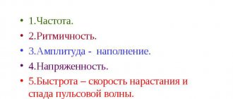
determine the correctness of the rhythm, the number of basic tones, their timbre, the integrity of the sound, the ratio of the volume of 1 and 2 tones. When additional tones are identified, their auscultatory features are noted: relation to the phases of the cardiac cycle, volume and timbre. To determine the melody of the heart, you should mentally reproduce it using syllabic phonation.
Difference between 1 and 2 heart sounds.
1st tone is longer and slightly lower than 2nd tone.
At the sites where the leaflet valves are auscultated, it is usually stronger than 2 tones. The 2nd tone, on the contrary, is somewhat shorter, higher and stronger than the 1st tone at the sites where the semilunar valves are heard. At the base of the heart, heart sounds are best conveyed by the syllables Bu' = tup' p,
and at the ventricles Bu' = tup.
It should be noted that in some completely healthy people the 2nd tone is stronger than the 1st and at the sites of auscultation of the leaflet valves. Sometimes, with rapid and, especially, irregular, arrhythmic heart activity, the 1st sound can be difficult to distinguish from the 2nd.
Change in the strength of heart sounds.
Heart sounds can change in strength, in character, bifurcate, additional tones may appear and peculiar heart rhythms may form. Changes in heart sounds may depend on the following main factors: 1. Changes in the contractile function of the ventricles, 2. Changes in the physical properties of the valves, 3. Changes in the level of blood pressure in the aorta and pulmonary artery, 4. From the non-simultaneous occurrence of individual components, 5. From external factors - changes in the properties of the sound-conducting medium - the lungs and chest wall, the state of the organs adjacent to the heart.
Decreased heart sounds
. The strength of heart sounds is weakened, first of all, in healthy people with a thick chest wall, with powerful muscle development and, especially, with excessive development of subcutaneous fatty tissue, in patients with edema, subcutaneous emphysema in the heart area. The development of pulmonary emphysema is even more important for weakening the volume of heart sounds, since emphysematous lung tissue has low sound conductivity. With severe pulmonary emphysema, heart sounds become barely audible. In patients with hydrothorax, pneumothorax, and hydropericardium, there is also a sharp decrease in the volume of heart sounds.
Weakening of heart sounds can be associated not only with causes external to the heart, but also with cardiac pathology. Heart sounds weaken with a decrease in the speed and force of contractions of the ventricles of the heart due to myocardial weakness. This can be observed in severe infectious diseases that occur with high myocardial intoxication, in myocarditis, in patients with hypertrophy and dilatation of the ventricles of the heart. Since the loudest component of any heart sound is the valvular component, if the closure of one or another heart valve is disrupted, the sound formed during the operation of the valve sharply weakens, until it disappears completely. In patients with mitral or tricuspid valve insufficiency, 1 tone sharply weakens. In patients with insufficiency of the aortic or pulmonary artery valves, weakening of the 2nd tone is observed. A weakening of the 2nd heart sound is observed in patients with a drop in blood pressure in the systemic or pulmonary circulation, when the semilunar valves close weaker than usual.
Get full text
Strengthening all heart sounds
observed with: 1) a thin chest wall, 2) when the heart is adjacent to the chest wall with a larger than usual area, for example, with shrinkage of the lungs, 3) with anemia, when, due to a decrease in blood viscosity, heart sounds become flapping, sharp, 4) in those cases when the speed and force of myocardial contraction increases, for example during physical activity, in patients with thyrotoxicosis, and during neuropsychic agitation. If the ventricles are insufficiently filled with blood, for example, with narrowing (stenosis) of the mitral orifice, the opening of the tricuspid valve, or with an extraordinary contraction of the heart (with extrasystole), contractions of the weakly filled ventricles of the heart with blood occur faster than usual. Therefore, in such patients there is also a sharp increase in tone 1.
Gain 2 tones
, or as they say more often, the accent of 2 tones over the aorta and pulmonary artery, is common and has significant diagnostic value. In children and people under 20 years of age, the 2nd sound over the pulmonary artery is normally louder than over the aorta. In older people, the 2nd tone above the aorta becomes louder than above the pulmonary artery. Strengthening of the 2nd tone above the aorta, its accent, is noted with an increase in blood pressure. When the aortic valve leaflets harden and, especially, when the aorta itself is sclerotic, tone 2 reaches significant strength and acquires a metallic tint. Similarly, an emphasis of the 2nd tone will appear on the pulmonary artery in patients with pulmonary hypertension of any origin - with heart defects, with acute or chronic pulmonary pathology, ranging from lobar pneumonia to pulmonary emphysema.
Split tones.
Split tones is a phenomenon when one of the two heart sounds is split into two parts, which are easily perceived by our ear as separate sounds. If this gap is very small and is not perceived by ear as separate sounds, then they speak of tone splitting. All transitions are possible between the splitting of a tone and its splitting, so there is no clear distinction between them.
Split 2 tones
. Non-simultaneous closure of the semilunar valves is the result of different durations of systole between the left and right ventricles. Systole ends the sooner the less blood the ventricle has to transfer to the aorta or pulmonary artery, the easier it is to fill them and the lower the blood pressure in them.
Above the base of the heart, bifurcation of 2 tones can occur in a healthy person at the end of inhalation and at the beginning of exhalation as a physiological phenomenon. As a pathological phenomenon, bifurcation is often observed with mitral valve defects, and especially often with mitral orifice stenosis. This bifurcation of the 2nd tone is best heard in the 3rd intercostal space on the left at the sternum. With mitral valve stenosis, the left ventricle is poorly filled with blood during the diastole phase and less blood than usual is ejected into the aorta. Consequently, the systole of the left ventricle of the heart decreases over time compared to the usual value. At the same time, these patients have high pulmonary hypertension, which means that the systole of the right ventricle lasts longer than usual. As a result of these changes in hemodynamics, non-simultaneous closure of the valves of the aorta and pulmonary trunk occurs, heard as a bifurcation of 2 tones. Thus, the bifurcation of 2 tones in the aorta and pulmonary artery is caused by the following conditions: 1) a rise in pressure in one of the vessels and normal pressure in the other, 2) low pressure in one of the vessels and normal in the other, 3) high pressure in one vessel and low in the other, 4) increased blood filling in one of the ventricles, 5) reduced blood filling of one of the ventricles, 6) increased filling of one of the ventricles and decreased filling of the other ventricle of the heart.
Split 1 tone
. It is heard when a normal tone is always followed by a weak abnormal tone. This phenomenon can occur in 10% of healthy people during auscultation in a supine position. As a pathological phenomenon, splitting of the 1st tone occurs with aortic sclerosis and with increased blood pressure in the systemic circulation.
Mitral valve opening tone.
In patients with mitral stenosis, with a normal heart rhythm (without atrial fibrillation), an increase in the number of heart sounds is observed, reminiscent of a split 2 heart sound, since the third additional sound quickly follows the 2nd normal heart sound.
This phenomenon is best heard over the apex of the heart. In healthy people, during the phase of rapid filling of the ventricles of the heart with blood, the mitral valve leaflets are silently pushed to the sides by blood. In patients with mitral valve stenosis, at the beginning of the diastole phase, when the ventricles begin to rapidly fill with blood, the shortened and sclerotic leaflets of the mitral valve form a funnel-shaped diaphragm. They cannot open freely and move towards the walls of the ventricle, they sharply tense up under the pressure of blood and generate the sound of the mitral valve opening. In this case, a unique three-membered heart rhythm is formed, called the quail rhythm.
The first component of this three-part rhythm is the first tone.
It is followed at the usual time interval by a second tone. Almost immediately after the second tone, the sound of the uterine valve opening follows after a short interval. A rhythm arises that can be transmitted by the sounds of Ta-tara
, reminiscent, in the figurative expression of old clinicians, of the cry of a quail “go to bed.” The rhythm of a quail is heard in normal or bradycardia. Only in the absence of tachycardia can one distinguish by ear the difference in intervals between the first - second and second - third components of the resulting three-part rhythm.
Gallop rhythm.
The splitting of the first tone is sometimes very sharp.
The part split off from the main tone is separated from it by some clearly perceptible interval and is heard as a separate independent tone. This phenomenon is no longer called a split tone, but a gallop rhythm, reminiscent of the clatter of the hooves of a galloping horse. This peculiar three-part rhythm appears against the background of tachycardia. The intervals between the first - second and second - third tones are perceived by ear as the same, the interval between the third and the following first sound of the next triad is perceived as somewhat larger. The resulting rhythm can be conveyed by sounds like ta-ra-ra, ta-ra-ra, ta-ra-ra.
The gallop rhythm is best determined above the apex of the heart and in the 3rd to 4th intercostal spaces to the left of the sternum. It can be heard better directly with the ear than with a phonendoscope. The gallop rhythm intensifies after light physical effort, when the patient moves from a vertical to a horizontal position, as well as at the end of inhalation - at the beginning of exhalation in a slowly and deeply breathing person.
The additional third tone during the gallop rhythm usually sounds dull and short. It can be positioned in relation to the fundamental tones as follows.
1. An additional tone can be heard during a long pause closer to the first tone. It is formed by the separation of the atrial and ventricular components of the first sound. It is called the presystolic gallop rhythm.
2. An additional tone can be heard in the middle of a long pause of the heart, i.e. in the middle of diastole. It is associated with the appearance of the 3rd heart sound and is called the diastolic gallop rhythm. Phonocardiography made it possible to distinguish protodiastolic (at the beginning of diastole) and mesodiastolic (mid-diastole) gallop rhythms. The protodiastolic gallop rhythm is caused by severe damage to the ventricular myocardium, most often by failure of the previously hypertrophied left ventricle. The appearance of an additional tone in diastole is caused by the rapid straightening of the flabby muscle of the left ventricle when it is filled with blood. This variant of the gallop rhythm can occur in normal and even bradycardia.
3. An additional tone can be heard immediately after the first tone. It is caused by different simultaneous excitation and contraction of the left and right ventricles of the heart due to conduction disturbances along the branches of the His bundle or along their branches. It is called the systolic gallop rhythm.
4. If, with high tachycardia, there are 3 and 4 heart sounds, then a short interval between them can lead to the fact that the four-member heart rhythm recorded on the phonocardiogram is perceived by ear as a three-member rhythm and a summed mesodiastolic gallop rhythm occurs (the summation of 3 and 4 sounds).
From a diagnostic point of view, the gallop rhythm is a very important symptom of heart weakness. According to the figurative expression, “The rhythm of a gallop is the cry of the heart for help.” It appears in patients with cardiac decompensation as a result of long-term arterial hypertension, with sclerosis of the heart muscle against the background of atherosclerosis, or a history of myocardial infarction. It is also detected in valvular heart defects, accompanied by damage to the heart muscle, in severe infections with toxic damage to the myocardium, for example, in diphtheria, and in acute myocarditis. Typically, the appearance of a gallop rhythm is a very unfavorable diagnostic sign.
Pendulum rhythm
- This is a two-part rhythm with equal pauses between the 1st and 2nd heart sounds. It occurs due to prolongation of ventricular systole during their hypertrophy, with cardiosclerosis and myocarditis.
Embryocardia
called the pendular rhythm heard during tachycardia. Normally, this rhythm is heard in the fetus. When it appears in an adult, embryocardia is evidence of severe myocardial damage, primarily due to an inflammatory process.
Characteristics of tone II
To listen to him, you need to place the bell of the phonendoscope over the base of the heart. This point is located slightly to the right of the xiphoid process of the sternum.
The volume and clarity of the second tone also depends on how tightly the valves, only now semilunar, close. In addition, the speed of their operation, that is, the closing and vibration of the risers, affects the sound produced. And additional qualities are the density of all structures involved in the formation of tone, as well as the position of the valves during the expulsion of blood from the heart.
Rules for listening to heart sounds
The sound of the heart is probably the most peaceful sound in the world, after white noise.
Scientists have a hypothesis that this is what the child hears during the prenatal period. But in order to identify damage to the heart, simply listening to how it beats is not enough. First of all, auscultation should be done in a quiet and warm room. The posture of the person being examined depends on which valve needs to be listened to more carefully. This could be a position lying on the left side, upright but with the body tilted forward, on the right side, etc.
The patient should breathe rarely and shallowly, and at the doctor’s request, hold his breath. In order to clearly understand where systole is and where diastole is, the doctor must, in parallel with listening, palpate the carotid artery, the pulse on which completely coincides with the systolic phase.
Procedure for auscultation of the heart
After a preliminary determination of absolute and relative cardiac dullness, the doctor listens to heart sounds.
It usually starts from the top of the organ. The mitral valve is clearly audible there. Then they move on to the valves of the main arteries. First to the aortic - in the second intercostal space to the right of the sternum, then to the pulmonary artery - at the same level, only on the left. The fourth listening point is the base of the heart. It is located at the base of the xiphoid process, but can move to the sides. So the doctor must check the shape of the heart and the electrical axis to accurately listen to the tricuspid valve.
Auscultation is completed at the Botkin-Erb point. The aortic valve can be heard here. It is located in the fourth intercostal space on the left of the sternum.
Heart sounds
The places of best detection of heart sounds - tones, as well as murmurs - do not always coincide with the anatomical localization of their sources - valves and the holes they close (Fig. 45). Thus, the mitral valve is projected at the site of attachment of the third rib to the sternum on the left; aortic - in the middle of the sternum at the level of the third costal cartilage; pulmonary artery - in the second intercostal space on the left at the edge of the sternum; tricuspid valve - in the middle of the line connecting the places of attachment to the sternum of the cartilages of the third left and fifth right ribs. Such proximity of the valve openings to each other makes it difficult to isolate sound phenomena at the place of their true projection onto the chest. In this regard, the locations of the best conduction of sound phenomena from each of the valves have been determined.
Rice. 45. Projection of heart valves onto the chest: A – aortic; L – pulmonary artery; D, T - two- and three-leaf.
The place for listening to the bicuspid valve (Fig. 46, a) is the area of the apical impulse, i.e., the 5th intercostal space at a distance of 1-1.5 cm inward from the left midclavicular line; aortic valve - II intercostal space on the right at the edge of the sternum (Fig. 46, b), as well as the 5th Botkin-Erb point (place of attachment of the III-IV rib to the left edge of the sternum; Fig. 46, c); pulmonary valve - II intercostal space on the left at the edge of the sternum (Fig. 46, d); tricuspid valve - the lower third of the sternum, at the base of the xiphoid process (Fig. 46, e).
Rice. 46. Listening to the heart valves: a - bicuspid at the apex; b, c — aortic, respectively, in the second intercostal space on the right and at the Botkin-Erb point; d — pulmonary valve; d - tricuspid valve; e - the order of listening to heart sounds.
Listening is carried out in a certain sequence (Fig. 46, e):
- apical beat area; II intercostal space on the right at the edge of the sternum;
- II intercostal space on the left at the edge of the sternum;
- lower third of the sternum (at the base of the xiphoid process);
- Botkin-Erb point.
This sequence is due to the frequency of damage to the heart valves.
The procedure for listening to heart valves:
In practically healthy individuals, when listening to the heart, two tones are usually detected - the first and second, sometimes the third (physiological) and even the fourth.
Normal heart sounds are I and II:
First tone
is the sum of sound phenomena occurring in the heart during systole. That's why it's called systolic. It occurs as a result of vibrations of the tense muscle of the ventricles (muscular component), closed leaflets of the bicuspid and tricuspid valves (valve component), the walls of the aorta and pulmonary artery during the initial period of blood entering them from the ventricles (vascular component), the atria during their contraction (atrial component).
Second tone
caused by the slamming and resulting vibrations of the aortic and pulmonary artery valves. Its appearance coincides with the beginning of diastole. That's why it's called diastolic.
Between the first and second tones there is a short pause (no sound phenomena are heard), and the second tone is followed by a long pause, after which the tone appears again. However, students beginning their studies often have great difficulty distinguishing between the first and second tones. To make this task easier, it is recommended to first listen to healthy people with slow heart rates. Normally, the first tone is heard louder at the apex of the heart and in the lower part of the sternum (Fig. 47, a). This is explained by the fact that sound phenomena from the mitral valve are better transmitted to the apex of the heart and the systolic tension of the left ventricle is more pronounced than that of the right. The second tone is heard louder at the base of the heart (at the sites where the aorta and pulmonary artery are heard; Fig. 47, b). The first tone is longer and lower than the second.
Rice. 47. Places of best listening to heart sounds: a – I tone; b – II tones.
By listening to alternately obese and thin people, one can be convinced that the volume of heart sounds depends not only on the condition of the heart, but also on the thickness of the tissues surrounding it. The greater the thickness of the muscle or fat layer, the lower the volume of tones, both the first and the second.
Rice. 48. Determination of the first heart sound by the apical impulse (a) and by the pulse of the carotid artery (b).
Heart sounds should be learned to be differentiated not only by the relative volume at the apex and base, by their different duration and timbre, but also by the coincidence of the appearance of the first tone and the pulse in the carotid artery or the first tone and the apical beat (Fig. 48). You cannot navigate by the pulse on the radial artery, since it appears later than the first tone, especially with a rapid rhythm. It is important to distinguish between the first and second tones not only due to their independent diagnostic significance, but also because they play the role of sound landmarks for identifying noise.
Third tone
caused by vibrations of the walls of the ventricles, mainly the left one (with their rapid filling with blood at the beginning of diastole). It is heard by direct auscultation at the apex of the heart or slightly inward from it, and is better with the patient lying down. This tone is very quiet and, in the absence of sufficient auscultation experience, may not be detected. It is better heard in young people (in most cases near the apex beat).
III heart sound (English):
Fourth tone
is the result of vibrations of the walls of the ventricles during their rapid filling at the end of diastole due to contraction of the atria. Rarely heard.
IV heart sound (English):
Next: change in heart sounds.
Additional tones
The sound of the heart does not always resemble rhythmic clicks.
Sometimes, more often than we would like, it takes on bizarre forms. Doctors have learned to identify some of them only by listening. These include: - Mitral valve click. It can be heard near the apex of the heart, it is associated with organic changes in the valve leaflets and appears only with acquired heart disease.
- Systolic click. Another type of mitral valve disease. In this case, its valves do not close tightly and seem to turn outward during systole.
- Recardton. Found in adhesive pericarditis. Associated with excessive stretching of the ventricles due to the moorings formed inside.
— Quail rhythm. Occurs with mitral stenosis, manifested by an increase in the first tone, an emphasis on the second tone on the pulmonary artery and a click of the mitral valve.
- Gallop rhythm. The reason for its appearance is a decrease in myocardial tone, which appears against the background of tachycardia.
I tone
I tone
is heard as a fairly intense sound over the entire surface of the heart. It is maximally expressed in the region of the apex of the heart and in the projection of the mitral valve. The main fluctuations of the first sound are associated with the closure of the atrioventricular valves, which was established by A. A. Ostroumov in his dissertation “On the origin of the first heart sound” (1873). On FCG, the following components are distinguished in the composition of the first tone: initial low-amplitude low-frequency oscillations associated with contraction of the ventricular muscles. The main one is the central segment of the first tone, consisting of oscillations of large amplitude and higher frequency, resulting from the closure of the mitral and tricuspid valves. The final part is low-amplitude oscillations associated with the opening of the semilunar valves of the aorta and pulmonary artery and the vibration of their walls. The total duration of the first tone ranges from 0.07 to 0.25 seconds. An assessment of the intensity of the first tone can be made during its graphical registration, however, an accurate measurement of the amplitude is not made due to the impossibility of accurately standardizing the amplitude characteristics during phonocardiography. At the apex of the heart, the amplitude of the first tone is 1.5 - 2 times greater than the amplitude of the second tone. Weakening of the first tone may be associated with a decrease in the contractile function of the heart muscle during myocardial infarction (see), myocarditis (see), rheumatic carditis (see Rheumatism) and with mitral valve insufficiency (see Acquired heart defects). The flapping nature of the first tone (an increase in both the amplitude and frequency of oscillations) is due to the compaction of the mitral valve leaflets and the shortening of their free edge while maintaining mobility with mitral stenosis (see Acquired heart defects). A very loud (“cannonball”) I tone occurs with complete atrioventricular block (see Heart block).
Intracardiac causes of increased and decreased heart sounds
Heart sounds are clear and rhythmic when a person is at rest or asleep. If he begins to move, for example, climbs the stairs to the doctor’s office, then this may cause an increase in the heart sound. Also, increased heart rate can be caused by anemia, diseases of the endocrine system, etc.
A dull heart sound is heard with acquired heart defects, such as mitral or aortic stenosis, or valve insufficiency. Aortic stenosis in the sections close to the heart makes its contribution: the ascending part, the arch, the descending part. Muffled heart sounds are associated with an increase in myocardial mass, as well as with inflammatory diseases of the heart muscle, leading to dystrophy or sclerosis.
Changes in heart sounds
Changes in the sound manifestation of the mechanical activity of the fibromuscular organ occur due to the pathology of cavities and septa. There are two reasons why waves are heard differently from the norm: pathological and physiological. If there are changes in the listening points, an extended examination is prescribed by the therapist or cardiologist. After a comprehensive examination, you may find:
- weakening of both sound manifestations. Usually the deviation is not associated with poor heart function, but is caused by: obesity, respiratory tract disease, myocardial inflammation;
- decline of the first tone. Occurs due to loose slamming of the valves of atypical muscle fibers, slowing down of contraction of the left cavity, with left ventricular hypertrophy, aortic stenosis;
- decline of the second tone. It is observed: when there is a violation of the closure of the connective tissue plates of the aorta and the pulmonary trunk, a decrease in blood pressure, a decrease in the mobility of the valves of the semilunar connective tissue plates. And also due to weakening of myocardial contractility, stagnation in the systemic and pulmonary circulation;
- increase in both sound manifestations. It is noted: in thin children, after severe physical stress, excessive activation of the nervous system, due to a violation of the diaphragm;
- increase of 1 tone at the peak. Occurs due to vibrations of the dense valves of the mitral connective tissue plates at the moment of their slamming;
- sharply increased first sound manifestation (Strazhesko). It is a sign of dysfunction in the conduction of electrical impulses from the atria into the cavity. And also arrhythmias, in which the systoles of the chambers and cavities coincide;
- increase in the second tone over the aorta. It is observed with hypertrophy of the left cavity of the heart. It also manifests itself with an increase in the speed of slamming of the valves of the connective tissue plates of a large azygos arterial vessel. It may be explained by the compaction of the valves of the aortic plates and walls while maintaining their mobility;
- accent of the second tone over the pulmonary trunk. It is a sign of hypertrophy of the right cavity. Occurs with a persistent increase in pressure in the pulmonary artery bed, narrowing of the left atrioventricular opening;
- decomposition of the first tone. Observed when intracavitary conduction through atypical cardiomyocytes is impaired;
- decomposition of the second tone. Occurs due to an increase in the time of expulsion of blood from the right cavity: anomaly of the interatrial wall, stenosis of the outflow tract of the right cavity, blockage of the orifice of the pulmonary trunk, blockade of atypical muscle fibers;
- three-part tone. It is heard against the background of a sharp rapid heartbeat;
- additional sound manifestation (4). The presystolic rhythm is heard before the first sound. Additional sound manifestation (3). Heard at the beginning of relaxation between contractions. Observed in pathologies: inflammation of the heart muscle, disruption of the anatomical structure of connective tissue plates and septa, hypertrophy of the fibromuscular organ;
- amplification of the first sound manifestation with decomposition of the second tone. It is heard at the apex of the fibromuscular organ and Botkin's point. It is a sign of narrowing of the left atrioventricular orifice. Caused by the appearance of a click when the mitral connective tissue plate opens;
- pendulum tone. Occurs when the duration of systole and diastole is the same, as well as the timbres of the 1st and 2nd sound manifestations do not differ in volume. It manifests itself due to decompensated dysfunction of the myocardium, arrhythmias with paroxysms, and a strong increase in temperature.
On a note! In addition to auscultation, graphic recording of sounds on paper tape is used to identify changes in sound manifestations. Waves are displayed as oscillations of varying amplitude and frequency.
Muffled tones
Deaf sound manifestations are also called weakened. They indicate weak activity of the fibromuscular organ. For example, when there is insufficiency of connective tissue plates or narrowing of a large unpaired arterial vessel, noisy sounds are clearly audible.
During auscultation, unclear, muffled heart sounds indicate a microscopic deviation of the middle cardiac muscle layer. Pathology occurs after: ischemic necrosis of areas of the fibromuscular organ, diffuse development of connective scar tissue in the myocardium, inflammation of the serous pericardium.
By listening to a dull sound manifestation at certain points, the doctor receives an accurate description of the changes occurring in the area of the main organ as:
- muffling of the first sound manifestation, heard at the apex of the fibromuscular organ, indicates inflammation of the heart muscle, cardiosclerosis or insufficiency of the atrioventricular plates;
- weakening of the second sound manifestation is heard on the right side of the ribs. Occurs due to insufficiency of the connective tissue plates of the aorta or narrowing of its mouth;
- the muffling of the second sound manifestation is heard on the left side of the ribs. Indicates insufficiency of the connective tissue plates of the pulmonary aorta or compression of its mouth.
Muffling of both sound manifestations can be caused by both pathological and physiological reasons. Pathological muting of tones may indicate causes that do not affect the heart. The reason for their appearance may be:
- violation of pulmonary ventilation and blood circulation;
- accumulation of fluid in one or both pleural cavities;
- accumulation of air or gases in the pleural cavities;
- inflammation of the serous pericardium.
Gallop rhythm
Pathological sumation pulse of the fibromuscular organ occurs with severe myocardial damage. Accompanied by a sharp rapid heartbeat. Acoustically, it resembles the clatter of a galloping horse. May be heard as early diastolic or presystolic.
The rhythm is characterized by the presence of 3 and 4 pathological sound manifestations, caused by heart failure and loss of muscle tone in the left cavity.
Heart murmurs
In addition to tones, the doctor may also hear other sounds, so-called noises. They are formed from the turbulence of the blood flow that passes through the cavities of the heart. Normally they shouldn't be there. All noise can be divided into organic and functional.
- Organic ones appear when anatomical, irreversible changes in the valve system occur in the organ.
- Functional noises are associated with disturbances in the innervation or nutrition of the papillary muscles, an increase in heart rate and blood flow speed, and a decrease in its viscosity.
Murmurs may accompany heart sounds or may be independent of them. Sometimes the pleural friction noise in inflammatory diseases is superimposed on the heartbeat, and then you need to ask the patient to hold his breath or lean forward and auscultate again. This simple trick will help you avoid mistakes. As a rule, when listening to pathological noises, they try to determine in what phase of the cardiac cycle they occur, find the place of best listening and collect the characteristics of the noise: strength, duration and direction.
Change in heart sounds
Previously: heart sounds.
A change in heart sounds can primarily be expressed in a weakening or strengthening of the sonority of one or both of them, in a change in timbre, duration, in their splitting or bifurcation, in some cases - in the appearance of additional tones. In this case, determining the place of best listening to pathological sound phenomena is of diagnostic importance. Strengthening of the second tone in the 2nd intercostal space on the left
speaks of its emphasis on the pulmonary artery (determined by comparing its volume and timbre on the pulmonary artery and aorta).
This indicates an increase in pressure in the pulmonary circulation, which can be observed in diseases of the heart, as well as the respiratory system (mitral defects, emphysema, pneumosclerosis, chronic pneumonia). The intensification of the second tone in the second intercostal space on the right
indicates its emphasis on the aorta, which is observed with an increase in blood pressure in the systemic circulation (arterial hypertension), as well as in the case of hardening of the wall and valve of the aorta in atherosclerosis and a number of other diseases.
Strengthening the first sound at the apex of the heart
most often occurs with narrowing of the left atrioventricular orifice (mitral stenosis), tachycardia. This is due to the fact that with this defect, during diastole, less blood flows into the left ventricle than normal, and it contracts more quickly (transition from a relaxed to a tense state). In addition, with mitral stenosis, the timbre of the first tone changes due to vibrations of the sclerotic cusps of the mitral valve. It takes on a crackling tone, reminiscent of the sound of a flag flapping in the wind. This sound at the apex of the heart with mitral stenosis is called “popping”.
Weakening of the first sound at the apex of the heart
can be observed during inflammatory processes of its muscles (myocarditis), cardiosclerosis (scar changes in the heart muscle), and damage to the valve apparatus (bicuspid and tricuspid, as well as aortic).
Weakening of the second sound on the aorta
possible with aortic defects (aortic valve insufficiency or stenosis of its mouth).
Weakening of the second tone on the pulmonary artery
occurs when the valve is insufficient or its opening is narrowed (stenosis).
If during auscultation of the heart, instead of one of the tones, two short ones are heard, following each other after a short period of time, then this indicates a split tone
.
If the difference in the time of occurrence of these components is insignificant and the impression of splitting is not created, we are talking about tone splitting
. Thus, there is no fundamental qualitative difference between bifurcation and splitting of tones. There is only some quantitative difference: splitting is the initial phase, and bifurcation is a more pronounced degree of disruption of the unity of tones.
Bifurcation and splitting of tones can be physiological and pathological. For example, the bifurcation of the first tone may depend on the non-simultaneous closure of the bicuspid and tricuspid valves as a result of changes in pressure in the chest during different phases of breathing. But more often, a split in the first tone indicates pathological changes in the heart. It occurs, as a rule, when one of the legs of the atrioventricular bundle (bundle of His) is blocked, which leads to non-simultaneous contraction of the right and left ventricles of the heart. This can occur with significant blockade of the atrioventricular (atrioventricular) node, with sclerosis of the initial part of the aorta.
In case of severe heart damage, a three-part rhythm can be heard. It is caused by weakening of the myocardium (inflammation, degenerative changes, toxic lesions) of the left ventricle and occurs as a result of rapid stretching of its walls under the pressure of blood flowing from the atrium. This creates the melody of a three-part rhythm (first, second and additional third tones), reminiscent of the clatter of a galloping horse - the “ gallop rhythm.”
" It is also figuratively called the “cry of the heart for help,” since it is a sign of severe heart damage. The rhythm of the gallop is best heard directly by the ear (along with the sound, a slight impulse is perceived, transmitted from the heart to the chest in the diastole phase) in the area of the apex of the heart or the third-fourth intercostal space on the left. It can be heard especially clearly when the patient is lying on his left side. But this creates inconvenience for direct listening with the ear. In such cases, a phonendoscope is used.
There are protodiastolic, mesodiastolic and presystolic gallop rhythms (depending on the diastole phase, during which the pathological third sound appears).
gallop rhythm
an auscultatory finding of three (triple r.) or four (quadruple r.) heart sounds;
the extra sounds occur in diastole and are related either to atrial contraction (S), to early rapid filling of a ventricle (S), or to concurrence of both events (summation gallop) . Translation: The gallop rhythm
is heard as a three-part or four-part rhythm. Additional sounds appear in diastole and are caused either by atrial contraction, or early rapid filling of the ventricle, or a combination of both mechanisms (summation gallop).
Much more common are bifurcation and splitting of the second tone, caused by non-simultaneous closure of the pulmonary artery and aortic valves due to increased pressure in the pulmonary or systemic circulation. Bifurcation and splitting of the second tone can also be physiological and pathological.
Physiological splitting of the second tone is heard exclusively at the base of the heart during inhalation and exhalation or during physical activity. At the end of a deep inhalation, when the chest expands due to a decrease in pressure in it, the blood is somewhat retained in the dilated vessels of the small circle and therefore flows in smaller quantities into the left atrium, and from there into the left ventricle. The latter, due to less blood filling, ends systole earlier than the right one, and the closure of the aortic valve precedes the closure of the pulmonary valve. During exhalation, the opposite conditions are created. In the case of increased pressure in the chest, blood, as if squeezed out of the vessels of the pulmonary circle, enters in large quantities into the left part of the heart, and the systole of the left ventricle, and therefore the beginning of its diastole, occurs later than the right.
At the same time, a split second tone may be a sign of serious pathological changes in the heart and its valves. Thus, a bifurcation of the second sound at the base of the heart (second intercostal space on the left) is heard with mitral stenosis. This is due to the fact that the hypertrophied and blood-filled right ventricle ends systole later than the left. Therefore, the aortic component of the second sound occurs earlier than the pulmonary one. Bifurcation or splitting of the second sound in bicuspid valve insufficiency is associated with greater blood filling of the left ventricle than normal, which leads to prolongation of its systole, and the diastole of the left ventricle begins later than the right. Because of this, the aortic valve closes later than the pulmonary valve.
One should distinguish from the true splitting of the second tone its sound melody, which only superficially resembles splitting. An example is the additional tone that occurs during the opening of the bicuspid (mitral) valve with mitral stenosis. It has a high-pitched clicking tone and is perceived as a loud echo following the second tone. The additional tone, together with the clapping first and second, form a peculiar melody, reminiscent of the cry of a quail. Hence the name of this sound phenomenon, heard with mitral stenosis at the apex of the heart - “ quail rhythm”
" Its distribution area is extensive - from the apex of the heart up and into the axillary fossa.
Sometimes, when listening to the heart, against the background of rare and dull tones, a lonely, very loud tone appears, the so-called “gun tone” Strazhesko
. It is caused by the simultaneous contraction of the atria and ventricles, which is observed with complete atrioventricular block, i.e. when impulses from the atria do not reach the ventricles and they each contract in their own rhythm (the atria contract more often), but in some cycle their contractions coincide.
Next: heart murmurs.
All heart sounds (English):