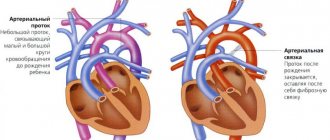
The examination method determines the degree of filling of the veins and their pulsation. In healthy animals, the veins are moderately filled; pulsation in the jugular vein can only be seen at the base of the neck, in the jugular groove. It is very well expressed in cattle, especially when the head is tilted.
Examination of the peripheral veins in case of weakness of the right half of the heart reveals a difficult outflow of blood from the entire venous network, which is expressed in a sharp overflow of the veins with blood, which is judged by the cyanotic appearance of the mucous membranes, edema, and increased relief of the pattern of the skin veins (they appear especially clearly on the facial parts of the head, ventral surface of the abdomen and inner thighs). Skin veins become elastic when blood stagnates in them. Venous stagnation is sharply reflected in the jugular veins. The latter increase in volume and stand out prominently in the jugular groove in the form of thick cords, their elasticity increases significantly. Very clearly indicated signs appear in traumatic pericarditis in cattle and tricuspid valve insufficiency. Changes are easier to detect in animals with delicate skin and short hair. Obstructed blood outflow in the pulmonary circulation can be caused by emphysema, pneumosclerosis, and chronic pneumonia. When examining peripheral veins, local diseases, inflammation of the veins - phlebitis, thrombophlebitis, can be identified.
Venous heart rate
Near the heart there are large hollow and jugular veins, in which the pressure fluctuates, the volume changes - a venous pulse appears. Vascular oscillations are inextricably linked with cardiac cycles and their causes are the cessation of the outflow of blood from the veins to the heart. Normally, the indicator should be negative. A positive test indicates pathological processes in the heart valves.
What does it represent?
In the large veins located next to the heart, pulse fluctuations are observed in the systolic phase of the atria and ventricles - when the myocardium contracts and blood is expelled from the heart into the vascular system.
In this case, the pressure in the veins increases, their walls begin to vibrate. Normally, the venous pulse is found only in the central, usually jugular veins, which are located closer to the heart.
In small blood vessels, pulse fluctuations are not detected.
Fluctuations are determined by visual signs using phlebography. Palpation is not used due to low pressure in the veins, as a result of which tension in the walls is not felt. On the phlebo-sphygmogram, the curve has a negative direction. Fluctuations in the walls of the veins are unsharp, sluggish, the rise of the pulse wave takes longer, unlike the walls of the arteries.
Non-closure of the tricuspid valve leaflets leads to pathological reflux of blood during systole from the right ventricle into the right atrium and affects the occurrence of a positive pulse.
The volume of the vein increases synchronously with systole. There is a rapid movement of blood in the direction opposite to normal.
A pulsation of the neck veins, noticeable to the naked eye, appears; the swelling corresponds to the systological phases.
Transfer pulsation of veins
This vital sign is often observed in people with a quick temper.
Occurs as a result of pulse fluctuations in the paired carotid artery, which originates in the chest cavity and passes to the neck.
Normally, such a pulse occurs in hot-tempered people; it appears during emotional stress, after physical exertion.
It is observed in neurocircular dystonia of the hypertensive type, hypertension, if there is aortic valve insufficiency.
Respiratory
It is diagnosed when a person suffers from diseases accompanied by high pressure in the chest area. This is due to changes in the lung tissue, appearing airy.
Such pulsation is found in people who suffer from bronchial asthma, pleurisy - if the circulatory system is oversaturated with blood. The process depends on inhalation and exhalation, as well as on the stage of development of the pathology.
During inspiration, the volume of venous blood decreases due to passive filling of the right atrium and the appearance of negative pressure in the chest. Exhalation is accompanied by dilation of blood vessels, due to the lack of opportunities for blood outflow.
Heart pulse
Corresponds to one contraction - systole and one relaxation - diastole of the heart. Divided into two types:
A negative indicator is formed when the veins collapse during ventricular contraction.
- Negative venous pulse. This type appears as a result of the collapse of the veins directly during the period of tension and expulsion of blood - into ventricular systole.
- Positive. Occurs due to filling of the jugular veins.
In a healthy person, only two types of venous pulse are possible - transmission and cardiac.
What is positive and negative pulse?
Positive pulsation is a consequence of dilation of the jugular vessels during the systolic period, which is part of the cardiac ventricular cycle.
It also occurs with acquired heart disease, due to incompetence of the tricuspid valve - when it does not close completely.
Through the open valves, blood flows from the ventricle towards the right atrium to the systemic circulation, where it stagnates. As a result, swelling, pain under the right rib, ascites, and yellowing of the skin appear.
Negative venous pulse is the compression of venous vessels during contractions of the ventricles and pumping of blood into the arteries. From the vein it is directed to the atrium under the influence of negative pressure of the thoracic cell and a decrease in its volume.
Negative pulsation is found in large vessels that are located near the heart. If a person is healthy, when lying down, the ventricles simultaneously contract and the jugular veins compress above the collarbone. A negative venous pulse is normal.
Phlebography
During diastole, accumulated blood flows into the atria and the pressure in them drops. Fluctuations in the venous pulse can be recorded using the following devices: phlebograph, mirror sphygmograph, electrosphygmograph.
Using the phlebography method, the venous pulse is recorded in the form of a curve - a phlebogram, which is recorded from the jugular veins, with the animal's head lowered. Vibrations of the veins reflect the work of the right atrium and ventricle. On the phlebogram, three waves are noted - a, c and V. Wave a coincides with atrial systole, due to the fact that at the moment of atrial systole, the place where the veins flow into them is clamped by a ring-shaped muscle, as a result of which the blood flow is suspended, the veins are stretched. Wave c appears at the beginning of ventricular systole, at the moment of closure of the leaflet valves. The portion of the decrease in the curve after wave C and the new rise to wave V correspond to atrial diastole and ventricular systole. During this period, blood first flows from the veins and atria, and then the atria and large veins (y wave) fill with blood. The downward section of the curve after wave V corresponds to the onset of total diastole, when blood from the atria and veins enters the ventricles.
The coincidence of the venous pulse waves with certain phases of cardiac activity is of interest to the veterinarian: from the recording of the venous pulse one can judge the duration of the cardiac phases, as well as the disturbance in the conduction of impulses from the atria to the ventricles. The time from o to c corresponds approximately to the time of conduction of the impulse from the atria to the ventricular muscles. Lengthening or loss of the ventricular cycle characterizes the slowdown and cessation of the conduction of excitation along the atrioventricular bundle, which occurs with partial and complete heart block.
Phlebography can be used to detect atrial fibrillation, tricuspid valve insufficiency, and oscillation of veins due to stagnation of blood in the venous system. Interpreting a venogram is quite difficult: it is easier to read with bradycardia and more difficult with tachycardia.
Positive venous pulse occurs
A rapid pulse is definitely a sign of increased stress on the heart, but in order to understand what to do to restore the normal heart rate, you must first identify the reasons causing the rapid heartbeat.
An increase in heart rate occurs under the influence of various factors and pathogens that force the heart muscle to increase the frequency of contractions.
In most cases, an increase in rhythm is associated with psycho-emotional stress or with an increase in physical stress on the body.
The normal heart rate for an adult is limited to 60 to 80 beats per minute. Even a slight increase of 5-10 beats above the upper norm can be a sign of serious pathology. Provided that the rapid heartbeat has become permanent and does not decrease and causes symptoms of malaise, dizziness, shortness of breath, having a bad effect on well-being.
What it is
Pulse reflects the rate of heart contraction. Its changes indicate malfunctions in the functioning of the main organ of the body. Increased rhythm is considered as one of the variants of tachycardia.
Increased heart rate is an increase in the frequency of contractions of the heart muscle. The pulse reflects the quality of myocardial function. With each heart contraction, the blood vessels undergo a shock. We feel it on the wrist when counting rhythmic waves.
It is impossible to have a low pulse with a fast heartbeat. This process is inextricably linked; as the pulse quickens, the heartbeat quickens. Therefore, all conditions associated with the load on the myocardium will affect the frequency of oscillations of the vascular walls.
The pulse rhythm serves us to timely detect a malfunction in the cardiovascular system of the body. An increase in heart rate without a physiological reason can be considered an important signal, after which you should undergo an examination to diagnose the pathology that causes an increase in heart rate.
Causes
The reasons for the development of a rapid pulse come down to insufficient training of the heart muscle. The disease is more common in people leading a passive lifestyle. The heart, accustomed to rest, can react sharply to physical activity. If the reason is more serious, then the pulse quickens to a critical level.
The reasons that can provoke increased heart rate and heart rate are divided into 2 categories:
- Physiological.
- Pathological.
Causes of the 1st category are safe; they manifest themselves when external conditions change, for example air temperature, body position, age, weight, hormonal levels or excessive stress on the body.
Category 2 should be taken very seriously, since pathological causes are associated with diseases and malfunctions in the body.
The pulse rhythm after a temporary functional increase (the physiological reason for the increase during exercise) to the maximum value should return to its initial state within five, maximum seven minutes.
If this does not happen, pay attention to pathological reasons.
Physiological
A simultaneous increase in blood pressure and increased heart rate during physical activity and stress is a natural reaction of the body to an irritant that causes mental arousal.
Provided that after 5-10 minutes. at rest, the pulse rate and blood pressure calmed down to normal.
Natural factors influencing increased heart rate occur regardless of the presence of common diseases.
Physiological tachycardia can be caused by:
- excessive emotions of a positive and negative nature;
- unusual physical work;
- increased hormonal levels due to stress or pregnancy;
- pain syndrome;
- sexual arousal;
- unfavorable temperature (heat);
- abuse of alcohol, coffee and strong tea;
- smoking;
- taking medications.
An increase in the rhythm of vascular oscillations for physiological reasons is a temporary phenomenon. Without concomitant diseases, the condition usually does not threaten life and health.
Pathological
Increased heart rate, which is pathological in nature, is usually a consequence of diseases such as heart disease, heart attack, and disruption of the cardiovascular system.
Pathological causes of rapid heart rate are manifestations of various diseases. The myocardium responds to them by increasing the number of heart contractions. This helps protect brain cells from hypoxia.
Pathological tachycardia may be a sign or consequence of:
- thyroid diseases;
- poisoning;
- injuries with massive bleeding;
- panic attacks;
- defects and other pathologies of the heart;
- damage to nerve fibers;
- shock states;
- disturbances of vascular tone;
- infectious diseases.
A rapid pulse can be called a protective reaction of the body against oxygen starvation. As soon as the heart senses a lack of circulating blood, it begins to work at an accelerated pace.
A frequent heart rate rhythm is called tachycardia, which manifests itself depending on the mechanism of its occurrence. The process of increased heart rate is quite complex and corresponds to a certain classification. There are three main types of tachycardia. Their manifestations can be seen using the table as an example.
Types of rapid pulse
Type of tachycardiaFeatures and symptoms
| Sinus (nodal) | More often it is a sign of a physiological increase in heart rate. Occurs with emotional excitability or stress. The number of heart contractions can exceed 100 beats per minute. The general condition is not impaired. The rhythm returns to normal when the provoking factor is eliminated. |
| Supraventricular (supra-ventricular) | It is characterized by a sharp increase in heart rate up to 200 or more beats per minute. To determine its quantity, it is enough to place your hand on any large vessel. The condition causes pain and a feeling of tightness in the chest. Characterized by anxiety and fear for one’s life. |
| Ventricular | This type of tachycardia can only be encountered in emergency situations. Disturbances in the functioning of the myocardial muscles cause an increase in heart rate of 250 beats and above. It is difficult for the heart to maintain such a rhythm. If emergency assistance is not provided, the condition can cause death. |
Important! If there is a sudden increase in readings to 100 beats per minute or higher, without the influence of external factors, you should seek professional help.
Norms for physiological indicators may differ among different categories of people. It depends on lifestyle, individual characteristics or the presence of bad habits. It happens that the number of beats per minute is increased, but the state of health is normal. This may be a consequence of the heart being well trained, for example in athletes.
But a constantly increased heart rate is sometimes a sign of hidden pathologies. Therefore, if the condition is accompanied by dizziness, shortness of breath or high blood pressure, you should undergo an examination. After making a medical diagnosis, it will be clear what to do if your heart rate increases in your case.
Possible complications
The process of increased heart rate is a gradual or sharp increase in the number of vibrations of the vascular walls, which indicates a lack of oxygen in the tissues. The heart, trying to increase blood discharge, works in increased wear and tear mode. Gradually, the myocardium loses the ability to perform qualitative contraction and conduction of impulses.
This can cause complications in the cardiovascular system, heart attack and even cardiac arrest. Sudden changes in tempo during increased heart rate can cause acute hypoxia of the brain and provoke fainting. The development of life-threatening conditions is the main reason why rapid rhythm is dangerous. Pathological tachycardia is caused by:
- vascular thrombosis;
- persistent arrhythmia;
- cardiac asthma;
- coronary heart disease.
Attention! Timely contact with a specialist reduces the risk of developing complications in the functioning of the heartbeat rhythm with a rapid pulse.
What to do for treatment
To get rid of pathological tachycardia, the causes of its occurrence must be eliminated. It will be necessary to undergo an examination and begin treatment for the underlying disease.
The need to measure heart rate at a doctor’s appointment is due to the ability to diagnose and treat diseases of the vascular system, as well as to find out about the presence or absence of abnormalities in the structure of organs, tissues, and body systems. For example, in this way a preliminary diagnosis of arrhythmia (a complete or partial disturbance of rhythm - the sequence of heartbeats) can be made, which requires drug treatment.
With the physiological increase in heart rate indicators it is a little easier. Here you can fight against risk factors with the help of doctors and on your own.
To treat increased myocardial excitability and normalize the rate, oral drops are prescribed:
- Valoserdin;
- Valerian;
- Motherwort;
- Valocordin;
- Corvalol.
These drugs have a sedative and calming effect. If desired, you can use herbal mixtures with chamomile, lemon balm, peony and hawthorn for treatment.
Emergency help
In severe conditions associated with a sudden strong increase in heart rate and deterioration in health, you should immediately call a doctor. Before the ambulance arrives, you should calm down, provide air access and take a strictly horizontal position.
Try to follow a few of our recommendations:
- take 5 or 6 deep breaths through your nose and exhale slowly through your mouth;
- inflate the children's balloon several times;
- massage the area of the eyeballs with your fingers;
- wash with cold water.
The emergency physician administers adrenergic blockers, which reduce the excitability of the heart. Subsequently, treatment with Anaprilin, Phenozepam or Phenobarbital is prescribed.
In rare cases, surgery may be used. The surgical method involves coagulation of the nerve endings of the myocardium and reducing the frequency of its contractions.
Increased heart rate is not a disease. The condition is a consequence of physiological or pathological changes in the functioning of the heart. As long as the organ functions correctly, the pulse will be normal, do not forget to monitor its rhythm and lead a healthy lifestyle.
Take the TEST: Do you have tachycardia?
Source: PulsNorma.ru
Arterial pulse
There are central arterial P. (P. of the aorta, subclavian and carotid arteries) and peripheral, determined on the arteries of the extremities.
Physiology
The origin of arterial P. is associated with the cyclic activity of the heart (see). The systolic volume of blood ejected into the aorta causes stretching of its initial part and an increase in pressure in it, which decreases during diastole. Pressure fluctuations propagate through the aorta and the arteries extending from it in the form of waves, stretching and lengthening the arterial walls. According to the pulsating changes in pressure, the movement of blood through the arteries acquires a pulsating character: blood flow accelerates during systole and slows down during diastole. The amplitude of oscillations and the shape of the pulse wave change as it moves from the center to the periphery, and the linear speed of blood flow gradually decreases due to resistance to blood flow, which increases as the diameter of the arteries decreases. The speed of propagation of the pulse wave (4-11 m/sec) significantly exceeds the linear speed of blood movement; edges in large arteries do not exceed 0.5 m/sec. Blood flow resistance has almost no effect on the speed of pulse wave propagation.
The pulsating nature of blood flow is important in the regulation of blood circulation (see) in general. The frequency and amplitude of pulsations affect vascular tone both through direct mechanical effects on the smooth muscles of the vascular wall and through afferent impulses from baroreceptor zones. In this case, the receptors can respond to changes in pulse blood volume and changes in pulse pressure.
Pulse volume is the amount of blood flowing through a given section of artery during each pulse period. Its value depends on the caliber of the artery, the degree of opening of its lumen, the volume of circulating blood, stroke volume, and blood flow speed. There is a direct relationship between the value of pulse volume and pulse pressure (the difference between systolic and diastolic pressure in the vessel).
Research methods
Rice.
1. Palpation of the pulse on the radial artery: a - II, III and IV fingers; b - III and IV fingers with fixation of the forearm with I and II fingers. Rice. 2. Sphygmogram of peripheral pulse: AB - anacrotic; BV - catacrota; The dicrotic wave is indicated by an arrow. In healthy people under conditions of physical rest, examination does not provide significant information about the nature of the P. In thin individuals, pulsation of the carotid arteries and transmitting pulsation of tissues in the jugular fossa may be noticeable. P. of the carotid and many peripheral arteries often becomes visible with significant physical exertion, with excitement, fever, severe anemia, thyrotoxicosis, and especially with aortic valve insufficiency. The main method of studying arterial P. is palpation. The brachial artery can be felt in the sulcus bicipitalis med. directly above the cubital fossa; axillary - at the bottom of the armpit on the head of the humerus after raising the straightened arm to a horizontal position. Palpation of the carotid arteries should be carried out carefully, taking into account the carotid reflex (see Autonomic reflexes), alternately on both sides. The femoral artery is palpated in the groin area with the thigh straightened and slightly rotated outward; popliteal - in the popliteal fossa with the patient lying on his stomach with the leg bent at the knee. The posterior tibial artery is identified in the condylar groove behind the medial malleolus; dorsalis pedis artery - in the proximal part of the first intermetatarsal space on the outer side of the long extensor of the big toe. Most often, P. is examined on the radial artery, the edges are located superficially and can be easily palpated between the styloid process of the radial bone and the tendon of the internal radial muscle. Having felt the artery, press it to the underlying bone (Fig. 1). In this case, the pulse wave is felt with the fingers as a push, movement, or as an increase in the volume of the artery. P.'s research must be carried out on both hands. In infants and hyperexcitable children, the superficial temporal arteries are palpated. Pulse fluctuations of peripheral arteries can be recorded using sphygmography (see); The graphic image of each pulse wave (Fig. 2) is characterized by its steep rise in the ascending part - anacrotic; the edges, having reached the top, turn into catacrotic - an oblique line going down, with an additional wave on it, called dicrotic. Graphic registration of P. makes it possible to establish such variants of its changes as anacrotic, asthenic, dicrotic, monocrotic P., as well as to carry out amplitude and chronometric analysis of pulse curves and measurement of pulse wave speed (see Sphygmography). Pulse fluctuations in the blood supply of small vessels are studied using plethysmography (see), rheography (see). To monitor P.'s frequency, special devices are used—pulse tachometers.
Clinical characteristics and diagnostic significance of changes in arterial pulse. During palpation examination of arteries, the characteristics of arterial P. are based on determining its frequency and assessing such qualities of P. as rhythm, filling, tension, height, speed.
Heart rate
count in no less than 0.5 minutes, and if the rhythm is incorrect, for a whole minute. In healthy adults, P.'s frequency in a horizontal position ranges from 60 to 80 per minute; in a vertical position, the frequency of P. is higher. In elderly people, the frequency of P/ is sometimes less than 60. In women, P. is on average 6-8 beats more often than in men of the same age.
An increase in P. is called tachyphygmia (pulsus frequens), a decrease is called bradysphygmia (pulsus rarus). Patol, increased P. occurs during fever: with an increase in body temperature by 1°, the pulse quickens by an average of 6-8 beats per minute. (in children by 15-20 beats). However, P.'s frequency does not always correspond to body temperature. Thus, with typhoid fever during fever, the increase in the frequency of P. lags behind the increase in temperature (relative bradysphygmia), and with peritonitis, a relative increase in P. is noted. Tachyphygmia as a reflection of tachycardia (see) is observed with autonomic dysfunction, heart failure, thyrotoxicosis, anemia. P.'s decrease occurs in trained athletes or is a constitutional feature. Patol, a decrease in P. is observed with obstructive jaundice, myxedema, and with increased intracranial pressure. A persistent and significant decrease in P. (40 or less per minute) occurs with complete transverse heart block (see). With extrasystole of the bigeminy type (see Extrasystole), if premature contractions of the ventricles are dynamically so weak that they do not cause a palpable pulse wave, a pronounced decrease in P is also noted.
In children
the heart rate is higher than in adults, which is due to a higher level of metabolism and the predominance of sympathetic nerve tone.
As the influence of the vagus nerve on the heart increases, P.'s frequency in children gradually decreases with age (table). PULSE RATE IN CHILDREN AND ADOLESCENTS OF DIFFERENT AGES AT REST
(according to A.F. Tour, 1967)
| Age | Pulse rate per minute | Age | Pulse rate per minute | Age | Pulse rate per minute | ||
| 6 months | 130—135 | 6 years | 90—95 | 12 years | 75—82 | ||
| 1 year | 120—125 | 7 years | 85—90 | 13 years | 72—80 | ||
| 2 years | 110—115 | 8 years | 80—85 | 14 years | 72—78 | ||
| 3 years | 105 — 110 | 9 years | 80—85 | 15 years | 70—76 | ||
| 4 years | 100—105 | 10 years | 78—85 |
In children of the same age, P.'s frequency is subject to large individual fluctuations. In girls it is 2-6 beats per minute. more than boys of the same age. These differences are revealed already in the neonatal period and are most pronounced in the prepubertal and pubertal periods. The maximum frequency of P. is observed in newborns; in the first hours of life, P. is relatively rare (up to 90-100 beats per minute); on days 2-3, P.’s frequency increases to 120-140 beats per minute. P. reduction in newborns to 100 beats per minute. and less should be considered as bradysphygmia, and its increase to 180 or more beats per minute - as tachyphygmia. When sucking, screaming, crying, P. can easily increase in frequency up to 180-200 beats per minute. P. is especially labile in premature infants; even at rest, its frequency fluctuates within . 120-160 beats per minute. P.'s frequency varies throughout the day. In children, the most frequent P. is observed in the morning; at night it decreases. This tendency is detected even in newborns, but in older children it is most pronounced. According to M. V. Rimsh (1971), the maximum frequency of P. in children under 7 years of age is recorded at 7-9 hours, in school-age children - at 10-12 hours; minimum - 1-3 hours (in children of the same age). The number of heartbeats in a sleeping child is 10-20 beats less than when awake. As the ambient temperature rises, P. becomes more frequent; P. in summer more often than in winter. In children, as in adults, the frequency of P. increases with physical activity, emotions, and after eating, especially hot dishes, spices, strong tea, and coffee. P.'s increase is proportional to the intensity of physical activity, but P.'s reaction to physical activity has age-related differences. Thus, according to V.M. Korol’s data (1969), in 8-year-old children the increase in the frequency of P. in the first minute of work is 50% relative to the initial one, and in 17-year-old boys it is 72%. The time for stabilization of the achieved level of heart rate also increases with age, and the restoration of the initial heart rate after stopping work at an older age occurs faster than at a younger age, which indicates a more perfect regulation of heart activity at an older age.
Rice. 3. Sphygmographic image of the pulse in atrial fibrillation: pulse waves follow randomly at different intervals.
Pulse rhythm
assessed by the regularity of pulse waves following one after another. In healthy people, pulse waves, like heart contractions, are observed at almost equal intervals, i.e. the pulse is rhythmic (pulsus regularis). In some heart rhythm disturbances (see Cardiac Arrhythmias), pulse waves follow at unequal intervals and P. becomes arrhythmic (pulsus irregularis). In healthy people, there may be an increase in P. during inhalation and a decrease in exhalation—respiratory arrhythmia; when holding the breath, P. becomes rhythmic. In case of bigeminy with hemodynamically effective extrasystoles, P. on the arteries is palpable as a pairwise alternation of waves of different strengths (the second wave is weakened) with an extended pause between these pairs of waves - bigeminy P. (pulsus bigeminus). Dicrotia P. should be distinguished from bigeminic P., or dicrotic P. (pulsus dicroticus), which is also palpated as a double beat, but this double beat corresponds to only one heartbeat. P.'s dicrotism is associated with changes in vascular tone and is caused by a sharp increase in the dicrotic wave of arterial P., which is clearly visible on the sphygmogram (see Sphygmography). With atrial fibrillation (see), pulse waves follow randomly at different intervals (Fig. 3). With sinoauricular block, incomplete atrioventricular block, and with early extrasystoles, loss of individual pulse waves is observed. If the number of heartbeats per unit time exceeds the number of pulse beats, they speak of P. deficiency. P. deficiency occurs with atrial fibrillation and extrasystole, it is caused by a sharp decrease in stroke output during some systoles of the left ventricle. Significant P. deficiency in patients with atrial fibrillation is one of the signs of heart failure.
Pulse filling
determined by the sensation of pulse changes in the volume of the palpated artery. The degree of arterial filling is influenced by the amount of blood ejected by the heart during systole (stroke volume), the total amount of blood in the body and its distribution. Under normal conditions, complete P. (pulsus plenus) is determined. With a decrease in stroke volume, blood loss, and a decrease in the volume of circulating blood, P.'s filling decreases. When there is a sharp decrease in P.'s filling, it is called empty (pulsus vacuus).
Pulse voltage
determined by the amount of force that must be applied to completely compress the pulsating artery. To do this, the radial artery is compressed with one of the fingers of the palpating hand and at the same time the P. is palpated distally with another finger, determining its decrease or disappearance. There are P. tense, or hard (pulsus durus), and P. soft (pulsus mollis).
The degree of P.'s tension depends on the level of blood pressure.
Rice. 4. Sphygmographic image of the alternating pulse: alternation of large and small pulse waves.
Pulse height
, or its value, gives an idea of the amplitude of oscillations of the arterial wall during the passage of the pulse wave. P.'s height is directly proportional to the magnitude of the pulse pressure and inversely proportional to the degree of tonic tension of the artery walls. High, or large, P. (pulsus altus, s. magnus) is observed with aortic valve insufficiency, thyrotoxicosis, physical stress, and fever. With a reduced or slow flow of blood into the aorta, as well as with an increase in the tension of the arterial wall, the height of the aorta decreases. Low, or small, P. (pulsus parvus, s. humilis) is observed with stenosis of the aortic orifice or left atrioventricular orifice, with tachycardia, with acute heart failure. With shock of various etiologies, P.'s value decreases sharply, the pulse wave is barely palpable. This kind of P. is called filamentous (pulsus filiformis). P.'s height can be reduced in case of hypertension, when exposed to cold due to increased tone of the arterial wall. Normally, the height of all pulse waves is the same (pulsus aequalis). With atrial fibrillation and extrasystole, the height of the pulse waves is different due to fluctuations in the stroke volume (Fig. 3). Sometimes alternation of large and small pulse waves is detected with the correct rhythm; this is the so-called intermittent or alternating (pulsus alternans) pulse (Fig. 4). Its occurrence is associated with the alternation of heart contractions of different strengths, observed with severe myocardial damage. So-called paradoxical P. (pulsus paradoxus) is characterized by a decrease in the amplitude of pulse waves during inspiration. It can be observed with exudative and adhesive pericarditis, mediastinal tumors, large pleural exudates, bronchial asthma, and pulmonary emphysema. Paradoxical P. occurs due to a decrease in the filling of the heart during inspiration. Sometimes the cause of paradoxical P. can be extracardiac: the chest, rising during inspiration, compresses the subclavian artery between 1 rib and the collarbone. In such cases, paradoxical P. is determined only on one or two arms, remaining normal on the legs.
If there is a difference in the height of the pulse wave on the left and right on symmetrical arteries, i.e., with P. asymmetry, it is called different (pulsus differens). P.'s asymmetry can be caused by an anomaly in the development and location of the artery on one side, congenital or acquired (for example, with atherosclerosis, Takayasu's disease) narrowing of the subclavian artery at the point of its origin from the aorta, as well as narrowing of the lumen of the artery due to its compression from the outside. An example is the weakening of P. on the left radial artery with mitral stenosis due to compression of the left subclavian artery by the enlarged left atrium. The complete disappearance of the pulse in the arteries is called acrotism.
Rice. 5. Sphygmographic image of a high fast (b) and small slow (c) pulse compared to a normal pulse (a).
Heart rate
assessed by the rapidity of changes in the volume of the palpated artery. On sphygmograms, fast, or short, P. (pulsus celer, s. brevis), which is usually high, is characterized by a rapid rise and sudden fall of the pulse wave (Fig. 5, b), due to this it is felt by the fingers as a blow or jump, from -for which it is also called galloping (pulsus saltans). It is observed with aortic valve insufficiency, with reduced resistance of the peripheral arteries in patients with thyrotoxicosis, with anemia, fever, and with arteriovenous aneurysms. Slow P. (pulsus tardus, s. longus), which is often small, is characterized by a long rise and slow decline of the pulse wave; on the sphygmogram (Fig. 5, c) the time of anacrosis is extended, the curve reaches the peak late, forming a relatively low plateau, and then slowly drops. Slow P. occurs with stenosis of the aortic mouth, with increased peripheral resistance to blood flow.
Venous pulse in an adult: variety, study
Venous pulse is the oscillation of the walls of large veins, inextricably linked with the cardiac cycle. Normally, this indicator should be negative. If the results are positive, they indicate the presence of pathological processes in the heart valves.
Concept of venous pulse
When the heart contracts, pressure readings in large arteries and veins fluctuate, causing the blood vessels to oscillate. Thanks to the devices, these movements can be accurately recorded, which will allow us to assess the condition of the heart and blood vessels. Indicators are taken into account in the process of diagnosing cardiac pathologies.
Venous pulse is determined using venography. It is easier to register over the jugular veins, which are located in the neck.
It is impossible to detect the presence of pulse fluctuations in small blood vessels. But in large veins that are located next to the heart, pulsation is clearly detected.
Its appearance is associated with the outflow of blood to the heart when the ventricles and atria relax. When these sections contract, pressure increases and the walls of the vessels pulsate. This happens not only with arteries, but also with veins. This pulse can be positive or negative.
In the first case, this indicates a dysfunction of the tricuspid valve, and the second value is normal. The nature of the vibrations of the venous walls has certain differences from the arterial ones. In this case, a longer rise in the pulse wave and a rapid decline are observed; the oscillations will be sluggish and unsharp.
Types of vein pulsation
There are three types of venous pulsation. Usually observed:
- Transmitted pulsation of veins. As a result of pulse oscillations of the carotid artery, this process spreads to the skin and neck muscles, causing the veins to move. Normally, transmission pulsation is observed in people who are easily irritated, under excessive emotional stress, and after physical exertion. This type is usually observed if a person suffers from hypertension or acquired heart disease.
- Respiratory pulsation. If there are no health problems, then this should not happen. This problem is diagnosed when a person suffers from diseases accompanied by increased pressure in the chest. Such processes are associated with emphysematous changes in the lung tissue, that is, with its increased airiness. Also, respiratory pulsation is detected if the circulatory system is oversaturated with blood in people suffering from bronchial asthma, pleurisy, and pneumothorax. This depends on inhalation and exhalation, as well as on the stage of development of the pathological process. When a person inhales, the volume of venous blood decreases, as there is passive filling of the right atrium and the appearance of negative pressure in the chest. Exhalation is accompanied by dilation of blood vessels, because the outflow of blood does not occur.
Also read: What is dicrotic pulse
There is also a third type of pulsation, which is called the cardiac pulse. It, in turn, is divided into two types:
- Negative venous pulse. If you lie down and lightly squeeze the vein, no pulsation will be observed. This is considered normal. Negative venous pulse refers to the construction or compression of the venous vessels. This occurs as the ventricles contract and blood is pumped into the arteries. In this case, blood from the vein is directed to the atrium under the negative pressure of the thoracic cell. Its volume decreases. If the condition of the body is normal, when in a horizontal position, the ventricles simultaneously contract and the jugular veins of the area above the collarbone are compressed. This is a completely normal phenomenon that does not indicate pathological processes in the body. Negative pulsation can be found in large vessels that are located near the heart.
- Positive venous pulse. In the absence of health problems, this phenomenon should not occur. This occurs when the jugular veins dilate significantly as the ventricles contract. Plus pulsation is detected in serious pathologies. Usually this phenomenon is characteristic of an acquired heart defect such as pathological processes in the tricuspid valve. At the same time, its complete closure does not occur during the systolic phase. Since the valves are not closed, blood from the ventricles enters the atrium cavity. From the atrium it spreads to the systemic circulation and causes the development of congestive processes and stasis. A similar problem can be recognized by swelling and pain in the area of the right hypochondrium, accumulation of fluid in the abdominal cavity, yellowing of the skin and mucous membranes.
If such symptoms appear, it is necessary to urgently visit a specialist, since this problem poses a serious danger to human life. The doctor will prescribe an examination and provide treatment.
Rhythmic beat detection parameters
Recording heartbeats is of great importance for determining the state of the cardiovascular system. By determining the pulse, you can find out the strength, frequency and rhythm of myocardial contractions.
The following properties of the pulse are distinguished:
- Frequency. The number of contractions the heart makes in 60 seconds. In an adult at rest, the norm is 60-80 heartbeats per minute.
- Rhythm. Regular repetition of pulse fluctuations and frequency of contractions of the heart muscle. In a state of health, pulse beats follow one after another at regular intervals.
- Filling. The characteristic depends on the pressure values, the amount of circulating blood and the elasticity of the arterial walls. Depending on the presented parameters, good, normal, satisfactory and insufficient pulse are distinguished.
- Voltage. It can be determined by the force that must be applied to stop the propagation of the pulse wave through the artery at the point of compression. When blood pressure is high, the pulse becomes tense and hard. At low blood pressure, the pulse can be assessed as soft.
- Speed. It is determined at the peak of pressure rise, when the artery wall reaches maximum pulse fluctuations. The speed depends on the increase in pressure during systole in the arterial system.
Venous pulse examination: types (positive and negative). Swelling and pulsation of the neck veins
When the heart contracts, pressure readings in large arteries and veins fluctuate, causing the blood vessels to oscillate. Thanks to the devices, these movements can be accurately recorded, which will allow us to assess the condition of the heart and blood vessels. Indicators are taken into account in the process of diagnosing cardiac pathologies.
Venous pulse is determined using venography. It is easier to register over the jugular veins, which are located in the neck.
It is impossible to detect the presence of pulse fluctuations in small blood vessels. But in large veins that are located next to the heart, pulsation is clearly detected.
Its appearance is associated with the outflow of blood to the heart when the ventricles and atria relax. When these sections contract, pressure increases and the walls of the vessels pulsate. This happens not only with arteries, but also with veins. This pulse can be positive or negative.
In the first case, this indicates a dysfunction of the tricuspid valve, and the second value is normal. The nature of the vibrations of the venous walls has certain differences from the arterial ones. In this case, a longer rise in the pulse wave and a rapid decline are observed; the oscillations will be sluggish and unsharp.
Jugular vein pulse (Table 1)
Jugular venous pulse assessment is one of the most challenging problems in cardiology. This venous pulse is formed as a result of the transmission of pulsation from the internal jugular vein to the surface of the skin and represents mainly the pulsation, and not the vein itself. It is necessary to evaluate two indicators - the degree of pulsation and its nature (waveform).
The pressure in the internal jugular vein is the same as in the RA. The pulsation is often invisible, especially if the patient is obese. If it really is not visible, you should write “not visible”, not “normal”. Swelling of the superficial veins may be due to the characteristics of the venous bed at the entrance to the chest, and not indicate the presence of a pulse in the jugular vein. Table 1.
Causes of high pulse in the jugular vein
| Right ventricular HF Right ventricular myocardial infarction Pulmonary hypertension Hypervolemia Compression of the superior vena cava TC stenosis Tricuspid regurgitation Decreased RV elasticity Constrictive pericarditis/pericardial tamponade |
The pulse in the jugular vein is assessed with the patient reclining at an angle of about 45°, the head should be partially turned to the side. Normally, the pulsation is visible at the level of the jugular notch or slightly above it behind the sternocleidomastoid muscle. It is necessary to evaluate the pulse on both sides.
It should be remembered that sometimes, normally, the pulse on the jugular vein on the left is higher than on the right, since when the innominate vein enters the chest, it may experience slight compression. If the pulsation is not visible, the patient should be asked to lie lower to increase the pulse on the jugular vein; Pressing on the liver area will lead to its further strengthening.
In our experience, the hepatojugular reflex has no other significance when examining a patient, although some authors believe that it contributes to the diagnosis of heart failure. The jugular pulse may not be visible due to its location behind the angle of the mandible, and visualization becomes possible only when the patient assumes a more upright position.
The pulsation of the carotid arteries can also be transmitted to the surface of the neck, which should be differentiated from the pulsation of the jugular veins. Firstly, they are distinguished by their waveform. The pulsation of the carotid arteries is usually represented by one wave.
Secondly, the venous pulse can be eliminated with light finger pressure, and no pulsation will be felt, whereas the pulsation of the carotid arteries is always felt and cannot be stopped with light pressure.
There is only one situation where it is possible to palpate the pulse in the jugular veins - in the presence of severe tricuspid regurgitation, leading to the formation of a V-wave due to the direct transmission of pressure at the moment of contraction of the RV to the RA, and then to the jugular vein. In this case, the pulsation can be felt in the neck area and is often transmitted to the liver area.
Which doctors should you contact?
If swelling and pulsation of the neck veins appear, you should consult a therapist or cardiologist. In the future, you may need to consult a rheumatologist, endocrinologist, pulmonologist, oncologist, or cardiac surgeon.
Symptom map
Select the symptoms that bother you, answer the questions. Find out how serious your problem is and whether you need
consult a doctor.
Start diagnostics Search by symptoms Dear users!
Before using the information provided by medportal.org, please read the terms of the user agreement.
Additional symptoms of NP
How can you tell if a person has heart rhythm problems? In addition to slowing down pulse activity, additional characteristics are distinguished. Externally they manifest themselves as the following symptoms:
- dizziness;
- weakness;
- lethargy;
- decreased performance;
- prostration;
- fainting state;
- pale skin (especially the face);
- the appearance of blueness of the nose and earlobes;
- cooling of hands and feet;
- loss of balance and orientation in space.
Such signs should raise concern about the patient's condition, since they indicate cardiac dysfunction. It is urgent to call a doctor to provide emergency care to the patient.
Normal limits
The normal pulse rate for a healthy person is 60-80 beats per minute. Deviation of these norms to a lesser extent is called bradycardia, and to a greater extent - tachycardia. These deviations indicate the development of pathological changes in the body and act as signs of various diseases. However, there are cases when situations arise that cause physiological acceleration of pulse impulses.
The pulse rate in women is slightly higher than in men, which is associated with instability of the nervous system
Conditions that cause physiological changes in heart rate:
Similar article:Heart pulse and its norm in women
- Sleep (in this state, all metabolic processes slow down, the heart does not experience additional stress, so the frequency of its contractions becomes less frequent).
- Daytime fluctuations (at night, the heart rate slows down, and in the afternoon it accelerates).
- Physical activity (hard physical labor provokes an increase in the frequency of the heart, mainly strengthening the work of the left ventricle).
- Emotional and mental stress (anxious states and periods of joy cause increased pulse fluctuations, which go away on their own after the restoration of normal emotional background).
- Fever (with each degree of increase in temperature, heart contractions accelerate by 10 beats per minute).
- Drinks (alcohol and caffeine speed up the heart rate).
- Medicines (taking libido enhancers and antidepressants can cause frequent pulse beats).
- Hormonal imbalance (women during menopause experience tachycardia caused by changes in hormonal levels).
- Athletes (the cardiovascular system of this category is trained, therefore it is not susceptible to sudden changes; they are characterized by a rare pulse).
Capillary pulse
Determination of capillary pulse in the nail bed area
Capillary pulse
(Quincke pulse) - a change in the color intensity of the nail bed, the lower lip pressed under the glass, and the hyperemic skin of the forehead, synchronous with the arterial pulse [9]. The presence of a capillary pulse is not normal, since in a healthy person the blood flow in the capillaries is continuous due to the activity of the precapillary sphincters. The appearance of a capillary pulse is associated with an increase in the difference between systolic and diastolic pressure, that is, with an increase in pulse pressure, because in this case the precapillary sphincters are not able to cope with their task. This is observed in many pathological conditions, primarily with aortic valve insufficiency.
Identification technique
There are three main methods for detecting capillary pulse:
- Light pressure on the end of the nail bed of any of the fingers. In a healthy person, the distal half of the pressed nail bed becomes pale; a clear boundary appears between it and the proximal unchanged half, which does not change its position until the pressure on the nail bed stops ( negative Quincke's symptom
).
In patients suffering from aortic valve insufficiency, rhythmic redness in the systole phase and blanching in the diastole phase of the pressed nail bed is observed ( positive Quincke's sign
). - There is a method that involves pressing a cover glass to the mucous membrane of the lower lip. In this case, the capillary pulse is detected in the form of rhythmic redness in systole and blanching in diastole of the area of the lip mucosa to which the cover glass was pressed.
- Capillary pulse is also detected by rubbing the skin of the forehead, while on the hyperemic area of the skin of the forehead, either redness or blanching, synchronous with the corresponding phases of the cardiac cycle, can also be detected.
Pulse rate
- Usually the pulse rate is counted for 15 seconds and multiplied by 4, but keep in mind that the pulse rate changes, because of this the result may differ, so it is better to count a full minute.
- During physical activity, changes in emotional state, as well as in cases of hemoglobin deficiency in the blood and other diseases, the pulse rate increases, since in response to the requirement of increased blood supply to organs and tissues, the human body typically responds by increasing heart contractions.