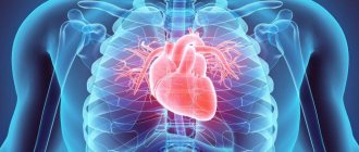
Heart
The heart is a muscular pumping organ located medially in the thoracic region. The lower end of the heart rotates to the left, so that about a little more than half the heart is on the left side of the body and the rest is on the right. The top of the heart, known as the base of the heart, connects the body's large blood vessels: the aorta, vena cava, pulmonary trunk, and pulmonary veins. There are 2 main circles of blood circulation in the human body: the lesser (pulmonary) circulation circle and the systemic circulation circle. The pulmonary circulation transports venous blood from the right side of the heart to the lungs, where the blood is oxygenated and returned to the left side of the heart. The pumping chambers of the heart that support the pulmonary circulation are the right atrium and the right ventricle. The systemic circulation carries highly oxygenated blood from the left side of the heart to all tissues of the body (except the heart and lungs). The systemic circulation removes waste from body tissues and removes venous blood from the right side of the heart. The left atrium and left ventricle of the heart are the pumping chambers for the Greater Circulation.
Circulation circles
These are closed pathways of vascular blood flow. There are two circuits of blood circulation that begin to function shortly after birth:
- The large circle connects the heart with all organs, ensuring metabolism;
- The small circle covers only the lungs and is the main link in a vital process - gas exchange.
Blood circulation begins with myocardial contraction, and gas exchange begins with inhalation.
Big circle
Contraction of the left ventricular chamber promotes the release of blood into the aorta. The branches of the aorta carry it to all tissues, branching all the way to the capillaries.
Here the blood delivers nutritional molecules of oxygen, proteins, fats and carbohydrates to the organs. Enriched with carbon dioxide from them, it becomes venous and enters the veins.
As they approach the heart, the veins unite into increasingly larger vessels until they form the last two venous trunks - the “vena cava”. From them, blood enters the right atrial chamber and descends into the ventricle of the same name.
Small circle
From the right ventricular chamber, blood moves to the pulmonary trunk, which is divided into two branches: right (goes to the right lung) and left (goes to the left lung). Exhalation removes carbon dioxide from the lungs.
Inhalation comes. The blood is again enriched with oxygen and moves to the left side of the heart. The left ventricle contracts - and the whole cycle repeats again.
The diagram of the systemic and pulmonary circulation of the heart is discussed in the video clip:
Normal values
- The time of blood movement (one blood circulation cycle) normally takes 25-30 seconds;
- A complete cardiac cycle occurs in 0.8 seconds, of which 0.45 seconds are contraction and 0.35 seconds are relaxation;
- The normal heart rate is 60-80 beats per minute;
- The average number of respiratory movements is normally 12-16 per minute. Moreover, for most people, exhalation is twice as long as inhalation;
- In one breath, the lungs absorb approximately 500 ml of air (100 ml of oxygen).
The heart is an organ that does not work continuously. Exactly half of its life the heart rests, and the organ that works non-stop is the brain.
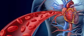
Blood vessels
Blood vessels are the body's highways that allow blood to flow quickly and efficiently from the heart to every area of the body and back. The size of blood vessels corresponds to the amount of blood that passes through the vessel. All blood vessels contain a hollow area called a lumen through which blood can flow in one direction. The area around the lumen is the vessel wall, which can be thin in the case of capillaries or very thick in the case of arteries. All blood vessels are lined by a thin layer of simple squamous epithelium known as endothelium , which keeps blood cells inside the blood vessels and prevents clots. The endothelium lines the entire circulatory system, all the pathways of the inside of the heart, where it is called endocardium .
Types of vasculitis
Vasculitis is classified according to the 2012 Chapel Hill Consensus Conference (CHCC) as follows:
By formation:
Primary - the development of the disease is caused by inflammation of the walls of the blood vessels themselves;
Secondary - the development of the disease is due to the reaction of blood vessels against the background of other diseases. Secondary can be:
- Vasculitis associated with hepatitis B virus (HBV);
- Cryoglobulinemic vasculitis associated with hepatitis C virus (HCV);
- Vasculitis associated with syphilis;
- ANCA vasculitis (ANCA) associated with medications;
- Drug-associated immune complex vasculitis;
- Vasculitis associated with cancer (syn. “paraneoplastic vasculitis”)
- Other vasculitis.
By localization:
Vasculitis of large blood vessels:
— Giant cell arteritis (GCA, Horton's disease, temporal arteritis, senile arteritis) is an autoimmune disease characterized by granulomatous inflammation of the main branches of the aorta, most often the branches of the carotid and temporal arteries. In many cases, it is combined with polymyalgia rheumatica, pain and some stiffness in the joints of the pelvic girdle and shoulders, as well as an increase in ESR. The cause is considered to be human infection with hepatitis, herpes, influenza and other viruses. It occurs mainly in people over 50 years of age.
Read also Zika virus. Symptoms, causes, infection and prevention
— Takayasu arteritis (nonspecific aortoarteritis) is an autoimmune disease in which a productive inflammatory process develops in the walls of the aorta and its branches, leading them to obliteration. As the disease progresses, pathological processes such as the formation of fibrous granulomas, destruction of elastic fibers, necrotization of smooth muscle cells of the blood vessel wall are observed, after which, after some time, thickening of the intima and medial tunic of the vessel is possible. Sometimes there may be no pulse in the hands, which is why the disease has another name - “pulseless disease.” According to statistics, Takayasu arteritis most often develops in women, in an approximate proportion of men 8 to 1, and patients are young people, from 15 to 30 years old.
Vasculitis of medium-sized vessels:
— Periarteritis nodosa (polyarteritis nodosa, periarteritis nodosa) is an inflammatory disease of the arterial wall of small and medium-sized blood vessels, leading to the development of aneurysms, thrombosis, and heart attacks. At the same time, there is no kidney damage (glomerulonephritis). The main causes are considered to be intolerance to certain medications, as well as persistence of the hepatitis B virus (HBV).
— Kawasaki disease is an acute and febrile disease characterized by inflammatory damage to the walls of small, medium and large-diameter veins and arteries, which is often combined with mucocutaneous lymphatic syndrome. Occurs mainly in children.
Vasculitis of small vessels:
— ANCA-associated vasculitis (AAV):
- Microscopic polyangiitis (MPA) is a not fully understood disease that is associated with the production of antibodies to the cytoplasm of neutrophils, which causes an inflammatory process to develop simultaneously in several organs (most often the lungs and kidneys become victims), and granulomas do not form. Doctors note the following features of the clinical course of GPA: the development of severe pulmonary-renal syndrome (about 50%), damage to the kidneys (about 90%), lungs (from 30 to 70%), skin (about 70%), and visual organs (about 30%). ), peripheral nervous system (about 30%), gastrointestinal tract (about 10%).
- Granulomatosis with polyangiitis (GPA, Wegener's granulomatosis) is a severe and rapidly progressive autoimmune disease characterized by granulomatous inflammation of the walls of small and medium-sized blood vessels (capillaries, venules, arterioles and arteries) involving the eyes, upper respiratory tract, and lungs in the pathological process , kidneys and other organs. If not adequately treated, it can lead to death within 1 year. Doctors note the following features of the clinical course of GPA: damage to the upper respiratory tract (90% or more), kidneys (about 80%), lungs (from 50 to 70%), visual organs (about 50%), skin (from 25 to 35% ), peripheral nervous system (20 to 30%), heart (20% or less), gastrointestinal tract (about 5%).
- Eosinophilic granulomatosis with polyangiitis (EGPA, Churg-Strauss syndrome) is an autoimmune disease caused by an excess of eosinophils in the blood and outside the bloodstream, characterized by granulomatous inflammation of the walls of small and medium-sized vessels involving the upper respiratory tract, lungs, kidneys and other organs in the pathological process. Often accompanied by bronchial asthma, runny nose and other sinusitis, elevated body temperature, shortness of breath, eosinophilia.
— Immune complex vasculitis of small vessels:
- Immunoglobulin-A associated vasculitis (hemorrhagic vasculitis, Henoch-Schönlein purpura, Henoch-Schönlein disease, allergic purpura);
- cryoglobulinemic vasculitis - characterized by damage to the walls of small vessels, mainly the kidneys and skin, the main cause of which is an excessive amount of cryoglobulins in the blood serum, due to which they first settle on the walls of the vessels and then modify them.
- Hypocomplementary urticarial vasculitis (anti-C1q vasculitis);
- Anti-GBM disease.
Vasculitis that can affect blood vessels of varying sizes:
- Behçet's disease is characterized by an inflammatory process in the arteries and veins of small and medium caliber, accompanied by frequent relapses of ulcerative formations on the mucous membranes of the mouth, eyes, skin, genitals, as well as damage to the lungs, kidneys, stomach, brain and other organs.
- Cogan syndrome.
Systemic vasculitis:
- Hemorrhagic vasculitis (Henoch-Schönlein purpura) is characterized by aseptic inflammation of the walls of small vessels (arterioles, venules and capillaries), multiple microthrombosis, developing mainly in the vessels of the skin, kidneys, intestines and other organs. Often accompanied by arthralgia and arthritis. The main cause of hemorrhagic vasculitis is the excessive accumulation of circulating immune complexes in the bloodstream, in which antigens predominate, due to which they settle on the inner surface of the blood wall (endothelium). After re-activation of proteins, the vascular wall changes;
- Lupus vasculitis;
- Behçet's disease;
- Rheumatoid vasculitis;
- Vasculitis in sarcoidosis;
- Takayasu arteritis;
- Other vasculitis.
Read also Endocarditis - symptoms, types, causes and treatment of endocarditis
Vasculitis of individual organs:
- Cutaneous arteritis;
- Cutaneous leukocytclastic angiitis - characterized by an isolated inflammatory process of blood vessels in the skin, without concomitant glomerulonephritis or systemic vasculitis;
- Primary angiitis of the central nervous system;
- Isolated aortritis;
- Other vasculitis.
Types of Blood Vessels
There are three main types of blood vessels: arteries, veins and capillaries . Blood vessels are often called so because they are located in an area of the body through which they carry blood or from structures adjacent to them. For example, the brachiocephalic artery carries blood to the brachial (arm) and forearm regions. One of its branches, the subclavian artery , passes under the collarbone: hence the name subclavian artery. The subclavian artery passes into the region of the axilla, where it becomes known as the axillary artery . Arteries and Arterioles: Arteries are blood vessels that carry blood away from the heart. Blood is carried through the arteries, usually highly oxygenated, leaving the lungs on its way to the body's tissues. The arteries of the pulmonary trunk and the arteries of the pulmonary circulation are an exception to this rule - these arteries carry venous blood from the heart to the lungs to saturate it with oxygen.
Structure and functions of the human venous system
The purpose of venules and veins is to return blood back to the heart. From tiny capillaries, blood flows into small venules, and from there into larger veins. Since the pressure in the venous system is much lower than in the arterial system, the walls of the vessels here are much thinner. However, the walls of the veins are also surrounded by elastic muscle tissue, which, by analogy with arteries, allows them to either strongly narrow, completely blocking the lumen, or expand greatly, in this case acting as a reservoir for blood. A feature of some veins, for example in the lower extremities, is the presence of one-way valves, the task of which is to ensure the normal return of blood to the heart, thereby preventing its outflow under the influence of gravity when the body is in an upright position.
Structure of the human venous system: 1-subclavian vein; 2-internal mammary vein; 3-axillary vein; 4-lateral vein of the arm; 5-brachial veins; 6-intercostal veins; 7-medial vein of the arm; 8-median ulnar vein; 9-sternoepigastric vein; 10-lateral vein of the arm; 11-ulnar vein; 12-medial vein of the forearm; 13-epigastric inferior vein; 14-deep palmar arch; 15-superficial palmar arch; 16 palmar digital veins; 17-sigmoid sinus; 18-external jugular vein; 19-internal jugular vein; 20-inferior thyroid vein; 21 pulmonary arteries; 22-heart; 23-inferior vena cava; 24 hepatic veins; 25 renal veins; 26-abdominal vena cava; 27-sperm vein; 28-common iliac vein; 29-perforating branches; 30-external iliac vein; 31-internal iliac vein; 32-external genital vein; 33-deep femoral vein; 34-great vein of the leg; 35-femoral vein; 36 accessory vein of the leg; 37-superior genicular veins; 38-popliteal vein; 39-inferior knee veins; 40-great vein of the leg; 41-small vein of the leg; 42-anterior/posterior tibial vein; 43-deep plantar vein; 44-dorsal venous arch; 45 dorsal metacarpal veins.
Arteries
The arteries face high blood pressure levels as they carry blood from the heart with great force. To withstand this pressure, the walls of the arteries are thicker, more elastic and more muscular than those of other vessels. The body's largest arteries contain a high percentage of elastic tissue, which allows them to stretch and accommodate the pressure of the heart. Smaller arteries are more muscular in the structure of their walls. Smooth muscle in the artery walls dilates the channel to regulate the flow of blood passing through their lumen. In this way, the body controls how much blood flow to different parts of the body under different circumstances. Regulating blood flow also affects blood pressure because smaller arteries provide less cross-sectional area, hence increasing the pressure of blood on the artery walls.
What is blood pressure?
Blood pressure is the force with which blood presses against the walls of the arteries. It increases when the heart contracts and pumps out blood, and decreases when the heart muscle relaxes. Blood pressure is stronger in the arteries and weaker in the veins.
Blood pressure is measured with a special device - a tonometer
.
Pressure readings are usually recorded in two numbers. Thus, normal blood pressure for an adult is considered to be 120/80
.
The first number is systolic pressure
- This is an indicator of pressure during heart contraction.
The second is diastolic pressure
- the pressure during relaxation of the heart.
Such dangerous excess weight
Professor Sergei Boytsov, chief specialist in preventive medicine at the Russian Ministry of Health and Social Development, talks about how extra pounds affect the heart and blood vessels.
Pressure is measured in the arteries and expressed in millimeters of mercury. In the capillaries, the pulsation of the heart becomes invisible and the pressure in them drops to approximately 30 mm Hg. Art.
A blood pressure reading can tell your doctor how your heart is working. If one or both numbers are higher than normal, this indicates high blood pressure. If it’s lower, it means it’s reduced.
High blood pressure indicates that the heart is working too hard: it requires more effort to push blood through the vessels.
It also indicates that a person has an increased risk of heart disease.
Capillaries
They are the smallest and thinnest blood vessels in the body and the most common. They can be found throughout almost all body tissues of the body. Capillaries connect to arterioles on one side and venules on the other side. Capillaries carry blood very close to the cells of the body's tissues for the purpose of exchanging gases, nutrients and waste products. The capillary walls consist of only a thin layer of endothelium, so this is the smallest possible size of the vessels. The endothelium acts as a filter to keep blood cells inside blood vessels while allowing fluids, dissolved gases, and other chemicals to diffuse along their concentration gradients out of the tissues. Precapillary sphincters are bands of smooth muscle found at the arteriole ends of capillaries. These sphincters regulate blood flow in the capillaries. Because there is a limited supply of blood and not all tissues have the same energy and oxygen requirements, precapillary sphincters reduce blood flow to inactive tissues and allow free flow in active tissues.
Veins and venules
Veins and venules are mostly return vessels of the body and act to ensure the return of blood to the arteries. Because the arteries, arterioles, and capillaries absorb most of the force of the heart's contractions, the veins and venules are subject to very low blood pressure. This lack of pressure allows the walls of the veins to be much thinner, less elastic, and less muscular than the walls of the arteries. Veins use gravity, inertia, and the strength of skeletal muscles to push blood toward the heart. To facilitate the movement of blood, some veins contain many one-way valves that prevent blood from flowing away from the heart. The body's skeletal muscles also squeeze the veins and help push blood through the valves closer to the heart.
When the muscle relaxes, the valve traps blood while the other pushes the blood closer to the heart. Venules are similar to arterioles in that they are small vessels that connect capillaries, but unlike arterioles, venules connect to veins instead of arteries. Venules take blood from many capillaries and place it in larger veins for transport back to the heart.
What have we learned about veins and arteries?
The veins and arteries of the human body are the basis of the vascular system. They have different structures because they must perform different functions. The artery carries oxygenated blood to the body's organs directly from the heart. To ensure the movement of blood, myocardial contractions are used, so the movement becomes intense and can reach a speed of 25 cm/sec.
In the veins, blood moves directly from the organs themselves to the heart. It is poor in oxygen, but it contains a lot of carbon dioxide and other decay products. The movement of the venous occurs due to the structure of the vessel. That is why the speed here is an order of magnitude lower. At the same time, the percentage of venous blood is 64%, while arterial blood accounts for only 14%.
Liver circulation
The veins of the stomach and intestines serve a unique function: instead of carrying blood directly back to the heart, they carry blood to the liver through the hepatic portal vein. The blood that passes through the digestive organs is rich in nutrients and other chemicals absorbed from food. The liver removes toxins, stores sugar, and processes digestive products before they reach other body tissues. Blood from the liver then returns to the heart through the inferior vena cava.
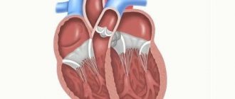
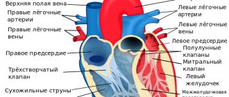
Structure and functions of the human heart
The heart is a kind of pump, consisting of two ventricles, which are interconnected and at the same time independent of each other. The right ventricle pumps blood through the lungs, the left ventricle pumps it through the rest of the body. Each half of the heart has two chambers: the atrium and the ventricle. You can see them in the image below. The right and left atria act as reservoirs from which blood flows directly into the ventricles. Both ventricles, at the moment of contraction of the heart, push out blood and drive it through the system of pulmonary and peripheral vessels.
Structure of the human heart: 1-pulmonary trunk; 2-pulmonary valve; 3-superior vena cava; 4th right pulmonary artery; 5th right pulmonary vein; 6-right atrium; 7-tricuspid valve; 8-right ventricle; 9-inferior vena cava; 10-descending aorta; 11-aortic arch; 12 left pulmonary artery; 13th left pulmonary vein; 14 left atrium; 15-aortic valve; 16-mitral valve; 17th left ventricle; 18-interventricular septum.
Blood
On average, the human body contains approximately 4 to 5 liters of blood. Acting as a fluid connective tissue, it transports many substances through the body and helps maintain the homeostasis of nutrients, wastes and gases. Blood consists of red blood cells, white blood cells, platelets and liquid plasma.
Erythrocytes , red blood cells, are by far the most common type of blood cell and make up about 45% of blood volume. Red blood cells are produced within the red bone marrow from stem cells at an astonishing rate of about 2 million cells every second. The shape of red blood cells is biconcave discs with a concave curve on both sides of the disc so that the center of the red blood cell is the thin part. The unique shape of red blood cells gives these cells a high surface area to volume ratio and allows them to fold to fit in thin capillaries. Immature red blood cells have a nucleus that is pushed out of the cell when it reaches maturity to provide it with its unique shape and flexibility. The absence of a nucleus means that red blood cells do not contain DNA and are unable to repair themselves once damaged. Red blood cells carry oxygen in the blood using the red pigment hemoglobin. Hemoglobin contains iron and proteins linked together, they can significantly increase the oxygen carrying capacity. The high surface area relative to the volume of red blood cells allows oxygen to be easily transferred into lung cells and from tissue cells into capillaries.
White blood cells, also known as leukocytes , make up a very small percentage of the total number of cells in the blood, but have important functions in the body's immune system. There are two main classes of white blood cells: granular leukocytes and agranular leukocytes.
Three types of granular leukocytes:
neutrophils, eosinophils and basophils. Each type of granular leukocyte is classified by the presence of vesicle-filled cytoplasms that give them their functions. Neutrophils contain digestive enzymes that neutralize bacteria that enter the body. Eosinophils contain digestive enzymes to digest specialized viruses that have been bound to antibodies in the blood. Basophils - amplifiers of allergic reactions - help protect the body from parasites. Agranular leukocytes: The two main classes of agranular leukocytes are lymphocytes and monocytes. Lymphocytes include T cells and natural killer cells, which fight viral infections, and B cells, which produce antibodies against pathogen infections. Monocytes develop into cells called macrophages, which capture and ingest pathogens and dead cells from wounds or infections.
Platelets are small cell fragments responsible for blood clotting and crust formation. Platelets are formed in the red bone marrow from large megakaryocyte cells that periodically rupture to release thousands of pieces of membrane that become platelets. Platelets do not contain a nucleus and only survive in the body for a week before being captured by macrophages, which digest them.
Plasma is the non-porous or liquid part of the blood that makes up about 55% of blood volume. Plasma is a mixture of water, proteins and solutes. About 90% of plasma consists of water, although the exact percentage varies depending on the individual's hydration level. Proteins within plasma include antibodies and albumin. Antibodies are part of the immune system and bind to antigens on the surface of pathogens that infect the body. Albumin helps maintain osmotic balance in the body, providing an isotonic solution for the body's cells. Many different substances can be found dissolved in plasma, including glucose, oxygen, carbon dioxide, electrolytes, nutrients, and cellular waste products. The function of plasma is to provide a transport medium for these substances as they move throughout the body.
Treatment of vasculitis
The effectiveness of treatment for vasculitis largely depends on timely and accurate diagnosis, treatment of affected organs and concomitant diseases.
In some cases, the disease goes away on its own, as is the case with primary allergic vasculitis. Complex therapy for vasculitis includes:
1. Drug treatment; 2. Physiotherapeutic methods of treatment; 3. Diet; 4. Preventive measures (at the end of the article).
Important! Before using medications, be sure to consult your doctor!
Drug treatment of vasculitis
Drug treatment of systemic vasculitis is aimed at the following goals:
- Suppression of immunopathological reactions that are the basis of the disease;
- Maintaining stable and long-term remission;
- Treatment of disease relapses;
- Preventing the development of secondary diseases and complications;
Medicines for vasculitis:
Glucocorticoids are a group of hormonal drugs that have anti-inflammatory, antiallergic, immunoregulatory, anti-stress, anti-shock and other properties. In this case, these hormones play one of the most important roles in the treatment of giant cell arteritis (GCA) and Takayasu arteritis, contributing in many cases to achieving stable and long-term remission. In the case of a very rapid response to glucocorticoids, the response can be considered as an additional diagnostic feature of GCA and polymyalgia rheumatica (PPR).
Among the glucocorticoids we can distinguish: Prednisolone, Hydrocortisone.
Cytostatic drugs (cytostatics) are a group of antitumor drugs that disrupt and slow down the mechanisms, division, growth and development of all cells of the body, which is especially important in the presence of malignant (cancerous) tumors. Also effective for nephritis.
Good effectiveness in therapy should be given to the simultaneous use of cytostatics with glucocorticoids, especially in cases of treatment of vasculitis such as ANCA, urticarial, hemorrhagic, cryoglobulinemic, giant cell arteritis, Takayasu arteritis. The duration of taking cytostatics ranges from 3 to 12 months.
Among the cytostatics we can highlight: Cyclophosphamide, Methotrexate, Doxorubicin, Fluorouracil.
Monoclonal antibodies are antibodies produced by immune cells that have immunosuppressive and antitumor properties and have been shown to be effective against skin cancer (melanoma), breast cancer and lymphatic leukemia. Medicines from the group of monoclonal antibodies are no less effective than cytostatics and are used in the treatment of ANCA vasculitis. The appointment is advisable in case of undesirable use of cytostatic drugs. Contraindications for use are the presence of hepatitis B virus (HBV), a positive intradermal tuberculin test, neutropenia, as well as low levels of IgG (immunoglobulins class G) in the blood.
Among the monoclonal antibodies against vasculitis are: Rituximab.
Immunosuppressants are a group of drugs that suppress the action of the immune system. Prescribed in combination with glucocorticoids.
Among the monoclonal antibodies against vasculitis are: “Azathioprine”, “Mycophenolate mofetil”.
If there are contraindications to Azathioprine, Leflunomide may be prescribed.
"Mycophenolate mofetil" is prescribed as an alternative treatment for patients with refractory or recurrent systemic vasculitis, for example, with kidney damage, however, with an increase in ALT and AST in the peripheral blood by 3 times or more, as well as a decrease in platelets (100×109/l ) and leukocytes (2.5×10⁹/l), the drug is stopped.
Normal human immunoglobulin - prescribed for severe kidney damage, infectious complications, hemorrhagic alveolitis.
Anti-infectious therapy – used for diseases of infectious etiology or concomitant infectious diseases.
If bacteria are present, antibacterial drugs are prescribed - Trimethoprim, Sulfamethoxazole.
Read also Vegetative-vascular dystonia (VSD). Symptoms, causes, types and treatment of VSD
If viruses are present, antiviral drugs are prescribed - Interferon alfa, Vidarabine, Lamivudine.
For hepatitis, drugs are prescribed by a hepatologist, depending on the type of hepatitis virus.
Detoxification therapy – used to bind toxins and biologically active substances, as well as remove them from the body.
The following detoxification drugs can be identified: Atoxil, Nutriclins, Polyphepan, Enterosgel.
Non-steroidal anti-inflammatory non-steroidal drugs (NSAIDs) - are prescribed for persistent and necrotic lesions, thrombophlebitis and nodular vasculitis.
Among NSAIDs one can highlight: Acetylsalicylic acid, Ibuprofen, Indomethacin, Phenylbutazone.
Anticoagulants are a group of drugs that prevent the formation of blood clots in blood vessels.
Anticoagulants include Warfarin and Heparin.
Antihistamine therapy is used if the patient is allergic to medications or foods.
Antihistamines include Diazolin, Claritin, Tavegil, Terfen, and Fenkarol.
Treatment of vasculitis in children also includes transfusion therapy.
Can also be used:
- Hemosorption;
- ACE inhibitors;
- Vasodilators.
2. Physiotherapeutic methods for treating vasculitis
Plasmapheresis – helps improve kidney function, reducing the risk of developing end-stage renal failure.
Diet for vasculitis
The diet for vasculitis is hypoallergenic, which is important if the disease is of an allergic nature.
Products from the highly allergenic group are excluded from the diet - eggs, whole milk, seafood (shellfish), caviar, strawberries, wild strawberries, bananas, melon, mango, persimmon, citrus fruits, tomatoes, carrots, bell peppers, nuts, coffee, cocoa, chocolate, baked goods , industrial canned food, as well as individually intolerable food products.
In case of severe nephritis, diet No. 7 is prescribed, with a further gradual transition to a hypochloride diet. For abdominal syndrome, the use of diet No. 1 is indicated.
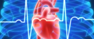
Functions of the cardiovascular system
The cardiovascular system has 3 main functions: transporting substances, protecting against pathogenic microorganisms and regulating the homeostasis of the body. Transport - it transports blood throughout the body. The blood delivers important substances with oxygen and removes waste with carbon dioxide, which will be neutralized and removed from the body. Hormones are carried throughout the body by liquid blood plasma. Protection - the vascular system protects the body with the help of its white blood cells, which are designed to cleanse cellular waste products. White cells are also designed to fight pathogenic microorganisms. Platelets and red blood cells form clots that can prevent the entry of pathogens and prevent fluid leaks. Blood carries antibodies that provide an immune response. Regulation is the body's ability to maintain control over several internal factors.
Functions
In the human body, the circulatory system performs several functions. The main one - transport - consists of the delivery of biological fluid to all organs and tissues, and the removal of metabolic products. Its functional purposes also include additional subfunctions:
- protective - blood components provide cellular and humoral protection against the penetration of foreign bodies;
- respiratory - thanks to blood, gas exchange occurs in tissues and organs;
- nutritional - the circulatory system is the main way of delivering nutrients to tissues and organs;
- excretory - delivery of metabolic products to the lungs and kidneys, where they are processed and excreted into the external environment;
- thermoregulatory - the circulatory system is capable of equalizing the temperature of the body to prevent hyper- and hypothermia of individual parts of the body or organs.
Another subfunction that determines the physiology of the cardiovascular system is the regulatory function. The circulatory system is considered the main transport route through which hormones, enzymes and other biological substances synthesized by internal organs, glands and tissues move. These compounds, in turn, can affect the functions of the cardiovascular system. For example, the release of adrenaline increases cardiac output, constricts peripheral blood vessels and directs major volumes of blood to vital organs: the heart, brain, and skeletal muscles.
Blood Pressure Regulation
The cardiovascular system can control blood pressure. Some hormones, along with autonomic nerve signals from the brain, affect the speed and force of heart contractions. An increase in contractile force and heart rate leads to an increase in blood pressure. Blood vessels can also affect blood pressure. Vasoconstriction reduces the diameter of the artery by contracting the smooth muscle in the artery walls. Sympathetic (fight or flight) activation of the autonomic nervous system causes blood vessels to constrict, resulting in increased blood pressure and decreased blood flow to the constricted area. Vasodilation is the expansion of smooth muscle in the walls of arteries. The volume of blood in the body also affects blood pressure. Higher blood volume in the body increases blood pressure by increasing the amount of blood pumped by each heartbeat. More viscous blood due to a clotting disorder can also increase blood pressure.
Hemostasis
Hemostasis, or blood clotting and crust formation, is controlled by blood platelets. Platelets usually remain inactive in the blood until they reach damaged tissue or begin to leak out of blood vessels through a wound. Once the active platelets become ball-shaped and very sticky, they cover the damaged tissue. Platelets begin to produce the protein fibrin to act as a structure for the clot. The platelets also begin to clump together to form a blood clot. The clot will serve as a temporary seal to keep blood in the vessel until the blood vessel cells can repair the damage to the vessel wall.
Some numbers
There are more than 95 thousand kilometers of blood vessels in the body of a healthy adult. More than seven thousand liters of blood are pumped through them every day.
The size of blood vessels varies from 25 mm
(aortic diameter)
to eight microns
(capillary diameter).
Down with atherosclerosis!
Atherosclerosis is one of the most common vascular diseases. It is characterized by the formation of fatty plaques on the walls of the arteries. Find out how to prevent atherosclerosis.